. 2010 Feb;67(4):655-69.
doi: 10.1007/s00018-009-0202-4. Epub 2009 Nov 24.

Intra-brain microinjection of human mesenchymal stem cells decreases allodynia in neuropathic mice

Affiliations

Intra-brain microinjection of human mesenchymal stem cells decreases allodynia in neuropathic mice

Dario Siniscalco et al. Cell Mol Life Sci. 2010 Feb.

Abstract

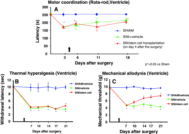
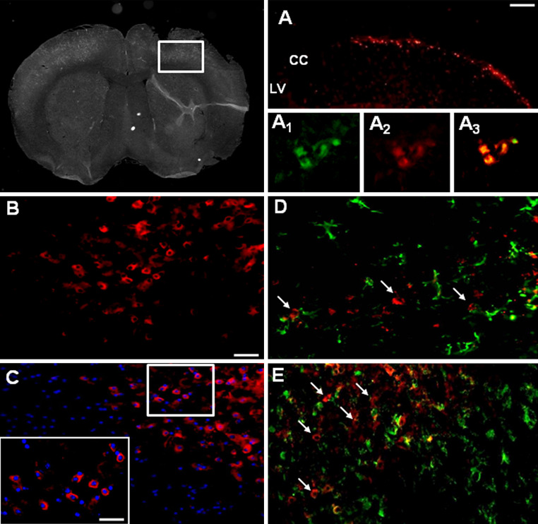
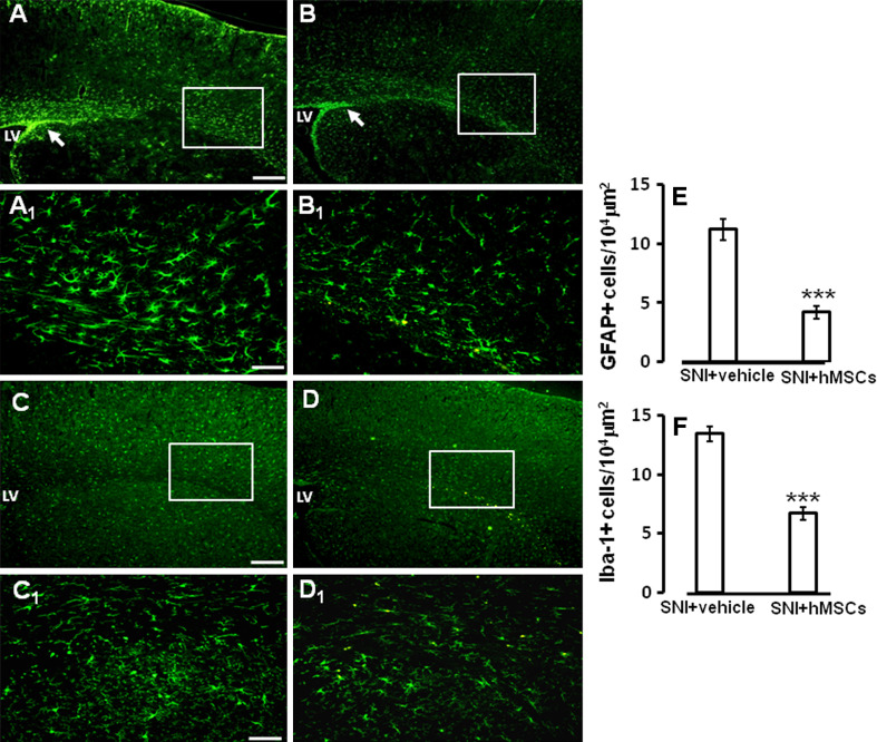
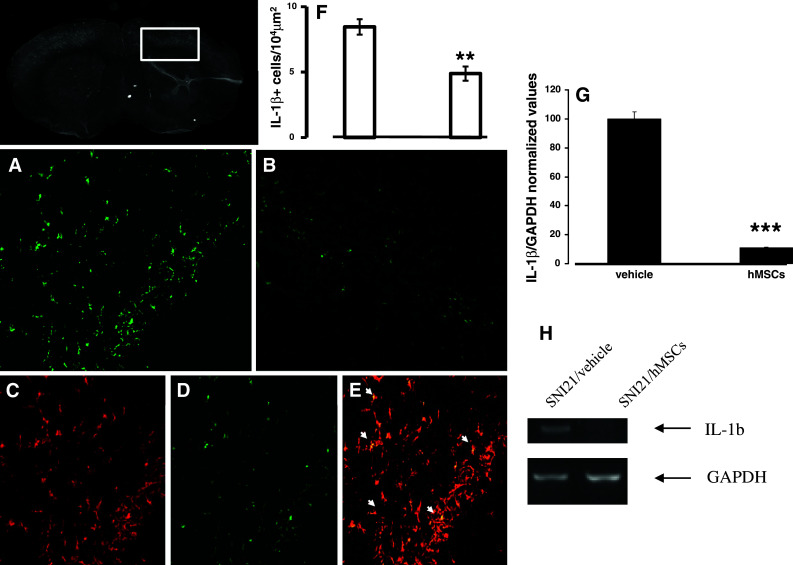
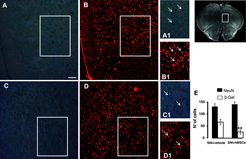
Neuropathic pain is a very complex disease, involving several molecular pathways. Current available drugs are usually not acting on the several mechanisms underlying the generation and propagation of pain. We used spared nerve injury model of neuropathic pain to assess the possible use of human mesenchymal stem cells (hMSCs) as anti-neuropathic tool. Human MSCs were transplanted in the mouse lateral cerebral ventricle. Stem cells injection was performed 4 days after sciatic nerve surgery. Neuropathic mice were monitored 7, 10, 14, 17, and 21 days after surgery. hMSCs were able to reduce pain-like behaviors, such as mechanical allodynia and thermal hyperalgesia, once transplanted in cerebral ventricle. Anti-nociceptive effect was detectable from day 10 after surgery (6 days post cell injection). Human MSCs reduced the mRNA levels of the pro-inflammatory interleukin IL-1beta mouse gene, as well as the neural beta-galactosidase over-activation in prefrontal cortex of SNI mice. Transplanted hMSCs were able to reduce astrocytic and microglial cell activation.


Figures

Fig. 1
Fig. 1
a Rotarod motor testing results. The effects of human mesenchymal stem cell injection on motor performance in the rotarod test is shown. Intra-brain administration of hMSCs in ventricle of SNI-mice had no effect on motor function compared with vehicle-treated SNI-mice. Human MSCs were transplanted on day 4 after the SNI surgery. Results are expressed as the mean ± SEM of the latency (s) (n = 5 mice/group). *P < 0.05 vs sham/vehicle. b Effects of human mesenchymal stem cells on reflex withdrawal responses (s, mean ± SEM) to thermal noxious stimuli in SNI mice. The onset of SNI-induced thermal hyperalgesia was evaluated in ipsilateral sides at 7, 10, 14, 17, and 21 days post-SNI. SNI mice showed a significant reduction in withdrawal latency to radiant heat in the ipsilateral paw (*P < 0.05 vs sham-operated mice). Human MSC treatment (on day 4 after SNI surgery, as indicated by the arrow) prevented the appearance of thermal hyperalgesia on day 21 after SNI (°P < 0.05 vs SNI mice). c Effects of human mesenchymal stem cells on reflex withdrawal responses (g, mean ± SEM) to mechanical noxious stimuli in SNI mice. The onset of SNI-induced mechanical allodynia was evaluated in ipsilateral sides at 7, 10, 14, 17, and 21 days post-SNI. Mice showed a significant reduction in the threshold to mechanical stimulation in the ipsilateral paw (*P < 0.05 vs sham-operated mice) after SNI surgery. Human MSC treatment (on day 4 after SNI surgery, as indicated by the arrow) prevented the appearance of mechanical allodynia at 7, 10, 14, 17, and 21 days post-SNI (°P < 0.05 vs SNI mice)
Fig. 2
Fig. 2
Representative cross-section of mouse whole midbrain area from hMSC-treated 21 days neuropathic mice. a DiI-labeled hMSCs homed at the injection site after transplantation, with a possible stem cell migration along the corpus callosum; LV lateral ventricle, CC corpus callosum. a1 Human MSCs expressed lineage-specific antigens at the time of injection in vivo. Representative fluorescent photomicrograph of hMSCs showing immunocytochemistry for CD73. CD73-positive hMSCs emitted green fluorescence. a2 DiI-labeled hMSCs emitted red fluorescence. a3 Pictures with both green and red fluorescence were merged. b Area in white rectangle inset is shown: CD73 positive hMSCs emitting red fluorescence. c The cell nuclei were counterstained with Hoechst 33258 (blue fluorescence), the inset represents ×40 magnification of the area in white rectangle (×20 magnification). d Double labeling of GFAP and CD73 positive profiles. Arrows indicate no merging between astrocytes and hMSCs. e Double labeling of Iba-1 and CD73 positive profiles. Arrows indicate no merging between microglial cells and hMSCs. Scale bars 100 μm (a), 50 μm (be) and 25 μm (inset in c)
Fig. 3
Fig. 3
a GFAP labeling of ventricle and somatosensory forelimb cortex from vehicle-treated 21 days-neuropathic mice. Notice the increased staining in the periventricular area, as indicated by the arrow; LV lateral ventricle. a1 High magnification of a, showing GFAP positive astrocytes. b GFAP labeling of ventricle and somatosensory forelimb cortex from hMSC-treated 21 days-neuropathic mice. Human MSC treatment was able to decrease GFAP staining in the periventricular area, as indicated by the arrow; LV lateral ventricle. b1 High magnification of b, showing reduced GFAP positive astrocytes. c Iba-1 labeling of ventricle and somatosensory forelimb cortex from vehicle treated neuropathic mice; LV lateral ventricle. c1 High magnification of c, showing Iba-1 positive microglial cells. d Iba-1 labeling of ventricle and somatosensory forelimb cortex from hMSC-treated neuropathic mice. Human MSC treatment was able to decrease Iba-1 staining in the periventricular area; LV lateral ventricle. d1 High magnification of d, showing reduced Iba-1 positive microglial cells. Scale bars 100 μm (a,b), 50 μm (a1, b1), 100 μm (c,d), and 50 μm (c1, d1). e The number of the GFAP positive profiles, indicating a strong reduction in the number of astrocytes after hMSC transplantation in neuropathic mice as compared to vehicle treated neuropathic animals. ANOVA followed by Tukey’s post hoc test, was used to determine the statistical significance among groups. ***P < 0.05 was considered statistically significant. f The number of the Iba-1 positive profiles, indicating a significant reduction in the number of microglial cells after hMSC transplantation in neuropathic mice as compared to vehicle treated neuropathic animals. ANOVA followed by Tukey’s post hoc test, was used to determine the statistical significance among groups. ***P < 0.01 was considered statistically significant
Fig. 4
Fig. 4
Representative cross-section of mouse whole midbrain area from hMSC-treated 21 days-neuropathic mice. a IL-1β positive profiles in the area enclosed by the white perimeter from vehicle treated 21 days-neuropathic mice. Scale bar 50 μm (a,b). b IL-1β positive profiles in the area enclosed by the white perimeter from hMSC-treated neuropathic mice, indicating a reduction in IL-1β profiles after hMSC treatment. c GFAP positive profiles in hMSC-treated neuropathic mice. d IL-1β positive profiles in hMSC-treated neuropathic mice. e Double labeling of GFAP and IL-1β positive profiles. Arrows indicate that the cells expressing IL-1β were astrocytes. f The number of the IL-1β positive profiles, indicating a significant reduction in the number of the cells expressing this interleukin after hMSC transplantation in neuropathic mice as compared to vehicle treated neuropathic animals. ANOVA followed by Tukey’s post hoc test, was used to determine the statistical significance among groups. **P < 0.05 was considered statistically significant. g IL-1β/GAPDH normalized values as obtained by RT-PCR analysis. Human MSC treatment (on day 4 after SNI surgery) reduced the mRNA levels of IL-1β gene in 21 days neuropathic mice (SNI21/hMSCs) respect to vehicle-treated 21 days-neuropathic mice (SNI21/vehicle). ANOVA, followed by Student–Neuman–Keuls post hoc test, was used to determine the statistical significance among groups. ***P < 0.01 was considered statistically significant. h Representative agarose gel blot analysis for IL-1β and housekeeping GAPDH mouse genes following RT-PCR. The semiquantitative analysis of mRNA levels was carried out by the “Gel Doc 2000 UV System” (Bio-Rad, Hercules, CA). Human MSC treatment (on day 4 after SNI surgery) reduced the mRNA levels of IL-1β gene in 21 days-neuropathic mice (SNI21/hMSCs) respect to vehicle-treated 21 days-neuropathic mice (SNI21/vehicle)
Fig. 5
Fig. 5
Representative cross-section of mouse whole prefrontal cortex area from hMSC-treated 21 days-neuropathic mice. a β-Gal positive profiles in the pre-limbic and infra-limbic cortex area from vehicle-treated 21 days-neuropathic mice. b NeuN positive profiles in the same area from vehicle-treated neuropathic mice (a1, b1). A detail enclosed in the white square of β-Gal and NeuN, respectively; arrows indicate the β-Gal positive profiles expressed in the NeuN-labeled neuronal cells. c β-Gal positive profiles in the pre-limbic and infra-limbic cortex area from hMSC-treated 21 days-neuropathic mice. d NeuN positive profiles in the same area from hMSC-treated neuropathic mice (c1, d1). A detail enclosed in the white square of β-Gal and NeuN, respectively; arrows indicate less β-Gal positive profiles expressed in the NeuN-labeled neuronal cells. e The number of the β-Gal positive profiles, indicating a significant reduction in the number of suffering, senescent neuronal cells after hMSC transplantation in neuropathic mice as compared to vehicle-treated neuropathic animals. ANOVA followed by Tukey’s post hoc test, was used to determine the statistical significance among groups. **P < 0.05 was considered statistically significant. Scale bar 50 μm

Similar articles

Cited by

References

    1. Merskey H, Bogduk N. Classification of chronic pain. Seattle: IASP; 1994.
    1. Siniscalco D, de Novellis V, Rossi F, Maione S. Neuropathic pain: is the end of suffering starting in the gene therapy? Curr Drug Targets. 2005;6:75–80. doi: 10.2174/1389450053344966. - DOI - PubMed
    1. de Novellis V, Siniscalco D, Galderisi U, Fuccio C, Nolano M, Santoro L, Cascino A, Roth KA, Rossi F, Maione S. Blockade of glutamate mGlu5 receptors in a rat model of neuropathic pain prevents early over-expression of pro-apoptotic genes and morphological changes in dorsal horn lamina II. Neuropharmacology. 2004;46:468–479. doi: 10.1016/j.neuropharm.2003.10.014. - DOI - PubMed
    1. Siniscalco D, Fuccio C, de Novellis V, Rossi F, Maione S. Molecular methods for neuropathic pain treatment. J Neuropathic Pain. 2005;1(3):35–42. doi: 10.1300/J426v01n03_04. - DOI
    1. Galluzi KE. Management of neuropathic pain. J Am Osteopath Assoc. 2005;105:12–19. - PubMed

LinkOut - more resources