

Impact of alpha-hydroxy-propanodeoxyguanine adducts on DNA duplex energetics: opposite base modulation and implications for mutagenicity and genotoxicity
- PMID: 19937758
- PMCID: PMC2891022
- DOI: 10.1002/bip.21355
Impact of alpha-hydroxy-propanodeoxyguanine adducts on DNA duplex energetics: opposite base modulation and implications for mutagenicity and genotoxicity
Abstract
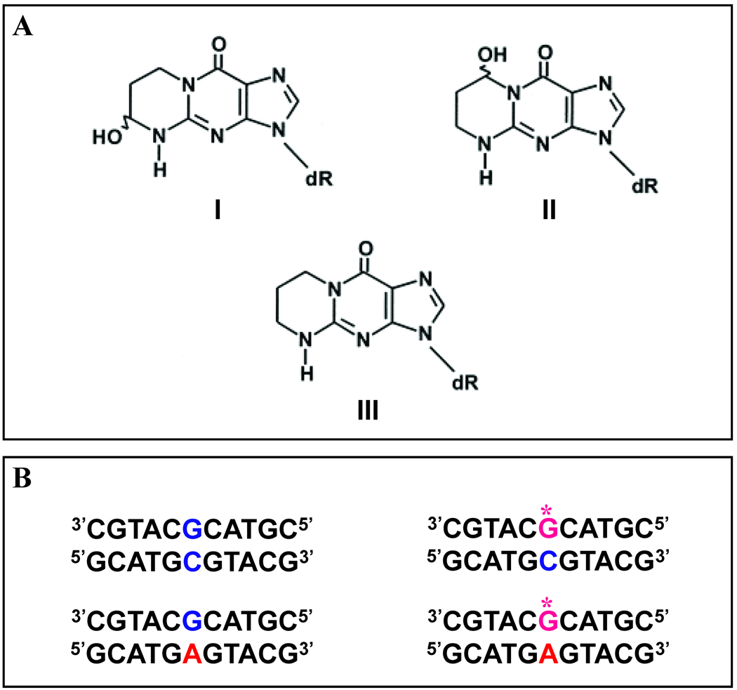
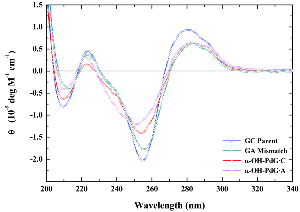
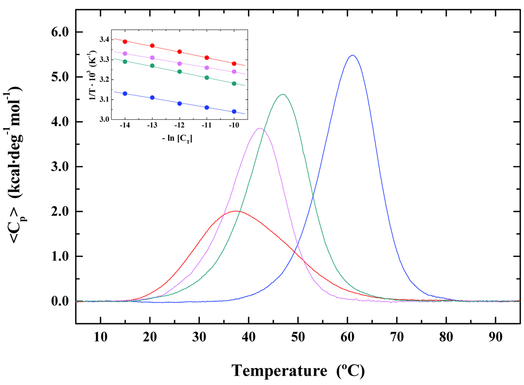
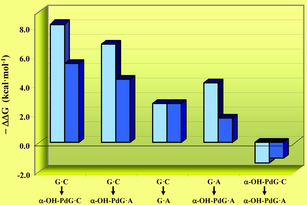
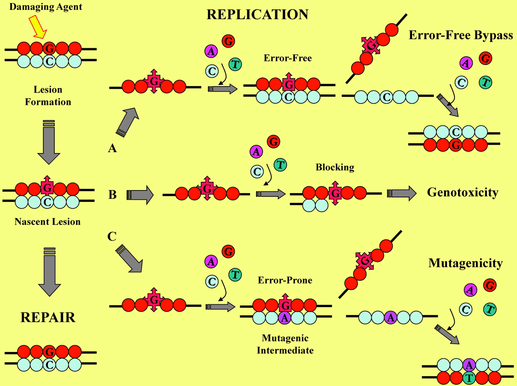
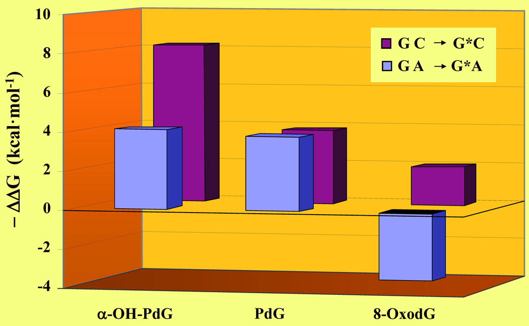
Acrolein is an alpha,beta-unsaturated aldehyde that is a major environmental pollutant, as well as a product of cellular metabolism. DNA bases react with acrolein to form two regioisomeric exocyclic guanine adducts, namely gamma-hydroxy-propanodeoxyguanosine (gamma-OH-PdG) and its positional isomer alpha-hydroxy-propanodeoxyguanosine (alpha-OH-PdG). The gamma-OH-PdG isomer adopts a ring-opened conformation with minimal structural perturbation of the DNA host duplex. Conversely, the alpha-OH-PdG isomer assumes a ring-closed conformation that significantly disrupts Watson-Crick base-pair alignments within the immediate vicinity of the damaged site. We have employed a combination of calorimetric and spectroscopic techniques to characterize the thermodynamic origins of these lesion-induced structural alterations. Specifically, we have assessed the energetic impact of alpha-OH-PdG centered within an 11-mer duplex by hybridizing the adduct-containing oligonucleotide with its complementary strand harboring a central base N [where N = C or A], yielding a pair of duplexes containing the nascent lesion (alpha-OH-PdG.C) or mismatched adduct (alpha-OH-PdG.A), respectively. Our data reveal that the nascent lesion is highly destabilizing, whereas its mismatched counterpart partially ameliorates alpha-OH-PdG-induced destabilization. Collectively, our data provide energetic characterizations of the driving forces that modulate error-free versus error-prone DNA translesion synthesis. The biological implications of our findings are discussed in terms of energetically probing acrolein-mediated mutagenicity versus adduct-induced genotoxicity.
(c) 2009 Wiley Periodicals, Inc.
Figures
Similar articles
-
Solution structure of DNA containing alpha-OH-PdG: the mutagenic adduct produced by acrolein.Nucleic Acids Res. 2009 Apr;37(7):2153-63. doi: 10.1093/nar/gkp076. Epub 2009 Feb 17. Nucleic Acids Res. 2009. PMID: 19223332 Free PMC article.
-
Spectroscopic characterization of interstrand carbinolamine cross-links formed in the 5'-CpG-3' sequence by the acrolein-derived gamma-OH-1,N2-propano-2'-deoxyguanosine DNA adduct.J Am Chem Soc. 2005 Dec 21;127(50):17686-96. doi: 10.1021/ja053897e. J Am Chem Soc. 2005. PMID: 16351098 Free PMC article.
-
NMR structure of duplex DNA containing the alpha-OH-PdG.dA base pair: a mutagenic intermediate of acrolein.Biopolymers. 2010 Apr;93(4):391-401. doi: 10.1002/bip.21366. Biopolymers. 2010. PMID: 20049919 Free PMC article.
-
Chemistry and biology of DNA containing 1,N(2)-deoxyguanosine adducts of the alpha,beta-unsaturated aldehydes acrolein, crotonaldehyde, and 4-hydroxynonenal.Chem Res Toxicol. 2009 May;22(5):759-78. doi: 10.1021/tx9000489. Chem Res Toxicol. 2009. PMID: 19397281 Free PMC article. Review.
-
Effects of 3,N4-ethenodeoxycytidine on duplex stability and energetics.IARC Sci Publ. 1999;(150):169-77. IARC Sci Publ. 1999. PMID: 10626218 Review.
Cited by
-
Novel post-synthetic generation, isomeric resolution, and characterization of Fapy-dG within oligodeoxynucleotides: differential anomeric impacts on DNA duplex properties.Nucleic Acids Res. 2011 Jul;39(13):5776-89. doi: 10.1093/nar/gkr082. Epub 2011 Mar 16. Nucleic Acids Res. 2011. PMID: 21415012 Free PMC article.
-
Forces Driving a Magic Bullet to Its Target: Revisiting the Role of Thermodynamics in Drug Design, Development, and Optimization.Life (Basel). 2022 Sep 15;12(9):1438. doi: 10.3390/life12091438. Life (Basel). 2022. PMID: 36143474 Free PMC article. Review.
-
PAM-OBG: A monoamine oxidase B specific prodrug that inhibits MGMT and generates DNA interstrand crosslinks, potentiating temozolomide and chemoradiation therapy in intracranial glioblastoma.Oncotarget. 2018 May 8;9(35):23923-23943. doi: 10.18632/oncotarget.25246. eCollection 2018 May 8. Oncotarget. 2018. PMID: 29844863 Free PMC article.
-
Aldehyde-Associated Mutagenesis─Current State of Knowledge.Chem Res Toxicol. 2023 Jul 17;36(7):983-1001. doi: 10.1021/acs.chemrestox.3c00045. Epub 2023 Jun 26. Chem Res Toxicol. 2023. PMID: 37363863 Free PMC article. Review.
-
Pathways for repairing and tolerating the spectrum of oxidative DNA lesions.Cancer Lett. 2012 Dec 31;327(1-2):61-72. doi: 10.1016/j.canlet.2012.02.001. Epub 2012 Feb 19. Cancer Lett. 2012. PMID: 22353689 Free PMC article. Review.
References
Publication types
MeSH terms
Substances
Grants and funding
LinkOut - more resources
Full Text Sources
Miscellaneous
