. 2010 May 1;88(6):1317-28.
doi: 10.1002/jnr.22295.

Calpain activation and Na+/Ca2+ exchanger degradation occur downstream of calcium deregulation in hippocampal neurons exposed to excitotoxic glutamate

Affiliations

Calpain activation and Na+/Ca2+ exchanger degradation occur downstream of calcium deregulation in hippocampal neurons exposed to excitotoxic glutamate

Tatiana Brustovetsky et al. J Neurosci Res. .

Abstract

Delayed calcium deregulation (DCD) plays an essential role in glutamate excitotoxicity, a major detrimental factor in stroke, traumatic brain injury, and various neurodegenerations. In the present study, we examined the role of calpain activation and Na(+)/Ca(2+) exchanger (NCX) degradation in DCD and excitotoxic cell death in cultured hippocampal neurons. Exposure of neurons to glutamate caused DCD accompanied by secondary mitochondrial depolarization. Activation of calpain was evidenced by detecting NCX isoform 3 (NCX3) degradation products. Degradation of NCX isoform 1 (NCX1) was below the detection limit of Western blotting. Degradation of NCX3 was detected only after 1 hr of incubation with glutamate, whereas DCD occurred on average within 15 min after glutamate application. Calpeptin, an inhibitor of calpain, significantly attenuated NCX3 degradation but failed to inhibit DCD and excitotoxic neuronal death. Calpain inhibitors I, III, and VI also failed to influence DCD and glutamate-induced neuronal death. On the other hand, MK801, an inhibitor of the NMDA subtype of glutamate receptors, added shortly after the initial glutamate-induced jump in cytosolic Ca(2+), completely prevented DCD and activation of calpain and strongly protected neurons against excitotoxicity. Taken together, our results suggest that, in glutamate-treated hippocampal neurons, the initial increase in cytosolic Ca(2+) that precedes DCD is insufficient for sustained calpain activation, which most likely occurs downstream of DCD.


Figures

Figure 1
Figure 1. Western blot analysis of proteolytic degradation of Na+/Ca2+ exchanger isoform 1 (NCX1) in cultured hippocampal neurons exposed to glutamate
In A, neurons were incubated without or with 100μM glutamate plus 10μM glycine (+Glu) for 20 minutes or an hour as indicated. The same blots were re-probed for glyceraldehyde-3-phosphate dehydrogenase (GAPDH) to ensure equal loading. In B, the columns show the densitometry data normalized by GAPDH loading control, averaged from three independent experiments (mean±SEM) and expressed in arbitrary units (a.u.) for the corresponding bands.
Figure 2
Figure 2. Western blot analysis of proteolytic degradation of Na+/Ca2+ exchanger isoform 3 (NCX3) in cultured hippocampal neurons exposed to glutamate
In A and B, neurons were incubated without or with 100μM glutamate plus 10μM glycine (+Glu) for 20 minutes, 1 hour, or 2 hours. A 55 kDa band represented a major degradation product of NCX3 (Bano et al., 2005). In A and B, protein loading was 5 or 50μg per lane respectively. Where indicated, neurons were pre-treated with 20μM calpeptin for 30 minutes prior to glutamate application. In addition, calpeptin (20μM) was present in the bath solution during incubation with glutamate. The same blots were re-probed for GAPDH to ensure equal loading. In C and D, the columns show the densitometry data normalized by GAPDH loading control, averaged from three independent experiments (mean±SEM) with low (5μg per lane) and high (50μg per lane) protein loading and expressed in arbitrary units (a.u.) for the corresponding bands obtained without and with calpeptin. *p<0.05, **p<0.01 comparing intensity of NCX3 degradation product bands without glutamate treatment and after one hour or two hours of incubation with glutamate respectively; #p<0.05, ##p<0.01 comparing intensity of NCX3 degradation product bands obtained without or with 20μM calpeptin after 1 or 2 hours of glutamate treatment respectively.
Figure 3
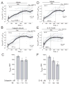
Figure 3. Calpeptin and Calpain Inhibitor-III (CI-III or MDL-28170) failed to protect cultured hippocampal neurons against DCD
In A, B and D, E, neurons were treated with 100μM glutamate (Glu) plus 10μM glycine as indicated. Here and in other similar Figures, thin grey traces show signals from individual neurons from the same dish, while thick black traces show averaged signals (mean±SEM) for Fura-2FF fluorescence ratio F340/F380. In A and D, neurons were pre-treated with a vehicle (1.5μl DMSO per 1.5 ml of the standard bath solution) for 30 minutes, and the vehicle was present in the bath solution during the experiment. Here and in other similar Figures, N is the number of neurons examined in each individual representative experiment. In B and E, neurons were pre-treated with 20μM calpeptin or 10μM CI-III respectively for 30 minutes prior to application of glutamate. In addition, calpeptin and CI-III were present in the bath solution as indicated. In C and F, statistical analysis of tDCD obtained without calpain inhibitors and in the presence of 5 and 20μM calpeptin or 1 and 10μM CI-III. tDCD was determined by finding the time between the beginning of glutamate exposure and the intersection point of two linear graphs approximating the uprising fragment of the averaged Fura-2FF ratio F340/F380 trace and the fragment corresponding to the elevated [Ca2+]c plateau. In C, *p<0.01 between control without inhibitors and experiments with calpeptin. In F, *p<0.01 between control without inhibitors and experiments with 1μM CI-III; **p<0.001 between control without inhibitors and experiments with 10μM CI-III. Here and in other similar panels with statistical analysis, N is the total number of neurons examined in these experiments. In A, B and D, E, data are mean±SEM, N=6 (6 independent experiments with neurons from 2 different platings). Here and in other similar experiments where indicated, the following agents were added to neurons: EGTA, 250μM; FCCP, 1μM.
Figure 4
Figure 4. Calpeptin and CI-III failed to improve survival rate of cultured hippocampal neurons exposed to glutamate
Neuronal death was evaluated using the Trypan Blue exclusion method (Dubinsky and Rothman, 1991). Neurons were exposed to 30 or 100μM glutamate (Glu) plus 10μM glycine for 10 minutes in the absence or presence of various concentrations of calpain inhibitors as indicated. Then, glutamate was removed and cell death was evaluated in a blind manner after 24 hours by unbiased observer counting Trypan Blue stained neurons. Where indicated, calpain inhibitors were added to neurons 30 minutes prior to glutamate application and were kept in the bath solution during and after incubation of neurons with glutamate. Data are mean±SEM, N=3 (3 independent experiments with neurons from 3 different platings).
Figure 5
Figure 5. Calpeptin failed to protect cultured hippocampal neurons against glutamate toxicity
Neurons were exposed to 100μM glutamate (Glu) plus 10μM glycine for 10 minutes in the absence or presence of calpeptin (20μM) as indicated. Then, glutamate was removed and cell death was evaluated by assessing propidium iodide (PI) nuclear staining (Li et al., 2009). In these experiments, calcein staining was used to visualize viable cells. Calpeptin was added to neurons 30 minutes prior to glutamate application and was kept in the bath solution during incubation of neurons with glutamate and after removal of glutamate. Where indicated, neurons were pre-treated with a vehicle (1.5μl DMSO per 1.5 ml of the standard bath solution) for 30 minutes, and the vehicle was present in the bath solution during the experiment. Bf, bright field.
Figure 6
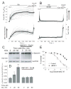
Figure 6. MK801 added shortly after the initial rise in [Ca2+]c prevented DCD, calpain activation, and significantly reduced neuronal death
In A and B, cultured hippocampal neurons (12 DIV) co-loaded with Fura-2FF and Rh123 were exposed to 100μM glutamate (Glu) plus 10μM glycine. N is the total number of neurons examined in these representative experiments. In B, MK801 (20μM) was added as indicated. In A and B, where indicated, the following agents were added to neurons: EGTA, 250μM; FCCP, 1μM. In C, western blot analysis of proteolytic degradation of α-spectrin in cultured hippocampal neurons exposed to glutamate with and without MK801. Neurons were incubated without or with 100μM glutamate plus 10μM glycine for 20 or 60 minutes. Where indicated, 20μM MK801 was added to neurons 90 s after glutamate application. The same immunoblots were re-probed for glyceraldehyde-3-phosphate dehydrogenase (GAPDH) to ensure equal loading. In D, the columns show the densitometry data normalized by GAPDH loading control, averaged from three independent experiments (mean±SEM) and expressed in arbitrary units (a.u.) for the corresponding bands. *p<0.05 in comparison with control without glutamate; **p<0.01 in comparison with control without glutamate. In E, a glutamate dose-dependence of neuronal death with and without MK801. Neurons were exposed to various concentrations of glutamate plus 10μM glycine for 10 minutes with or without MK801 treatment (20μM, added 90 s after glutamate application). Cell death was evaluated by the Trypan Blue exclusion method 24 hours after glutamate removal (Dubinsky and Rothman, 1991).
Figure 7
Figure 7. MK801 added shortly after glutamate protected cultured hippocampal neurons against glutamate toxicity
Neurons were exposed to 100μM glutamate (Glu) plus 10μM glycine for 10 minutes in the absence or presence of MK801 (20μM) added 90 s after glutamate. Then, glutamate was removed and cell death was evaluated by assessing propidium iodide (PI) nuclear staining (Li et al., 2009). In these experiments, calcein staining was used to visualize viable cells. Where indicated, neurons were pre-treated with a vehicle (1.5μl DMSO per 1.5 ml of the standard bath solution) for 30 minutes, and the vehicle was present in the bath solution during the experiment. Bf, bright field.

References

    1. Adamec E, Beermann ML, Nixon RA. Calpain I activation in rat hippocampal neurons in culture is NMDA receptor selective and not essential for excitotoxic cell death. Brain Res Mol Brain Res. 1998;54:35–48. - PubMed
    1. Araujo IM, Carreira BP, Pereira T, Santos PF, Soulet D, Inacio A, Bahr BA, Carvalho AP, Ambrosio AF, Carvalho CM. Changes in calcium dynamics following the reversal of the sodium-calcium exchanger have a key role in AMPA receptor-mediated neurodegeneration via calpain activation in hippocampal neurons. Cell Death Differ. 2007;14:1635–1646. - PubMed
    1. Bano D, Young KW, Guerin CJ, Lefeuvre R, Rothwell NJ, Naldini L, Rizzuto R, Carafoli E, Nicotera P. Cleavage of the plasma membrane Na+/Ca2+ exchanger in excitotoxicity. Cell. 2005;120:275–285. - PubMed
    1. Bartus RT, Elliott PJ, Hayward NJ, Dean RL, Harbeson S, Straub JA, Li Z, Powers JC. Calpain as a novel target for treating acute neurodegenerative disorders. Neurol Res. 1995;17:249–258. - PubMed
    1. Bevers MB, Lawrence E, Maronski M, Starr N, Amesquita M, Neumar RW. Knockdown of m-calpain increases survival of primary hippocampal neurons following NMDA excitotoxicity. J Neurochem. 2009;108:1237–1250. - PMC - PubMed

Publication types

MeSH terms

LinkOut - more resources