Comparative Study
. 2009 Nov;66(5):617-29.
doi: 10.1002/ana.21802.

Intrathecal pathogenic anti-aquaporin-4 antibodies in early neuromyelitis optica

Affiliations
Comparative Study

Intrathecal pathogenic anti-aquaporin-4 antibodies in early neuromyelitis optica

Jeffrey L Bennett et al. Ann Neurol. 2009 Nov.

Abstract

Objective: The serum of most neuromyelitis optica (NMO) patients contains autoantibodies (NMO-IgGs) directed against the aquaporin-4 (AQP4) water channel located on astrocyte foot processes in the perivessel and subpial areas of the brain. Our objectives were to determine the source of central nervous system (CNS) NMO-IgGs and their role in disease pathogenesis.

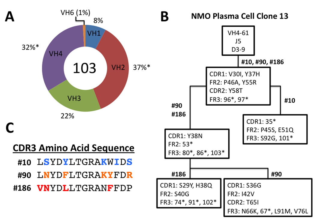
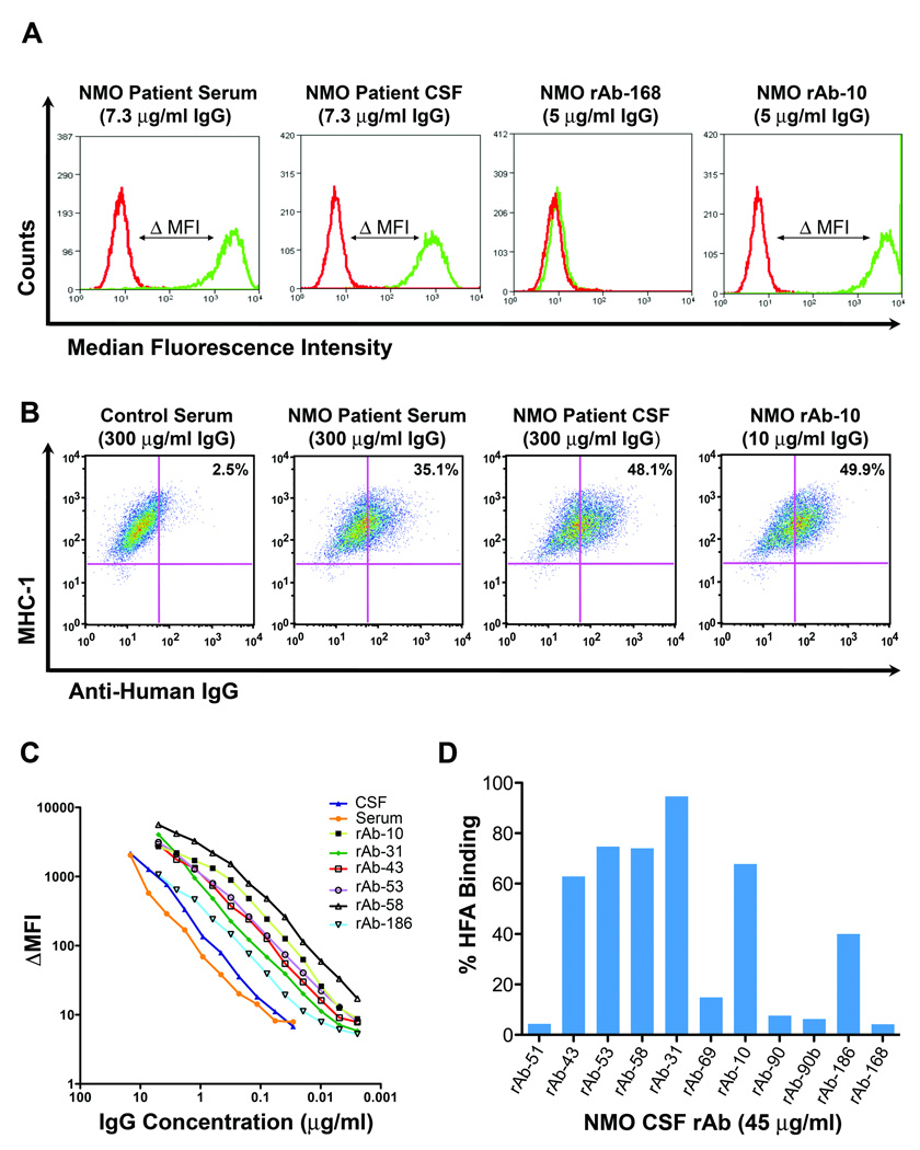
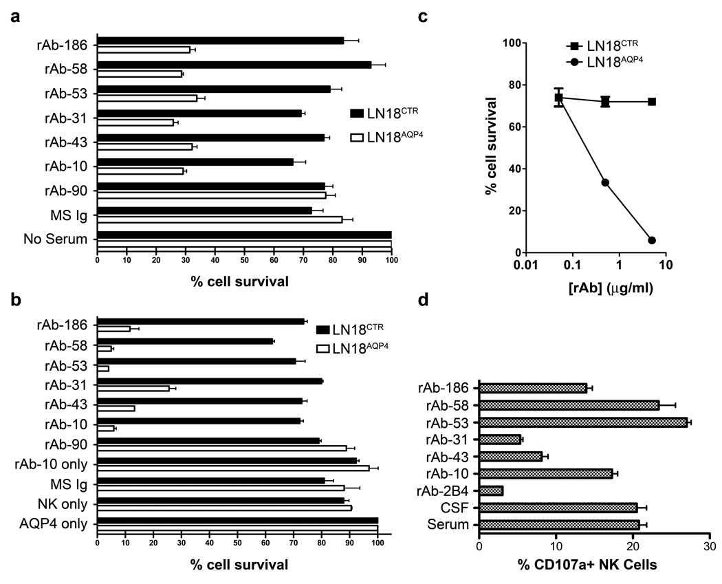
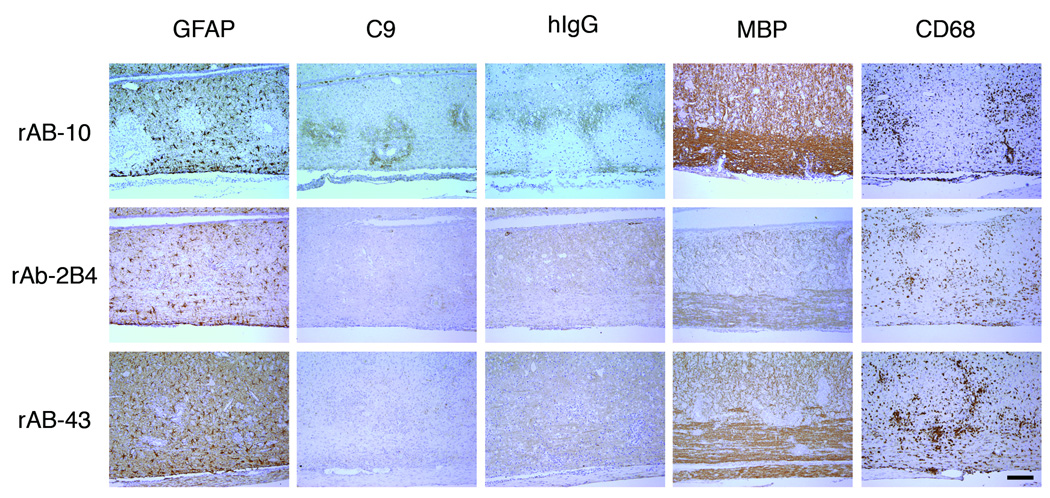
Methods: Fluorescence-activated cell sorting and single-cell reverse transcriptase polymerase chain reaction were used to identify overrepresented plasma cell immunoglobulin (Ig) sequences in the cerebrospinal fluid (CSF) of an NMO patient after a first clinical attack. Monoclonal recombinant antibodies (rAbs) were generated from the paired heavy and light chain sequences and tested for target specificity and Fc effector function. The effect of CSF rAbs on CNS immunopathology was investigated by delivering single rAbs to rats with experimental autoimmune encephalomyelitis (EAE).

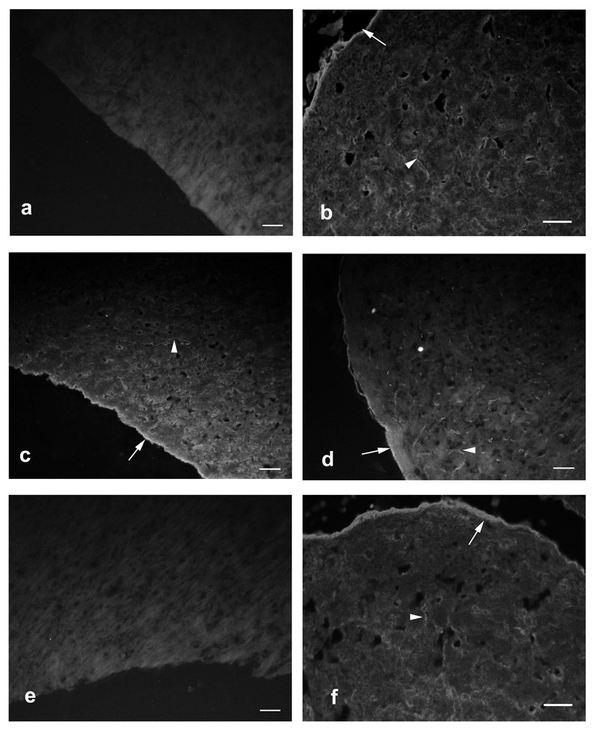
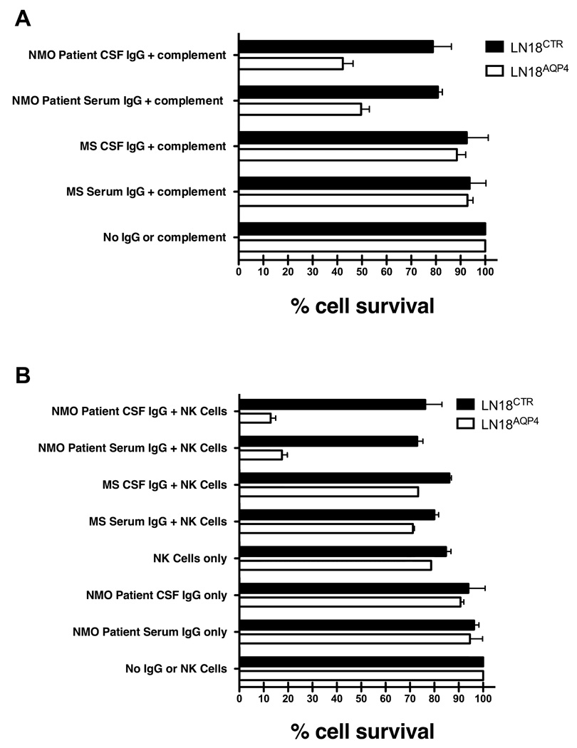
Results: Repertoire analysis revealed a dynamic, clonally expanded plasma cell population with features of an antigen-targeted response. Using multiple independent assays, 6 of 11 rAbs generated from CSF plasma cell clones specifically bound to AQP4. AQP4-specific rAbs recognized conformational epitopes and mediated both AQP4-directed antibody-dependent cellular cytotoxicity and complement-mediated lysis. When administered to rats with EAE, an AQP4-specific NMO CSF rAb induced NMO immunopathology: perivascular astrocyte depletion, myelinolysis, and complement and Ig deposition.

Interpretation: Molecular characterization of the CSF plasma cell repertoire in an early NMO patient demonstrates that AQP4-specific Ig is synthesized intrathecally at disease onset and directly contributes to CNS pathology. AQP4 is now the first confirmed antigenic target in human demyelinating disease.


Conflict of interest statement

Conflict of interest: All authors report no conflict of interest.

Figures

Fig 1
Fig 1
The CD138+ NMO CSF plasma cell repertoire shows VH2 family germline bias and extensive intraclonal diversity. (A) The percentage of VH usage in 103 CD138+ CSF plasma cells is indicated. *Statistically significant difference (binomial distribution, P <0.01). (B) Lineage analysis of the three plasma cell VH sequences comprising plasma cell clone 13 demonstrates intraclonal diversity. The topmost box contains the germline V, D, and J segments used in the clonal heavy chain rearrangement. The numbers above each box indicate the plasma cells (Supplemental Table) that contain the mutations listed inside. The location of each mutation within the FR or CDR region of VH sequence is noted on the left. Replacement mutations are demarcated by the initial amino acid, amino acid position, and the new amino acid resulting from the mutation (e.g., Y37H). Silent mutations are demarcated by the amino acid position followed by an asterisk (e.g., 96*). (C) CDR3 amino acid sequence of each plasma cell VH in clone 13. Bolded, colored amino acids denote somatic hypermutations resulting in amino acid substitutions within the germline V(D)J rearrangement.
Fig 2
Fig 2
Most of the NMO CSF rAbs were directed against AQP4. (A) Histograms demonstrate staining of LN18CTR control (red line) and LN18AQP4 (green line) cells with serum, CSF, or rAb from our NMO patient. Antibody binding to AQP4 was detected using an anti-human IgG secondary antibody and quantitated by flow cytometry. The difference between the median fluorescence intensity observed with LN18CTR and LN18AQP4 (ΔMFI) corresponds to the amount of IgG bound to AQP4 on the surface of cells. (B) Dot plots show combined staining of MHC class I human fetal astrocytes (HFAs) with control serum, NMO patient serum, NMO patient CSF, or NMO CSF rAb. Serum, CSF, or rAb binding to HFAs was detected with PE-conjugated anti-human IgG and quantified by flow cytometry. Control serum was routinely diluted to 1:50. Patient CSF was diluted to the identical IgG concentration of the matched patient serum (300 µg/ml). (C) Change in median fluorescence intensity (ΔMFI) is plotted against IgG concentration for the serum, CSF, and AQP4-specific CSF rAbs of our NMO patient. (D) The percentage HFA binding is plotted for each NMO CSF rAb. 6 of 11 rAbs bound HFAs (mean = 69 ± 17%; range, 40 to 94.6%).
Fig 3
Fig 3
CSF NMO rAbs bind murine cerebellar tissue. Control NMO serum, NMO patient serum, NMO patient CSF and CSF rAb-10 display the same pattern of binding to murine cerebellum by indirect immunofluorescence microscopy: linear staining of pia (arrows) and white matter microvessels (arrowheads). (A) Negative control MS patient serum (1:60 dilution). (B) Positive control NMO serum (1:60 dilution). (C) Our NMO patient’s serum (1:60 dilution). (D) CSF rAb-10 (50 µg/ml). (E) CSF rAb-43 (50 µg/ml). (F) Our NMO patient’s CSF (1:6 dilution). Scale bars indicate 50µm.
Fig 4
Fig 4
Serum and CSF IgG from our NMO patient directs complement-mediated lysis and antibody-dependent cell-mediated cytotoxicity (ADCC) of target cells expressing AQP4. (A) AQP4-specific complement-mediated killing of LN18AQP4 and LN18CTR cells by CSF rAbs was quantitated using FACS analysis. The experiment was performed in duplicate, and the percentage cell survival was normalized to the negative control sample (No IgG or complement). Mean and standard deviation are shown. IgG purified from the serum (NMO Patient Serum IgG + complement) or CSF (NMO Patient CSF IgG + complement) significantly reduced the viability of LN18AQP4 target cells (p < 0.001, paired t-test). Serum and CSF IgG purified from an MS patient resulted in no significant change in LN18AQP4 viability (p = 0.34, paired t-test). (B) ADCC of LN18AQP4 and LN18CTR cells in the presence of human NK cells and NMO patient serum or CSF IgG was evaluated by FACS analysis. The number of viable cells in culture was quantitated and normalized to the number of surviving cells in the negative control (No IgG or NK cells) sample. In the presence of human NK cells, NMO patient serum (NMO Patient Serum IgG + NK Cells) and CSF IgG (NMO Patient CSF IgG + NK Cells) resulted in a specific loss of more than 80% of LN18AQP4 target cells (p < 0.001, paired t-test). ADCC of LN18AQP4 target cells with NMO patient serum and CSF IgG was significantly greater than that resulting from AQP4-negative MS serum (MS Serum IgG + NK Cells) and CSF IgG (MS CSF IgG + NK Cells) (p < 0.001, t-test).
Fig 5
Fig 5
AQP4-specific CSF rAbs are functional IgG1 antibodies. (A) AQP4-specific complement-mediated killing of LN18AQP4 and LN18CTR cells by CSF rAbs was quantitated using FACS analysis. The experiment was performed in duplicate, and the percentage cell survival was normalized to the negative control (No Serum) sample. The loss of LN18AQP4 cell viability was greater than 75% in the presence of AQP4-specific rAb (rAbs-10, -31, -43, -53, -58, and -186) and complement. Non-specific reduction in cell viability rarely exceeded 20% in the absence of serum (No Serum) or the presence of nonspecific serum (MS Ig) or CSF rAb (rAb-90). Mean and standard deviation are shown. (B) The induction of antibody-dependent cell-mediated cytotoxicity (ADCC) of LN18AQP4 and LN18CTR cells in the presence of human NK cells and CSF rAbs was evaluated by FACS analysis. After 12 hrs of cocultivation, the number of viable LN18 cells in culture was quantitated and normalized to the number of surviving cells in the negative control (AQP4 only) sample that contained NMO patient serum in the absence of human NK cells. AQP4-specific rAbs-10, -53, and -58 resulted in a specific loss of more than 94% of the target LN18AQP4 cells; AQP4-specific rAbs-43, -186, and -31 caused a slightly lower amount of LN18AQP4-specific target cell death. No impact on LN18AQP4 cell survival was observed in the absence of rAb (NK only), the absence of NK cells (rAb-10 only), the presence of nonspecific serum (MS Ig) or CSF rAb (rAb-90). The experiment was performed in duplicates; mean and standard deviation are shown. (C) Serial dilution of CSF rAb-10 demonstrates that NK-mediated lysis of LN18AQP4 cells is dose dependent. AQP4-specific rAb-10 (5 µg/ml, 0,5 µg/ml and 0,05 µg/ml) was preincubated with either LN18AQP4 or LN18CTR target cells and the level of ADCC in the presence of human NK cells was quantitated by FACS analysis. Cell survival was determined after 10 hr cocultivation and normalized to the negative control (LN18CTR) cell line. (D) NK cell surface mobilization of CD107a was quantified by FACS analysis after coincubation of HFAs with NMO patient serum, NMO patient CSF, or AQP4-specific NMO CSF rAb. NMO patient serum, NMO patient CSF, and AQP4-specific CSF rAbs-10, --53, -58, and -186 resulted in comparable elevations in CD107a surface expression on human NK cells. CSF rAbs-43 and -31 triggered only modest CD107a surface mobilization compared to a measles virus nucleocapsid-specific control rAb (rAb-2B4). The experiment was performed in duplicates; mean and standard deviation are shown.
Fig 6
Fig 6
AQP4-specific CSF rAb-10 induced prototypic NMO pathology in guinea pig MBP72–85-induced EAE rats. The rows are arranged by NMO CSF rAb, and the columns are ordered by immunohistochemical stain. In rats injected with rAb-10, glial fibrillary acidic protein (GFAP) immunohistochemistry revealed prominent perivascular loss of astrocyte cell bodies and processes. Staining for complement C9 (C9) and human Ig (hIg) demonstrated significant deposits of perivascular complement protein and human Ig deposition beyond the zone of perivascular astrocyte depletion. Intravenous transfer of negative control measles virus-specific rAb-2B4 or human AQP4-specific rAb-43 produced no additional immunopathology. Myelin basic (MBP) immunohistochemistry demonstrated some perivascular myelin vacuolization in rAb-10-treated animals, but myelin remained largely intact. There was no difference in the extent of CNS tissue macrophage infiltration (CD68) induced by rAbs-10, -43, and -2B4. Scale bar: 200 µM.

Comment in

Similar articles

Cited by

References

    1. Wingerchuk DM, Lennon VA, Lucchinetti CF, et al. The spectrum of neuromyelitis optica. Lancet neurology. 2007;6:805–815. - PubMed
    1. Lennon VA, Kryzer TJ, Pittock SJ, et al. IgG marker of optic-spinal multiple sclerosis binds to the aquaporin-4 water channel. J Exp Med. 2005;202:473–477. - PMC - PubMed
    1. Lennon VA, Wingerchuk DM, Kryzer TJ, et al. A serum autoantibody marker of neuromyelitis optica: distinction from multiple sclerosis. Lancet. 2004;364:2106–2112. - PubMed
    1. Roemer SF, Parisi JE, Lennon VA, et al. Pattern-specific loss of aquaporin-4 immunoreactivity distinguishes neuromyelitis optica from multiple sclerosis. Brain. 2007;130:1194–1205. - PubMed
    1. Takahashi T, Fujihara K, Nakashima I, et al. Anti-aquaporin-4 antibody is involved in the pathogenesis of NMO: a study on antibody titre. Brain. 2007;130:1235–1243. - PubMed

Publication types

MeSH terms

LinkOut - more resources