. 2010 Mar;78(4):917-31.
doi: 10.1002/prot.22617.

The structure and NO binding properties of the nitrophorin-like heme-binding protein from Arabidopsis thaliana gene locus At1g79260.1

Affiliations

The structure and NO binding properties of the nitrophorin-like heme-binding protein from Arabidopsis thaliana gene locus At1g79260.1

Christopher M Bianchetti et al. Proteins. 2010 Mar.

Abstract

The protein from Arabidopsis thaliana gene locus At1g79260.1 is comprised of 166-residues and is of previously unknown function. Initial structural studies by the Center for Eukaryotic Structural Genomics (CESG) suggested that this protein might bind heme, and consequently, the crystal structures of apo and heme-bound forms were solved to near atomic resolution of 1.32 A and 1.36 A, respectively. The rate of hemin loss from the protein was measured to be 3.6 x 10(-5) s(-1), demonstrating that it binds heme specifically and with high affinity. The protein forms a compact 10-stranded beta-barrel that is structurally similar to the lipocalins and fatty acid binding proteins (FABPs). One group of lipocalins, the nitrophorins (NP), are heme proteins involved in nitric oxide (NO) transport and show both sequence and structural similarity to the protein from At1g79260.1 and two human homologues, all of which contain a proximal histidine capable of coordinating a heme iron. Rapid-mixing and laser photolysis techniques were used to determine the rate constants for carbon monoxide (CO) binding to the ferrous form of the protein (k'(CO) = 0.23 microM(-1) s(-1), k(CO) = 0.050 s(-1)) and NO binding to the ferric form (k'(NO) = 1.2 microM(-1) s(-1), k(NO) = 73 s(-1)). Based on both structural and functional similarity to the nitrophorins, we have named the protein nitrobindin and hypothesized that it plays a role in NO transport. However, one of the two human homologs of nitrobindin contains a THAP domain, implying a possible role in apoptosis. Proteins 2010. (c) 2009 Wiley-Liss, Inc.


Figures
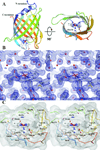
Figure 1
Figure 1
A. Cartoon representation of the overall structure of nitrobindin is shown going from blue at the N-terminus to red at the C-terminus. The bound heme is shown as sticks with carbon in white, nitrogen in blue, oxygen in red, and iron in orange. A view along the barrel axis (right) shows the cavity formed by the β-barrel. His158 coordinates the heme iron and is shown in the same color scheme as above. B. Stereo view of the 2Fo–Fc map contoured at 1.3 σ of the heme pocket. nitrobindin and the bound heme are shown as sticks with the same color scheme as above with waters represented as red spheres. C. Stereo view of the hydrophobic cavity. The Cα ribbon diagram is shown going from blue at the N-terminus to red at the C-terminus. Amino acids that are within 4 Å of the heme are shown as sticks and labeled.
Figure 2
Figure 2
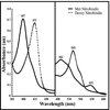
UV-Vis spectrum of nitrobindin in 5 mM MES-HCl buffer at pH 6.0 containing NaCl (50mM), and TCEP (0.3mM). The solid line is for protein exposed to air and the dotted line is after nitrobindin is reduced with sodium dithionite.
Figure 3
Figure 3
Time courses for CO association and dissociation. A. Absorbance trace for bimolecular CO rebinding measured at 436 nm after flash photolysis of the Fe-CO complex. The CO concentration for this trace was 1 atmosphere (1000 µM), and the protein concentration was approximately 100 µM. For all CO association and dissociation experiments nitrobindin was in 100 mM potassium phosphate buffer at pH 7 containing EDTA (1 mM) at 20 °C. The overall association rate (k’CO) was computed as the slope of a linear fit to a plot of kobs versus the CO concentration. B. Measurement of the CO dissociation constant by stopped-flow spectrophotometry. CO–nitrobindin was mixed with 100 mM potassium phosphate buffer at pH 7 containing EDTA (1 mM), and 1 atmosphere (2000 µM) of NO at 20 °C. The displacement of the CO–nitrobindin complex was measured by monitoring the absorbance decrease at 424 nm.
Figure 4
Figure 4
Iron-carbonyl IR spectrum in the range of 1900–2000 cm−1 for nitrobindin, compared to wild-type sperm whale myoglobin, and a distal histidine variant myoglobin (H64L). The IR spectra were measured at room temperature in 100 mM potassium phosphate buffer at pH 7 containing EDTA (1 mM).
Figure 5
Figure 5
Time course for NO rebinding to ferric nitrobindin after flash photolysis. Absorbance changes were measured at 405nm. The NO concentration for this trace was 1 atmosphere (2000 µM) of NO, and the protein concentration was approximately 100 µM. For all NO association experiments nitrobindin was in 100 mM potassium phosphate buffer at pH 7 containing EDTA (1 mM) at 20 °C. The overall association rate (k`NO) is taken from the slope of a linear fit of the rate of absorbance change versus the NO concentration.
Figure 6
Figure 6
Stereo view of a secondary structural alignment of nitrobindin in red and residues 13–139 of NP4 in blue (PDB ID 1YWD). The bound heme of nitrobindin is shown in red and the bound heme of NP4 is shown in blue.

Similar articles

Cited by

References

    1. Gray HB, Winkler JR. Electron transfer in proteins. Annu Rev Biochem. 1996;65:537–561. - PubMed
    1. Ortiz de Montellano PR. Cytochrome P450: Structure, Mechanism, and Biochemistry. New York: Plenium press; 1995.
    1. Rodgers KR. Heme-based sensors in biological systems. Curr Opin Chem Biol. 1999;3(2):158–167. - PubMed
    1. Chan MK. Recent advances in heme-protein sensors. Curr Opin Chem Biol. 2001;5(2):216–222. - PubMed
    1. Olson JS, Phillips GN., Jr Kinetic pathways and barriers for ligand binding to myoglobin. J Biol Chem. 1996;271(30):17593–17596. - PubMed

Publication types

MeSH terms