

Endoscopic ultrasound and magnetic resonance imaging for re-staging rectal cancer after radiotherapy
- PMID: 19938195
- PMCID: PMC2785059
- DOI: 10.3748/wjg.15.5563
Endoscopic ultrasound and magnetic resonance imaging for re-staging rectal cancer after radiotherapy
Abstract
Aim: To compare the sensitivity and specificity of two imaging techniques, endoscopic ultrasound (EUS) and magnetic resonance imaging (MRI), in patients with rectal cancer after neoadjuvant chemoradiation therapy. And we compared EUS and MRI data with histological findings from surgical specimens.
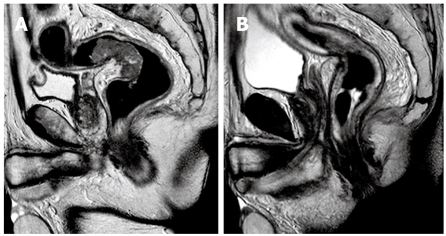
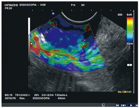
Methods: Thirty-nine consecutive patients (51.3% Male; mean age: 68.2 +/- 8.9 years) with histologically confirmed distal rectal cancer were examined for staging. All patients underwent EUS and MRI imaging before and after neoadjuvant chemoradiation therapy.
Results: After neoadjuvant chemoradiation, EUS and MRI correctly classified 46% (18/39) and 44% (17/39) of patients, respectively, in line with their histological T stage (P > 0.05). These proportions were higher for both techniques when nodal involvement was considered: 69% (27/39) and 62% (24/39). When patients were sorted into T and N subgroups, the diagnostic accuracy of EUS was better than MRI for patients with T0-T2 (44% vs 33%, P > 0.05) and N0 disease (87% vs 52%, P = 0.013). However, MRI was more accurate than EUS in T and N staging for patients with more advanced disease after radiotherapy, though these differences did not reach statistical significance.
Conclusion: EUS and MRI are accurate imaging techniques for staging rectal cancer. However, after neoadjuvant RT-CT, the role of both methods in the assessment of residual rectal tumors remains uncertain.
Figures

References
-
- Vanagunas A, Lin DE, Stryker SJ. Accuracy of endoscopic ultrasound for restaging rectal cancer following neoadjuvant chemoradiation therapy. Am J Gastroenterol. 2004;99:109–112. - PubMed
-
- Savides TJ, Master SS. EUS in rectal cancer. Gastrointest Endosc. 2002;56:S12–S18. - PubMed
-
- Improved survival with preoperative radiotherapy in resectable rectal cancer. Swedish Rectal Cancer Trial. N Engl J Med. 1997;336:980–987. - PubMed
-
- Harewood GC. Assessment of clinical impact of endoscopic ultrasound on rectal cancer. Am J Gastroenterol. 2004;99:623–627. - PubMed
MeSH terms
LinkOut - more resources
Full Text Sources
