

Asymptomatic lymphangioma involving the spleen and retroperitoneum in adults
- PMID: 19938204
- PMCID: PMC2785067
- DOI: 10.3748/wjg.15.5620
Asymptomatic lymphangioma involving the spleen and retroperitoneum in adults
Abstract
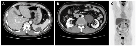
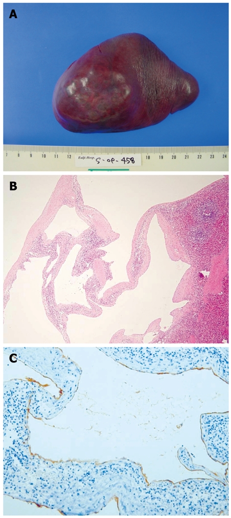
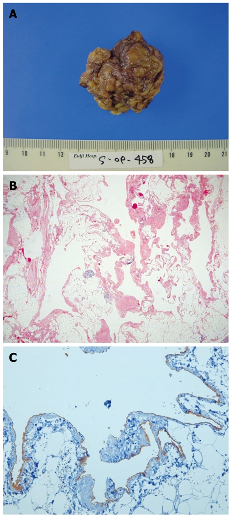
Lymphangioma, a benign neoplasm of the lymphatic system, is common in children but rare in adults. Its clinical manifestations include abdominal pain, nausea, vomiting and a palpable mass. However, abdominal sonography or abdominal computed tomography (CT) scan can also incidentally reveal lymphangioma. A larger or symptomatic lymphangioma is treated with total resection to prevent recurrence, infection, torsion and enlargement. Although lymphangioma rarely becomes malignant, its prognosis is generally good. We report a cystic lymphangioma of the spleen and retroperitoneum, which was incidentally found in a 56-year-old man who was hospitalized due to a colon mass. Physical examination showed no specific findings. Abdominal CT revealed a 5.7 cm, non-enhanced multilobulated cystic mass with multiple septa in the spleen and a 10 cm lobulated cystic mass in the paraaortic area. Splenectomy and retroperitoneal resection of the cystic mass were conducted. The endothelium of splenic and retroperitoneal cyst was immunohistochemically stained with D2-40 antibody. The patient was finally diagnosed with splenic cystic and retroperitoneal cavernous lymphangioma.
Figures
Similar articles
-
Splenic lymphangioma that manifested as a solid-cystic mass: a case report.World J Gastroenterol. 2013 Feb 7;19(5):781-3. doi: 10.3748/wjg.v19.i5.781. World J Gastroenterol. 2013. PMID: 23429434 Free PMC article.
-
Retroperitoneal lymphangioma.Asian J Surg. 2006 Jan;29(1):51-4. doi: 10.1016/S1015-9584(09)60297-9. Asian J Surg. 2006. PMID: 16428102
-
Giant retroperitoneal lymphangioma in a 70-year-old male: a case report.Pan Afr Med J. 2022 Jun 24;42:153. doi: 10.11604/pamj.2022.42.153.34175. eCollection 2022. Pan Afr Med J. 2022. PMID: 36187030 Free PMC article.
-
Pediatric cystic lymphangioma of the retroperitoneum: A case report and review of the literature.Medicine (Baltimore). 2020 Jul 10;99(28):e20827. doi: 10.1097/MD.0000000000020827. Medicine (Baltimore). 2020. PMID: 32664076 Free PMC article. Review.
-
[Retroperitoneal cystic lymphangioma: about 5 cases and review of the literature].Pan Afr Med J. 2016 Oct 6;25:73. doi: 10.11604/pamj.2016.25.73.10002. eCollection 2016. Pan Afr Med J. 2016. PMID: 28292036 Free PMC article. Review. French.
Cited by
-
Splenic Lymphangioma Mimicking Lymphomatous Involvement: A Case Report with Review of the Literature.Case Rep Med. 2023 Jun 20;2023:9969213. doi: 10.1155/2023/9969213. eCollection 2023. Case Rep Med. 2023. PMID: 37383046 Free PMC article.
-
Case report and literature review: Giant retroperitoneal cystic lymphangioma.Front Surg. 2023 Jan 17;10:1074067. doi: 10.3389/fsurg.2023.1074067. eCollection 2023. Front Surg. 2023. PMID: 36733888 Free PMC article.
-
Splenic lymphangioma in adult patient treated with laparoscopic splenectomy: A rare case report.SAGE Open Med Case Rep. 2023 Jan 3;11:2050313X221147196. doi: 10.1177/2050313X221147196. eCollection 2023. SAGE Open Med Case Rep. 2023. PMID: 36636099 Free PMC article.
-
Cystic pancreatic lymphangioma.Rare Tumors. 2012 Apr 12;4(2):e27. doi: 10.4081/rt.2012.e27. Epub 2012 May 17. Rare Tumors. 2012. PMID: 22826784 Free PMC article.
-
Splenic cystic lymphangioma with atypical ultrasound findings.J Med Ultrason (2001). 2016 Jan;43(1):99-105. doi: 10.1007/s10396-015-0659-8. Epub 2015 Aug 27. J Med Ultrason (2001). 2016. PMID: 26703174
References
-
- de Perrot M, Rostan O, Morel P, Le Coultre C. Abdominal lymphangioma in adults and children. Br J Surg. 1998;85:395–397. - PubMed
-
- Kim DH, Byun JN, Jang JY. Cystic lymphangioma involving the mesentery and the retroperitoneum: A case report. J Korean Radiol Soc. 2005;52:347–350.
-
- Takayama A, Nakashima O, Kobayashi K, Kojiro M. Splenic lymphangioma with papillary endothelial proliferation: a case report and review of the literature. Pathol Int. 2003;53:483–488. - PubMed
-
- Cherk M, Nikfarjam M, Christophi C. Retroperitoneal lymphangioma. Asian J Surg. 2006;29:51–54. - PubMed
-
- Chang CH, Hsieh CB, Yu JC, Jan CI. A case of lymphangioma of the spleen. J Med Sci. 2004;24:109–112.
Publication types
MeSH terms
LinkOut - more resources
Full Text Sources
