Review
. 2010 Jul 15;13(2):157-92.
doi: 10.1089/ars.2009.2657.

Interactions of multiple gas-transducing systems: hallmarks and uncertainties of CO, NO, and H2S gas biology

Affiliations
Review

Interactions of multiple gas-transducing systems: hallmarks and uncertainties of CO, NO, and H2S gas biology

Mayumi Kajimura et al. Antioxid Redox Signal. .

Abstract

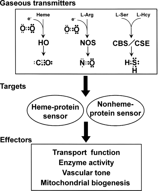
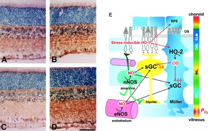
The diverse physiological actions of the "biologic gases," O2, CO, NO, and H2S, have attracted much interest. Initially viewed as toxic substances, CO, NO, and H2S play important roles as signaling molecules. The multiplicity of gas actions and gas targets and the difficulty in measuring local gas concentrations obscures detailed mechanisms whereby gases exert their actions, and many questions remain unanswered. It is now readily apparent, however, that heme-based proteins play central roles in gas-generation/reception mechanisms and provide a point where multiple gases can interact. In this review, we consider a number of key issues related to "gas biology," including the effective tissue concentrations of these gases and the importance and significance of the physical proximity of gas-producing and gas-receptor/sensors. We also take an integrated approach to the interaction of gases by considering the physiological significance of CO, NO, and H2S on mitochondrial cytochrome c oxidase, a key target and central mediator of mitochondrial respiration. Additionally, we consider the effects of biologic gases on mitochondrial biogenesis and "suspended animation." By evaluating gas-mediated control functions from both in vitro and in vivo perspectives, we hope to elaborate on the complex multiple interactions of O2, NO, CO, and H2S.


Figures

FIG. 1.
FIG. 1.
Scheme to illustrate pathways of gas signal-transducing systems by the four gases, O2, CO, NO, and H2S. Valence electrons in each gas molecule are shown as dots and crosses. HO = heme oxygenase; NOS = nitric oxide synthase; CBS = cystathionine β-synthase; and CSE =cystathionine γ-lyase.
FIG. 2.
FIG. 2.
(A) The reaction catalyzed by heme oxygenase (HO). The HO reaction consists of three oxidation steps, where one molecule of O2 is used in each reaction step. The reaction starts with the formation of the Fe3+ heme–HO complex. Then Fe3+ heme is reduced to the Fe2+ state by the first electron donated from NADPH-cytochrome P450 reductase. O2 binds with the heme of the complex. The iron-bound O2 converts to a peroxy intermediate (Fe3+-OOH). Subsequently, the terminal O2 of Fe3+-OOH attacks the α-meso-carbon of the porphyrin ring to form Fe3+ α-meso-hydroxylheme. A subsequent conversion of Fe3+ α-meso-hydroxylheme to verdoheme requires another O2, and this step produces CO by the regiospecific cleavage of the porphyrin ring of the heme at the α-meso carbon atom. The rate-determining step is O2 binding to verdoheme, which is much slower than O2 binding to the heme complex (324). This step produces Fe2+ and biliverdin. Adapted by permission from Macmillan Publishers Ltd: Schuller et al. Nat Struct Biol 6: 860–867, 1999 (270). (B) The reaction catalyzed by nitric oxide synthase (NOS). NO is synthesized from the guanido nitrogen atom(s) of l-arginine by the action of NOS. The process involves the incorporation of an O2 into the unstable intermediate Nω-hydroxy-l-arginine and subsequently into l-citrulline (250, 257).
FIG. 3.
FIG. 3.
Biosynthesis of H2S. H2S is synthesized mainly by three enzymes: cystathionine β-synthase (CBS), cystathionine γ-lyase (CSE), and mercaptopyruvate sulfurtrans-ferase (MPST). Cysteine is converted by aspartate aminotransferase (AAT) to 3-mercaptopyruvate, which subsequently gives off H2S by the action of MPST.
FIG. 4.
FIG. 4.
Catabolism of H2S and oxidation states of the sulfur atoms of various compounds. The sulfur atom of H2S (reduced divalent) is oxidized in mitochondria to a fully oxidized state as a sulfate (hexavalent). The reduced sulfur atoms can be stored as labile sulfur species that can be released as H2S in response to a signal. Dotted lines, Different catabolic pathways. *Sulfane sulfur. TST = thiosulfate:cyanide sulfurtransferase; SO =sulfite oxidase; TSMT =thiol S-methyltransferase. Adapted by permission from Elsevier Ltd: Lloyd, Trends Microbiol 14: 456–462, 2006 (185).
FIG. 5.
FIG. 5.
Ligand discrimination by the oxidative state of the iron center of prosthetic heme. The oxidative state of the iron of heme in hemoglobin shifts between Fe2+ and Fe3+ states. The ferrous form (Fe2+) of hemoglobin prefers to bind ligands such as O2, CO, and NO. Conversely, ferric heme (Fe3+) prefers to bind water, H2S, and anions such as CN, N3−, and OH. L = ligands.
FIG. 6.
FIG. 6.
Distinct positional changes at the distal histidine induced by different ligands: O2, CO, and NO. Ligand geometry in the heme pocket of sperm whale myoglobin is shown. The heme iron is coordinated to a proximal histidine (His93), making the iron five-coordinate with a free binding site for O2. The distal histidine (His64) located above the free binding site can form a hydrogen bond to the sixth iron ligand. The heme is ligated by O2 (1MBO, blue), CO (1VXF, green), or NO (1HJT, red). Each structure was determined by x-ray crystallography, and each structure is superposed on another. Hydrogen bonds, interatomic distances <3.0 Å, are represented by lines. Adapted by permission from Wiley-Blackwell Ltd: from Brucker et al. Proteins 30: 352–356, 1998 (35).
FIG. 7.
FIG. 7.
Difference in transaxial effects of CO and NO ligation on the prosthetic heme of hemoglobin. Ligand affinities of the Fe2+-heme–hemoglobin complex and the binding constants for proximal base are compared between CO and NO. For the gas–ligand affinity, NO binds better without the proximal base, which sets NO apart from CO. Correspondingly the binding constant for the proximal base is decreased by ∼103-fold; weakening the bonding of the heme iron to the imidazole base of the histidine residue. KE, equilibrium constant in M−1. Adapted from Yonetani et al., Journal of Biological Chemistry 273: 20323–20333, 1998 (364).
FIG. 8.
FIG. 8.
(A) Schematic presentation of structural changes in the immediate vicinity of the heme of α-NO hemoglobin. NO forms a five-coordinated nitrosyl–heme complex in which the α-heme Fe-His (F8) bond is weakened and cleaved, causing a shift in the quaternary structural equilibrium from the R- toward the extreme T-state. In this form, the O2 affinity in the β-subunit is substantially reduced, making it an extreme low-affinity O2 carrier. (B) Comparison of O2-delivery capacities of untreated RBCs (grey line) and α-NO RBCs (black line) at pH 7.4 and 15°C. Normal hemoglobin can carry four O2 per tetramer, whereas α-NO hemoglobin can only carry two, because the α-subunits are ligated by NO. Under normal conditions, RBCs unload ∼11% of total O2, whereas α-NO RBCs can deliver 29%. Adapted by permission from Taylor & Francis Ltd.: Tsuneshige et al., Artif Cells Blood Substit Immobil Biotechnol 29: 347–357, 2001 (322).
FIG. 9.
FIG. 9.
Simplified drawing to show the proposed mechanism of NO sensing by sGC. NO binds and unbinds from the Fe2+ heme in the regulatory domain, which transduces the signal to the functional domain, where a conformational change is transduced to the functional unit, and the cyclase activity is turned on. For a more complete scheme, readers are referred to the articles by Marletta and his co-workers (73, 354). The equilibrium constants, KNO, KCO, are calculated based on rate constants from the literature.
FIG. 10.
FIG. 10.
Effects of CO and NO on the activity and structure of the prosthetic heme of rat recombinant full-length CBS. (A) Sodium sulfate polyacrylamide gel electrophoresis for purification of rat recombinant CBS. Lane 1, crude extract; lane 2, purified CBS. (B) Effects of CO and NO on the Fe2+-CBS activity under optimal substrate conditions at pH 7.4. CO, but not NO (100 μM), significantly attenuated the activities of the ferrous enzyme. Data indicate mean ± SEM of four experiments. The activities were measured by determining the conversion of homocysteine and serine to cystathionine. *p < 0.05 versus the group treated with vehicle (V). The concentration of CBS-heme was 10 μM. (C) Stopped-flow visible spectrophotometry for Fe2+-CBS to examine temporal transitional changes after mixing with CO. Data exhibited a decrease at 448 nm and a reciprocal elevation at 422 nm, demonstrating stabilization of the six-coordinated CO-Fe2+-histidine complex. (D) Electron spin resonance spectroscopy indicating the five-coordinate NO-Fe2+ complex of the CBS-heme. Arrow, g value = 2.008. Adapted from Shintani et al., Hepatology 49: 141–150, 2009. (E) Crystal structure of the human CBS (drawn from PDB 1JBQ). The heme and PLP are presented in a stick model.
FIG. 11.
FIG. 11.
Vasodilatory response on reducing endogenous CO generation associated with augmented NO generation under a CO-suppressed condition in the rat brain. (A) Changes in arteriolar diameter at 60 min after the superfusion of various reagents. Supplementation of CO (10 μM) significantly reduces the vasodilatory response induced by HO inhibition. Inhibition of NOS by L-NAME (1 mM) abolishes this vasodilatation. Diameters are standardized as a percentage of baseline diameters before applying the reagents. *p < 0.05; an increase as compared with the vehicle-treated control. #p < 0.05; as compared with the ZnPP-treated group. Values are expressed as mean ± SEM. (B) Time-dependent elevation of NO production in the pial microcirculation. In the control group, NO-associated fluorescence is faint at 20 min. At 60 min, it becomes obvious at the vascular walls and at the cells located in extravascular space. Conversely, in the ZnPP-treated group, fluorescence is evident even at 20 min, and it increases further at 60 min. Adapted from Ishikawa et al., Circ Res 97: e104–e114, 2005 (135).
FIG. 12.
FIG. 12.
Three different routes by which CO controls sGC activities and vascular tone. (A) CO modestly stimulates sGC, thereby reducing the tonic contractile tension of vascular walls. For this action to take place, the local amount of NO must be low, as reported in the liver microcirculation, where constitutive NO appears to be cancelled by basal superoxide. (B) In contrast, when a sufficient amount of NO exists, CO partially inhibits sGC, enhancing its tonic contractile actions. (C) CO interferes with NOS activities as a first step, and it subsequently reduces NO, resulting in sGC inhibition. Here, the gas acts as a tonic vasoconstrictor.
FIG. 13.
FIG. 13.
Visualization of sGC activities with alterations in NO and CO generation. Monoclonal antibody against sGC, mAb3221, makes it possible to examine the activation state of sGC. Shown here is the immunoreactivity to mAb3221, which acquires a greater binding affinity through its recognition of a regiospecific structure determined by the two subunits of sGC. (A) Vehicle-treated control. (B) L-Arg treatment. (C) L-NAME treatment. (D) ZnPP treatment. Scale bar, 50 μm. (E) Schematic drawing of the relation between gas-producing enzymes and sGC in the rat retina. It is proposed that NO is the dominant activator of sGC, but endogenous CO produced in Muller glia cells plays a role in refining the NO-mediated regulation of sGC function. Blue, green, yellow, and orange represent increasing partial O2 pressure (po2). OS, outer segment; ONL, outer nuclear layer; INL, inner nuclear layer; IPL, inner plexiform layer; OpFL, optic fiber layer. Adapted by permission from The FASEB Journal: Kajimura et al., FASEB J 17: 506–508, 2003 (142).

References

    1. Abu-Soud HM. Wang J. Rousseau DL. Fukuto JM. Ignarro LJ. Stuehr DJ. Neuronal nitric oxide synthase self-inactivates by forming a ferrous-nitrosyl complex during aerobic catalysis. J Biol Chem. 1995;270:22997–23006. - PubMed
    1. Acevedo CH. Ahmed A. Hemeoxygenase-1 inhibits human myometrial contractility via carbon monoxide and is upregulated by progesterone during pregnancy. J Clin Invest. 1998;101:949–955. - PMC - PubMed
    1. Adachi T. Ishikawa K. Hida W. Matsumoto H. Masuda T. Date F. Ogawa K. Takeda K. Furuyama K. Zhang Y. Kitamuro T. Ogawa H. Maruyama Y. Shibahara S. Hypoxemia and blunted hypoxic ventilatory responses in mice lacking heme oxygenase-2. Biochem Biophys Res Commun. 2004;320:514–522. - PubMed
    1. Ahmad AS. Zhuang H. Dore S. Heme oxygenase-1 protects brain from acute excitotoxicity. Neuroscience. 2006;141:1703–1708. - PubMed
    1. Alderton WK. Cooper CE. Knowles RG. Nitric oxide synthases: structure, function and inhibition. Biochem J. 2001;357:593–615. - PMC - PubMed

Publication types

MeSH terms

LinkOut - more resources