. 2009 Nov 26:2:35.
doi: 10.1186/1757-1146-2-35.

Foot posture influences the electromyographic activity of selected lower limb muscles during gait

Affiliations

Foot posture influences the electromyographic activity of selected lower limb muscles during gait

George S Murley et al. J Foot Ankle Res. .

Abstract

Background: Some studies have found that flat-arched foot posture is related to altered lower limb muscle function compared to normal- or high-arched feet. However, the results from these studies were based on highly selected populations such as those with rheumatoid arthritis. Therefore, the objective of this study was to compare lower limb muscle function of normal and flat-arched feet in people without pain or disease.

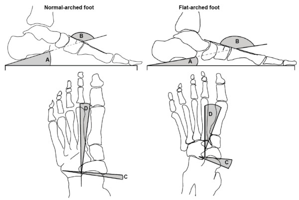
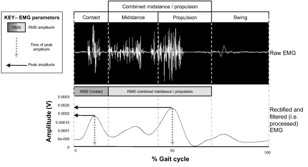
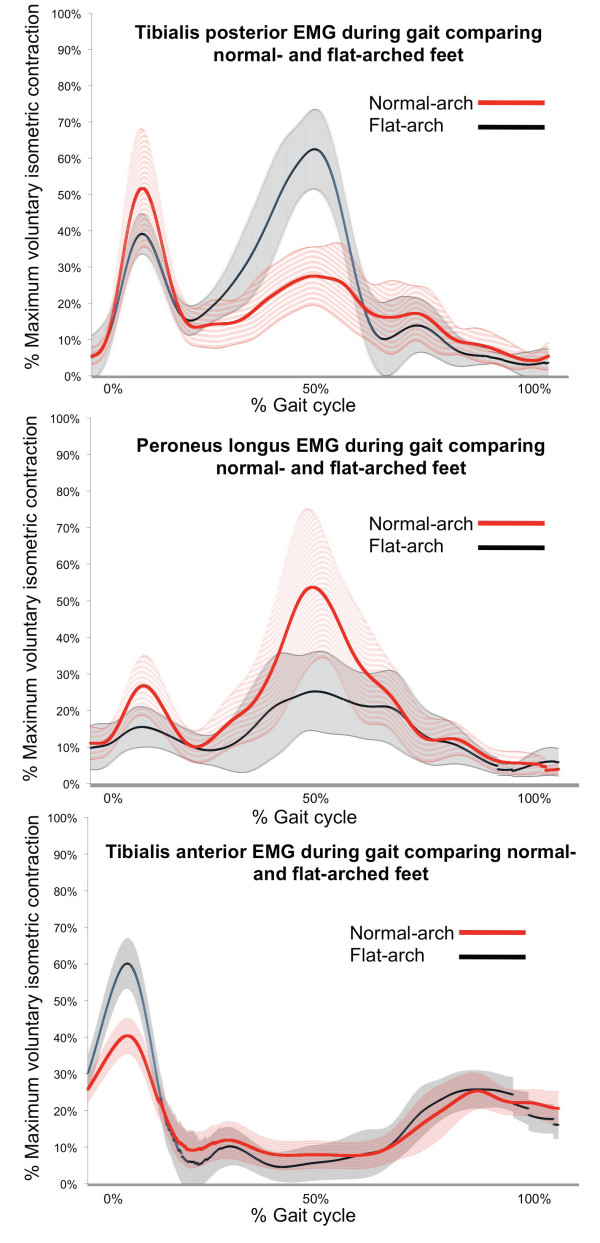
Methods: Sixty adults aged 18 to 47 years were recruited to this study. Of these, 30 had normal-arched feet (15 male and 15 female) and 30 had flat-arched feet (15 male and 15 female). Foot posture was classified using two clinical measurements (the arch index and navicular height) and four skeletal alignment measurements from weightbearing foot x-rays. Intramuscular fine-wire electrodes were inserted into tibialis posterior and peroneus longus under ultrasound guidance, and surface EMG activity was recorded from tibialis anterior and medial gastrocnemius while participants walked barefoot at their self-selected comfortable walking speed. Time of peak amplitude, peak and root mean square (RMS) amplitude were assessed from stance phase EMG data. Independent samples t-tests were performed to assess for significant differences between the normal- and flat-arched foot posture groups.

Results: During contact phase, the flat-arched group exhibited increased activity of tibialis anterior (peak amplitude; 65 versus 46% of maximum voluntary isometric contraction) and decreased activity of peroneus longus (peak amplitude; 24 versus 37% of maximum voluntary isometric contraction). During midstance/propulsion, the flat-arched group exhibited increased activity of tibialis posterior (peak amplitude; 86 versus 60% of maximum voluntary isometric contraction) and decreased activity of peroneus longus (RMS amplitude; 25 versus 39% of maximum voluntary isometric contraction). Effect sizes for these significant findings ranged from 0.48 to 1.3, representing moderate to large differences in muscle activity between normal-arched and flat-arched feet.

Conclusion: Differences in muscle activity in people with flat-arched feet may reflect neuromuscular compensation to reduce overload of the medial longitudinal arch. Further research is required to determine whether these differences in muscle function are associated with injury.


Figures

Figure 1
Figure 1
Footprint with reference lines for calculating the arch index. The length of the foot (excluding the toes) is divided into equal thirds to give three regions: A -- forefoot; B -- midfoot; and C -- heel. The arch index is then calculated by dividing the midfoot region (B) by the entire footprint area (i.e. Arch index = B/[A+B+C]).
Figure 2
Figure 2
Calculating normalised navicular height truncated. The distance between the supporting surface and the navicular tuberosity is measured. Foot length is truncated by measuring the perpendicular distance from the 1st metatarsophalangeal joint to the most posterior aspect of the heel. Normalised navicular height truncated is calculated by dividing the height of the navicular tuberosity from the ground (H) by the truncated foot length (L) (i.e. Normalised navicular height truncated = H/L).
Figure 3
Figure 3
Traces from two representative participants illustrate x-ray angular measurements from normal (left) and flat-arched (right) foot posture. Lateral views (top) show: calcaneal inclination angle; calcaneal-first metatarsal angle; anterior posterior views (bottom) show: talonavicular coverage angle; talus second metatarsal angle. A - calcaneal inclination angle, B - calcaneal-first metatarsal angle, C - talo-navicular coverage angle, D - talus-second metatarsal angle. Angle A decreases with flat-arched foot posture; angle B, C and D increase with flat-arched foot posture, compared to the normal-arched foot posture.
Figure 4
Figure 4
A single gait cycle showing raw and processed EMG for tibialis posterior from a single participant. Time of peak amplitude, peak amplitude and RMS amplitude (root mean square) were derived from the linear envelope (processed curve).
Figure 5
Figure 5
Ensemble averaged EMG curves for tibialis posterior, peroneus longus and tibialis anterior for 30 participants with normal-arch and 30 participants with flat-arch feet. The curves differ slightly to the actual results (Table 2), as these curves are derived from a single gait cycle for each participant to illustrate the main findings. Solid lines -- mean amplitude; shaded area surrounding solid line -- 95% confidence interval. Significant differences are generally indicated where 95% confidence intervals separate between groups. HC - heel contact.

References

    1. Redmond AC, Crane YZ, Menz HB. Normative values for the Foot Posture Index. J Foot Ankle Res. 2008;1:6. doi: 10.1186/1757-1146-1-6. - DOI - PMC - PubMed
    1. Hunt AE, Smith RM. Mechanics and control of the flat versus normal foot during the stance phase of walking. Clin Biomech. 2004;19:391–397. doi: 10.1016/j.clinbiomech.2003.12.010. - DOI - PubMed
    1. Redmond AC, Crosbie J, Ouvrier RA. Development and validation of a novel rating system for scoring standing foot posture: The Foot Posture Index. Clin Biomech. 2006;21:89–98. doi: 10.1016/j.clinbiomech.2005.08.002. - DOI - PubMed
    1. Burns J, Keenan A-M, Redmond A. Foot Type and overuse onjury in triathletes. J Am Podiatr Med Assoc. 2005;95:235–241. - PubMed
    1. Yates B, White S. The incidence and risk factors in the development of medial tibial stress syndrome among naval recruits. Am J Sports Med. 2004;32:772–780. doi: 10.1177/0095399703258776. - DOI - PubMed