Hemagglutinin-pseudotyped green fluorescent protein-expressing influenza viruses for the detection of influenza virus neutralizing antibodies
- PMID: 19939917
- PMCID: PMC2812370
- DOI: 10.1128/JVI.01433-09
Hemagglutinin-pseudotyped green fluorescent protein-expressing influenza viruses for the detection of influenza virus neutralizing antibodies
Abstract
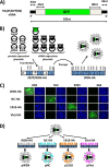
Influenza virus is a highly contagious virus that causes yearly epidemics and occasional pandemics of great consequence. Influenza virus neutralizing antibodies (NAbs) are promising prophylactic and therapeutic reagents. Detection of NAbs in serum samples is critical to evaluate the prevalence and spread of new virus strains. Here we describe the development of a simple, sensitive, specific, and safe screening assay for the rapid detection of NAbs against highly pathogenic influenza viruses under biosafety level 2 (BSL-2) conditions. This assay is based on the use of influenza viruses in which the hemagglutinin (HA) gene is replaced by a gene expressing green fluorescent protein (GFP). These GFP-expressing influenza viruses replicate to high titers in HA-expressing cell lines, but in non-HA-expressing cells, their replication is restricted to a single cycle.
Figures


Similar articles
-
Competitive detection of influenza neutralizing antibodies using a novel bivalent fluorescence-based microneutralization assay (BiFMA).Vaccine. 2015 Jul 9;33(30):3562-70. doi: 10.1016/j.vaccine.2015.05.049. Epub 2015 Jun 1. Vaccine. 2015. PMID: 26044496 Free PMC article.
-
Measurement of neutralizing antibody responses against H5N1 clades in immunized mice and ferrets using pseudotypes expressing influenza hemagglutinin and neuraminidase.Vaccine. 2009 Nov 12;27(48):6777-90. doi: 10.1016/j.vaccine.2009.08.056. Epub 2009 Sep 2. Vaccine. 2009. PMID: 19732860 Free PMC article.
-
Potential Role of Nonneutralizing IgA Antibodies in Cross-Protective Immunity against Influenza A Viruses of Multiple Hemagglutinin Subtypes.J Virol. 2020 Jun 1;94(12):e00408-20. doi: 10.1128/JVI.00408-20. Print 2020 Jun 1. J Virol. 2020. PMID: 32269119 Free PMC article.
-
Realities and enigmas of human viral influenza: pathogenesis, epidemiology and control.Vaccine. 2002 Aug 19;20(25-26):3068-87. doi: 10.1016/s0264-410x(02)00254-2. Vaccine. 2002. PMID: 12163258 Review.
-
Recent advances in "universal" influenza virus antibodies: the rise of a hidden trimeric interface in hemagglutinin globular head.Front Med. 2020 Apr;14(2):149-159. doi: 10.1007/s11684-020-0764-y. Epub 2020 Apr 1. Front Med. 2020. PMID: 32239416 Free PMC article. Review.
Cited by
-
Genetically engineered, biarsenically labeled influenza virus allows visualization of viral NS1 protein in living cells.J Virol. 2010 Jul;84(14):7204-13. doi: 10.1128/JVI.00203-10. Epub 2010 May 12. J Virol. 2010. PMID: 20463066 Free PMC article.
-
A Novel Fluorescent and Bioluminescent Bireporter Influenza A Virus To Evaluate Viral Infections.J Virol. 2019 May 1;93(10):e00032-19. doi: 10.1128/JVI.00032-19. Print 2019 May 15. J Virol. 2019. PMID: 30867298 Free PMC article.
-
Activity of human serum antibodies in an influenza virus hemagglutinin stalk-based ADCC reporter assay correlates with activity in a CD107a degranulation assay.Vaccine. 2020 Feb 18;38(8):1953-1961. doi: 10.1016/j.vaccine.2020.01.008. Epub 2020 Jan 17. Vaccine. 2020. PMID: 31959425 Free PMC article.
-
Visualizing influenza virus infection in living mice.Nat Commun. 2013;4:2369. doi: 10.1038/ncomms3369. Nat Commun. 2013. PMID: 24022374 Free PMC article.
-
Epistatic Interactions within the Influenza A Virus Polymerase Complex Mediate Mutagen Resistance and Replication Fidelity.mSphere. 2017 Aug 16;2(4):e00323-17. doi: 10.1128/mSphere.00323-17. eCollection 2017 Jul-Aug. mSphere. 2017. PMID: 28815216 Free PMC article.
References
-
- de Jong, M. D., T. T. Tran, H. K. Truong, M. H. Vo, G. J. Smith, V. C. Nguyen, V. C. Bach, T. Q. Phan, Q. H. Do, Y. Guan, J. S. Peiris, T. H. Tran, and J. Farrar. 2005. Oseltamivir resistance during treatment of influenza A (H5N1) infection. N. Engl. J. Med. 353:2667-2672. - PubMed
-
- Garten, R. J., C. T. Davis, C. A. Russell, B. Shu, S. Lindstrom, A. Balish, W. M. Sessions, X. Xu, E. Skepner, V. Deyde, M. Okomo-Adhiambo, L. Gubareva, J. Barnes, C. B. Smith, S. L. Emery, M. J. Hillman, P. Rivailler, J. Smagala, M. de Graaf, D. F. Burke, R. A. Fouchier, C. Pappas, C. M. Alpuche-Aranda, H. Lopez-Gatell, H. Olivera, I. Lopez, C. A. Myers, D. Faix, P. J. Blair, C. Yu, K. M. Keene, P. D. Dotson, Jr., D. Boxrud, A. R. Sambol, S. H. Abid, K. St. George, T. Bannerman, A. L. Moore, D. J. Stringer, P. Blevins, G. J. Demmler-Harrison, M. Ginsberg, P. Kriner, S. Waterman, S. Smole, H. F. Guevara, E. A. Belongia, P. A. Clark, S. T. Beatrice, R. Donis, J. Katz, L. Finelli, C. B. Bridges, M. Shaw, D. B. Jernigan, T. M. Uyeki, D. J. Smith, A. I. Klimov, and N. J. Cox. 2009. Antigenic and genetic characteristics of swine-origin 2009 A(H1N1) influenza viruses circulating in humans. Science 325:197-201. - PMC - PubMed
-
- Johnson, N. P., and J. Mueller. 2002. Updating the accounts: global mortality of the 1918-1920 “Spanish” influenza pandemic. Bull. Hist. Med. 76:105-115. - PubMed
-
- Kashyap, A. K., J. Steel, A. F. Oner, M. A. Dillon, R. E. Swale, K. M. Wall, K. J. Perry, A. Faynboym, M. Ilhan, M. Horowitz, L. Horowitz, P. Palese, R. R. Bhatt, and R. A. Lerner. 2008. Combinatorial antibody libraries from survivors of the Turkish H5N1 avian influenza outbreak reveal virus neutralization strategies. Proc. Natl. Acad. Sci. U. S. A. 105:5986-5991. - PMC - PubMed
Publication types
MeSH terms
Substances
Grants and funding
LinkOut - more resources
Full Text Sources
Other Literature Sources

