. 2010 Feb;84(4):2047-62.
doi: 10.1128/JVI.01984-09. Epub 2009 Nov 25.

Cellular corepressor TLE2 inhibits replication-and-transcription- activator-mediated transactivation and lytic reactivation of Kaposi's sarcoma-associated herpesvirus

Affiliations

Cellular corepressor TLE2 inhibits replication-and-transcription- activator-mediated transactivation and lytic reactivation of Kaposi's sarcoma-associated herpesvirus

Zhiheng He et al. J Virol. 2010 Feb.

Abstract

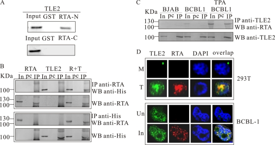
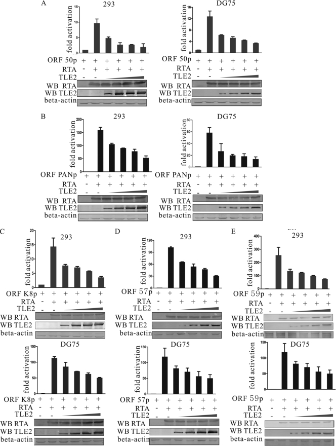
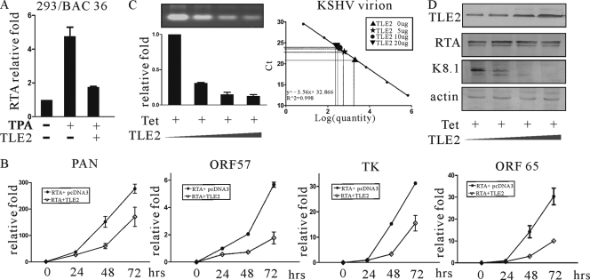
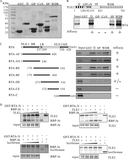
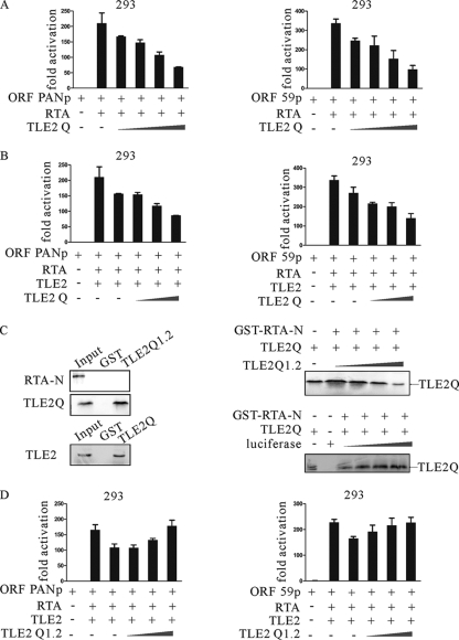
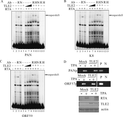
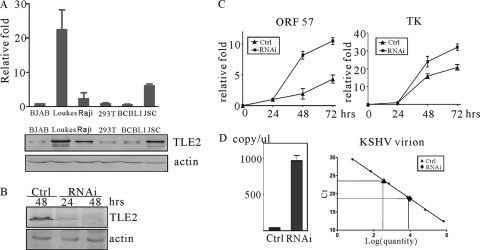
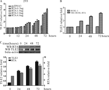
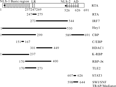
Replication and transcription activator (RTA) encoded by open reading frame 50 (ORF50) of Kaposi's sarcoma-associated herpesvirus (KSHV) is essential and sufficient to initiate lytic reactivation. RTA activates its target genes through direct binding with high affinity to its responsive elements or by interaction with cellular factors, such as RBP-Jkappa, Ap-1, C/EBP-alpha, and Oct-1. In this study, we identified transducin-like enhancer of split 2 (TLE2) as a novel RTA binding protein by using yeast two-hybrid screening of a human spleen cDNA library. The interaction between TLE2 and RTA was confirmed by glutathione S-transferase (GST) binding and coimmunoprecipitation assays. Immunofluorescence analysis showed that TLE2 and RTA were colocalized in the same nuclear compartment in KSHV-infected cells. This interaction recruited TLE2 to RTA bound to its recognition sites on DNA and repressed RTA auto-activation and transactivation activity. Moreover, TLE2 also inhibited the induction of lytic replication and virion production driven by RTA. We further showed that the Q (Gln-rich), SP (Ser-Pro-rich), and WDR (Trp-Asp repeat) domains of TLE2 and the Pro-rich domain of RTA were essential for this interaction. RBP-Jkappa has been shown previously to bind to the same Pro-rich domain of RTA, and this binding can be subject to competition by TLE2. In addition, TLE2 can form a complex with RTA to access the cognate DNA sequence of the RTA-responsive element at different promoters. Intriguingly, the transcription level of TLE2 could be upregulated by RTA during the lytic reactivation process. In conclusion, we identified a new RTA binding protein, TLE2, and demonstrated that TLE2 inhibited replication and transactivation mediated by RTA. This provides another potentially important mechanism for maintenance of KSHV viral latency through interaction with a host protein.


Figures

FIG. 1.
FIG. 1.
GST binding assay, coimmunoprecipitation, and immunofluorescence indicate that TLE2 interacts with RTA in vitro and in vivo. (A) GST-fused RTA protein (aa 1 to 530) bound to in vitro transcribed and translated 35S-labeled TLE2 protein. In vitro transcribed and translated TLE2 was added to immobilized GST-RTA or GST control. A control lane indicates the size of the labeled in vitro transcribed and translated TLE2, using as input counts per minute about 20% of those used in the pulldown assay with GST-RTA. (B) HEK 293T cells were transfected with expression constructs of His-tagged TLE2 and/or full-length RTA. Following extract preparation, complexes were immunoprecipitated with His or RTA MAb and analyzed by Western blotting. (C) BCBL1 cells were mock induced or TPA induced. Cell extracts were immunoprecipitated with anti-TLE2 antibody and analyzed by Western blotting with anti-RTA MAb. (D) Immunofluorescence showed that RTA was localized to the same nuclear compartment as TLE2 in different cells. HEK 293T cells were transfected with 20 μg of pcDNA3.1 (M, mock) or cotransfected with 10 μg of pCR3.1-RTA and 10 μg of pcDNA-TLE2 expression vectors (T, transfection). At 24 h posttransfection, cells were harvested for immunofluorescence analysis. Uninduced (Un) or induced (In) BCBL1 cells were also used for immunofluorescence analysis. In, input; Pc, preclear; IP, immunoprecipitation; WB, Western blotting.
FIG. 2.
FIG. 2.
TLE2 represses RTA-mediated auto-activation (A) and transactivation (B to E). Ten million HEK 293 cells or DG75 lymphoma cells were transfected with 1 μg of luciferase reporter construct, 1 μg of pCR3.1-RTA, and 2.5, 5, 10, or 20 μg of pcDNA-TLE2 expression construct. Total transfected DNA was normalized with pcDNA3.1. Promoter activity was expressed as the fold activation relative to activity with reporter alone (control). Means and standard deviations (SDs) from three independent transfections are shown. Protein lysates were analyzed by Western blotting (WB) for expression of transfected protein with His MAb to detect TLE2 and the RTA MAb to detect RTA protein and for levels of internal control with the β-actin polyclonal antibody.
FIG. 3.
FIG. 3.
TLE2 downregulates RTA-mediated lytic gene transactivation and KSHV virion production through repression of RTA. (A) Real-time PCR analysis of RTA transcripts in 293/Bac36 cells induced by TPA. 293/Bac36 cells were transfected with TLE2 expression vector or control vector. At 12 h posttransfection, cells were treated with TPA at a final concentration of 20 ng/ml. Real-time PCR was performed at 24 h postinduction. (B) Real-time PCR analysis of PAN, ORF57, TK, and ORF65 transcripts in 293/Bac36 cells. 293/Bac36 cells were transfected with an RTA expression vector to reactivate the virus. A TLE2 expression construct or pcDNA3 was cotransfected. Real-time PCR was performed at 12, 24, 48, and 72 h posttransfection. (C) Ten million BCBL1 cells were transfected with 5, 10, or 20 μg of pcDNA-TLE2 expression vector. At 24 h posttransfection, cells were induced with TPA. At 72 h postinduction, supernatants of transfected cells were harvested for PCR to check the virion production level of KSHV. A standard curve for virion quantification was used to calculate the number of KSHV virions in the supernatants. (D) Cell lysates were analyzed by Western blotting for expression of transfected protein with His MAb to detect TLE2, K8.1 MAb to detect KSHV ORF K8.1, RTA MAb to detect RTA protein, and the β-actin polyclonal antibody for levels of internal control. Lanes 1, 20.0 μg of pcDNA3.1; lanes 2, 5 μg of pcDNA-TLE2 and 15 μg of pcDNA3.1; lanes 3, 10 μg of pcDNA-TLE2 and 10 μg of pcDNA3.1; lanes 4, 20 μg of pcDNA-TLE2.
FIG. 4.
FIG. 4.
Mapping the RTA interaction domains in TLE2 and TLE2 interaction domains in RTA. (A) SDS-PAGE was used to analyze the purified GST-fused TLE2 domains expressed in E. coli. (B) Truncated versions of TLE2 (TLE2-Q, TLE2-GP, TLE2-CcN, TLE2-SP, and TLE2-WDR) are shown schematically, along with the positions of the start and end residues. All TLE2 truncated constructs (directed under T7 promoter) were 35S labeled by in vitro translation (1/10 loading), bound to GST or GST-RTA-N beads, washed with NETN buffer, and separated by SDS-PAGE. (C) The conserved domains of RTA and truncated versions of RTA (RTA-AF, RTA-AG, RTA-BE, RTA-BF, RTA-DE, and RTA-CE) are shown schematically, along with the positions of the start and end residues: the basic region (aa 1 to 237), Leu repeats (aa 247 to 269), activation domain (AD), and two nuclear localization sites (NLS-1 and NLS-2). All RTA truncated constructs (directed under T7 promoter) were 35S labeled by in vitro translation (1/10 loading), bound to GST or GST-TLE2-Q/SP/WDR beads, washed with NETN buffer, and separated by SDS-PAGE. (D) TLE2/RBP-Jκ competition for RTA binding is shown in the top panel. TLE2 and RBP-Jκ were in vitro transcribed and translated. The 35S-labeled products were incubated with GST-RTA-N. A fixed amount of RBP-Jκ and increasing amounts of TLE2 (left) or a fixed amount of TLE2 and increasing amounts of RBP-Jκ (right) were used. Pulldown products were electrophoresed on 8% SDS-PAGE gels, dried, and exposed to a PhosphorImager. Input controls of 10% for TLE2 and RBP-Jκ were also run. In the bottom panel, a fixed amount of RBP-Jκ (left) or TLE2 (right) and increasing amounts of luciferase were used. Pulldown products were electrophoresed on 8% SDS-PAGE gels, dried, and exposed to a PhosphorImager.
FIG. 5.
FIG. 5.
Q domain is indispensable for repression mediated by TLE2. (A) Ten million HEK 293 cells were transfected with 1 μg of luciferase reporter construct, 1 μg of pCR3.1-RTA, and 2.5, 5, 10, or 20 μg of pcDNA-TLE2-Q expression construct. (B) Ten million HEK 293 cells were transfected with 1 μg of luciferase reporter construct, 1 μg of pCR3.1-RTA, 10 μg of TLE2, and 2.5, 5, or 10 μg of pcDNA-TLE2-Q expression construct. Total transfected DNA was normalized with pcDNA3.1. Promoter activity was expressed as the fold activation relative to activity with reporter alone (control). Means and standard deviations (SDs) from three independent transfections are shown. (C) TLE2-Q and RTA constructs (directed under T7 promoter) were 35S labeled by in vitro translation (1/10 loading), bound to GST or GST-TLE2-Q1.2 beads, washed with NETN buffer, and separated by SDS-PAGE (upper panel). TLE2 (directed under T7 promoter) were 35S labeled by in vitro translation (1/10 loading), bound to either GST or GST-TLE2-Q beads, washed with NETN buffer, and separated by SDS-PAGE (middle panel). TLE2-Q1.2-interfered tetramerization of TLE2. TLE2-Q and TLE-Q1.2 were in vitro transcribed and translated (bottom panel). The 35S-labeled products were incubated with GST-RTA-N. A fixed amount of TLE2 and increasing amounts of TLE2-Q1.2 were used. Pulldown products were electrophoresed on 8% SDS-PAGE gels, dried, and exposed to a PhosphorImager. Input controls of 10% for TLE2 and TLE2-Q1.2 were also run. A fixed amount of TLE2 and increasing amounts of luciferase were used. Pulldown products were electrophoresed on 15% SDS-PAGE gels, dried, and exposed to a PhosphorImager. (D) Ten million HEK 293 cells were transfected with 1 μg of luciferase reporter construct, 1 μg of pCR3.1-RTA, 10 μg of TLE2, and 2.5, 5, or 10 μg of pcDNA-TLE2-Q1.2 expression construct. Total transfected DNA was normalized with pcDNA3.1. Promoter activity was expressed as the fold activation relative to activity with reporter alone (control). Means and SDs from three independent transfections are shown.
FIG. 6.
FIG. 6.
TLE2 and RTA form a complex when recruited to RRE. (A, B, and C) 32P-labeled oligonucleotides that corresponded to the RREs identified in ORF57, ORF K8, and ORF59 promoters were incubated with nuclear extract from HEK 293 cells transfected with RTA, TLE2, or both. After incubation, complexes were analyzed by EMSA and autoradiography. The RTA-bound complexes were supershifted by anti-RTA MAb (lanes 3). Complex formation was augmented by incubation with increasing amounts of TLE2 (lanes 6 to 8), and the band representing this complex could be supershifted by anti-RTA or anti-His MAb (lanes 9 and 10). No shift was observed by addition of RTA or His MAb alone (lanes 12 and 13). R, anti-RTA MAb; H, anti-His MAb; N, nonspecific antibody. (D) TLE2 did not diminish the association between RTA and the RRE in 293/Bac36 cells. ChIP assays were carried out on nonreactivated and reactivated 293/Bac36 cells transfected with pcDNA-TLE2. Reactivated cells were treated with 20 ng/ml TPA. Endogenous RTA and associated DNA fragments were coimmunoprecipitated using an RTA-specific polyclonal IgG and protein A-agarose alongside agarose only and IgG controls. PCR amplification using primers against the PAN or ORF57 promoter (bottom). Immunoblotting was carried out with an RTA-specific IgG to confirm RTA expression following reactivation and with an actin-specific IgG to demonstrate equal loading. P, positive control; N, negative control.
FIG. 7.
FIG. 7.
TLE2 knockdown enhances RTA-mediated transactivation and lytic replication. (A) The expression level of TLE2 was assessed by quantitative reverse transcription-PCR and Western blotting. In the upper panel, the data shown are an average of the results obtained with triplicate reactions. Error bars indicate the standard deviations of the three averaged replicates. In the bottom panel, Western blotting was performed for analysis of TLE2 expression in various cell lines. Beta-actin was used as internal control. (B) 293/Bac36 cells were transfected with TLE2-specific RNAi, and cells were harvested at 24 and 48 h posttransfection. TLE2 was detected by Western blotting. (C) 293/Bac36 cells transfected with TLE2-specific RNAi were treated by TPA and harvested at 24 and 48 h posttreatment. ORF57 and TK transcripts were analyzed by real-time PCR. (D) Supernatants of transfected cells were harvested at 48 h postinduction for PCR to check the virion production level of KSHV (left). A standard curve for virion quantification was used to calculate the number of KSHV virions in the supernatants (right).
FIG. 8.
FIG. 8.
RTA upregulates TLE2 expression. Real-time PCR was used to calculate TLE2 transcripts in HEK 293 (A) and TRE-BCBL1-RTA (B) cells. HEK 293 cells were transfected with RTA expression vector. TRE-BCBL1-RTA cells were treated with tetracycline. Real-time PCR was performed at 12, 24, 48, and 72 h posttransfection or postinduction. (C) Cell lysates of TRE-BCBL1-RTA cells treated by tetracycline were analyzed by Western blotting (WB) for levels of expression of TLE2 with the TLE2 polyclonal antibody and for levels of internal control with the beta-actin polyclonal antibody. The relative densities of the bands were measured with ImageQuant software (Molecular Dynamics).
FIG. 9.
FIG. 9.
Summary of RTA binding proteins with confirmed binding regions within RTA: TRAP/mediator and SWI/SNF (27), CREB-binding protein and HDAC (28), STAT3 (29), RBP-Jκ (37), interferon regulatory factor 7 (69, 79), and CCAAT/enhancer-binding protein-alpha (75). Other RTA binding proteins with no RTA binding region defined, such as LANA (33), Oct-1 (56), are not shown here.

Similar articles

Cited by

References

    1. Ashraf, S. I., and Y. T. Ip. 1998. Transcriptional control: repression by local chromatin modification. Curr. Biol. 8:R683-R686. - PubMed
    1. Ballestas, M. E., P. A. Chatis, and K. M. Kaye. 1999. Efficient persistence of extrachromosomal KSHV DNA mediated by latency-associated nuclear antigen. Science 284:641-644. - PubMed
    1. Bernard, S., B. Cajavec, L. Pujo-Menjouet, M. C. Mackey, and H. Herzel. 2006. Modelling transcriptional feedback loops: the role of Gro/TLE1 in Hes1 oscillations. Philos. Transact. A Math. Phys. Eng. Sci. 364:1155-1170. - PubMed
    1. Brantjes, H., J. Roose, M. van De Wetering, and H. Clevers. 2001. All Tcf HMG box transcription factors interact with Groucho-related co-repressors. Nucleic Acids Res. 29:1410-1419. - PMC - PubMed
    1. Buscarlet, M., and S. Stifani. 2007. The “Marx” of Groucho on development and disease. Trends Cell Biol. 17:353-361. - PubMed

Publication types

MeSH terms

LinkOut - more resources