. 2010 Feb;84(4):1881-90.
doi: 10.1128/JVI.01856-09. Epub 2009 Nov 25.

Tpl2/AP-1 enhances murine gammaherpesvirus 68 lytic replication

Affiliations

Tpl2/AP-1 enhances murine gammaherpesvirus 68 lytic replication

Xudong Li et al. J Virol. 2010 Feb.

Abstract

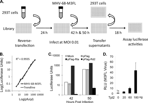
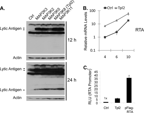
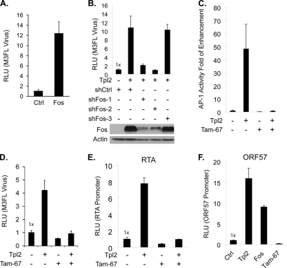
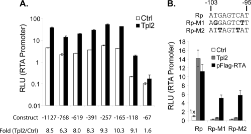
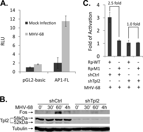
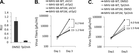
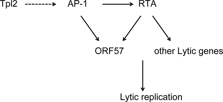
How cellular factors regulate gammaherpesvirus lytic replication is not well understood. Here, through functional screening of a cellular kinase expression library, we identified mitogen-activated protein kinase kinase kinase 8 (MAP3K8/Tpl2) as a positive regulator of murine gammaherpesvirus 68 (MHV-68 or gammaHV-68) lytic gene expression and replication. Tpl2 enhances MHV-68 lytic replication by upregulating lytic gene expression and promoter activities of viral lytic genes, including RTA and open reading frame 57 (ORF57). By screening a cellular transcription factor library, we identified the Fos AP-1 transcription factor as a downstream factor that is both necessary and sufficient for mediating the enhancement of MHV-68 lytic replication by Tpl2. In addition, Tpl2 stimulates the promoter activities of key viral lytic genes, including RTA and ORF57, in an AP-1-dependent manner. We identified an AP-1-responsive element on the MHV-68 RTA promoter as the cis element mediating the upregulation of RTA promoter activity by Tpl2. MHV-68 lytic infection upregulates Fos expression, AP-1 activity, and RTA promoter activity in a Tpl2-dependent manner. We constructed a mutant MHV-68 virus that abolished this AP-1-responsive element. This mutant virus exhibited attenuated lytic replication kinetics, indicative of a critical role of this AP-1-responsive element during lytic replication. Moreover, Tpl2 knockdown inhibited the lytic replication of wild-type MHV-68 (MHV-68-WT) but not that of the MHV-68 mutant virus, indicating that endogenous Tpl2 promotes efficient virus lytic replication through AP-1-dependent upregulation of RTA expression. In summary, through tandem functional screens, we identified the Tpl2/AP-1 signaling transduction pathway as a positive regulator of MHV-68 lytic replication.


Figures

FIG. 1.
FIG. 1.
Tpl2 enhances MHV-68 lytic replication. (A) Overall scheme of the screen. The individual kinase expression plasmids predotted on a 384-well plate were reverse transfected into 293T cells. Cells were infected with MHV-68-M3FL at an MOI of 0.01 at 24 h posttransfection. At 42 and 50 h postinfection, 10 μl of supernatants was transferred to naive 293T cells that were seeded on 96-well plates (referred to as destination plates) 1 day prior to transfer. Luciferase activities of 293T cells on the destination plates were measured at 18 h posttransfer. (B) Luciferase activities as indicators of MHV-68-M3FL titers. 293T cells were infected with serially diluted MHV-68-M3FL viruses. Luciferase activities were determined at 18 h postinfection and plotted against titers determined by plaque assays. Representative results are presented as the means for triplicates. (C) Effects of RTA and a dominant negative mutant of RTA on MHV-68-M3FL lytic replication. 293T cells were mock transfected or transfected with the indicated plasmids, followed by MHV-68-M3FL infection, supernatant transfer, and luciferase assays of cells on the destination plate as described for panel A. Error bars indicate standard deviations. (D) Dose response of MHV-68-M3FL lytic replication to MAP3K8 transfection. Virus production at 42 h postinfection from 293T cells transfected with the indicated amounts of the MAP3K8 plasmid was determined as for panel C.
FIG. 2.
FIG. 2.
Tpl2 upregulates viral lytic gene expression. (A) Western blotting analysis of MHV-68 lytic protein expression in cells expressing various kinases. 293T cells were transfected with individual kinase expression plasmids and infected with MHV-68 at an MOI of 1. At 12 or 24 h postinfection, whole-cell lysates were analyzed by Western blotting for MHV-68 lytic antigen expression using rabbit hyperimmune serum against MHV-68-infected rabbit cells. An actin immunoblot is shown as a loading control. (B) Tpl2 increases the mRNA levels of RTA. 293T cells were transfected with Tpl2 or the backbone control plasmid and infected with MHV-68 at an MOI of 1 at 24 h posttransfection. The relative mRNA levels of RTA normalized to actin were determined by reverse transcription-quantitative PCR at the indicated time points postinfection. (C) Tpl2 activates the MHV-68 RTA promoter. 293T cells were cotransfected with pBLRP, a luciferase reporter construct containing approximately 1.1 kb of upstream sequence of RTA, the control plasmid SV40-RL (Renilla luciferase), and Tpl2, pFlag-RTA, or the control backbone plasmid. Luciferase activity was determined at 24 h posttransfection. Results are presented as the means for triplicates with their standard deviations.
FIG. 3.
FIG. 3.
Tpl2 enhances MHV-68 lytic replication through upregulation of AP-1 activities. (A) Effects of Fos on MHV-68-M3FL replication. 293T cells were transfected with a Fos expression plasmid or control plasmid, followed by MHV-68-M3FL infection, supernatant transfer at 42 h postinfection, and luciferase assays of cells on the destination plate as described for Fig. 1C. Error bars indicate standard deviations. (B) shRNA against Fos inhibits Fos upregulation and MHV-68 lytic replication induced by Tpl2. 293T cells were cotransfected with Tpl2 or the control backbone plasmid with shRNA vectors targeting Fos or a control shRNA vector, shCtrl. For Western blotting analysis, whole-cell lysates were subjected to SDS-PAGE followed by Western blotting and analyzed for Fos expression at 48 h posttransfection. An actin immunoblot is shown as a loading control. For MHV-68 lytic replication assay, transfected cells were infected with MHV-68-M3FL, followed by supernatant transfer to fresh 293T cells and luciferase analysis. (C) Effects of Tpl2 or Tam-67 on AP-1 activities. 293T cells were cotransfected with an AP-1 reporter construct, SV40-RL, and Tpl2, Tam-67, or Tpl2 plus Tam-67. Luciferase activities were determined at 24 h posttransfection. (D) Tam-67 inhibits the enhancement of virus replication by Tpl2. 293T cells were transfected with the indicated plasmids, followed by MHV-68-M3FL infection, supernatant transfer at 42 h postinfection, and luciferase assays of cells on the destination plate as described for Fig. 1C. (E) Tam-67 inhibits the activation of the RTA promoter by Tpl2. The RTA promoter reporter plasmid, pBLRP, and SV40-RL were cotransfected with the indicated expression plasmids into 293T cells. Luciferase activities were determined at 24 h posttransfection. (F) Tpl2 and Fos activate the ORF57 promoter. 57pLuc and SV40-RL were cotransfected with the indicated expression plasmids into 293T cells. Luciferase activities were determined at 24 h posttransfection.
FIG. 4.
FIG. 4.
Tpl2 activates the MHV-68 RTA promoter via an AP-1-responsive element. (A) 293T cells were cotransfected with RTA promoter luciferase construct pBLRP with serial 5′ deletions and SV40-RL, a control Renilla luciferase plasmid, and Tpl2 or the control backbone plasmid. Luciferase activity was determined at 24 h posttransfection. (B) 293T cells were cotransfected with mutant pBLRP with nucleotides in the AP-1 elements mutated and SV40-RL, a control Renilla luciferase plasmid, and Tpl2, pFlag-RTA, or the control backbone plasmid. Luciferase activity was determined at 24 h posttransfection. The upper panel shows the mutations introduced into pBLRP. Results are presented as the means for triplicates with their standard deviations.
FIG. 5.
FIG. 5.
Endogenous Tpl2 promotes AP-1-dependent activation of the RTA promoter during virus lytic replication. (A) 293 cells were transfected with AP-1 luciferase reporter plasmid AP1-FL or the parental vector pGL2-basic. At 12 h posttransfection, cells were mock infected or infected with MHV-68; the cells were subjected to luciferase reporter assay at 24 h postinfection. Error bars indicate standard deviations. (B) 293 cells were transduced with shCtrl or shTpl2. At 3 days posttransduction, cells were infected with MHV-68; cells were collected at different time points postinfection for Western blotting analysis. (C) 293 cells were transduced with shCtrl or shTpl2. At 3 days posttransduction, cells were transfected with Rp-WT or Rp-M1 reporter plasmid. At 12 h posttransfection, cells were mock infected or infected with MHV-68; cells were subjected to luciferase reporter assay at 24 h postinfection. The fold activation (infection/mock infection) was calculated.
FIG. 6.
FIG. 6.
Endogenous Tpl2 promotes MHV-68 lytic replication through facilitating AP-1-dependent activation of the RTA promoter. (A) 293 cells were pretreated with Tpl2 kinase inhibitor (Tpl2Inh) or dimethyl sulfoxide (DMSO) vehicle control for an hour and were then infected with MHV-68-M3FL in the presence of Tpl2Inh or DMSO, followed by supernatant transfer to fresh 293T cells and luciferase analysis. Error bars indicate standard deviations. (B) 293 cells were transduced with shCtrl or shTpl2. At 3 days posttransduction, cells were infected with the indicated viruses at an MOI of 0.01. Cell lysates and supernatants were collected at day 1 and day 3 after infection. Virus titers were determined by plaque assays on Vero cells. (C) 293 cells were pretreated with 5 μM Tpl2Inh or DMSO vehicle control for an hour. Cells were then infected with the indicated viruses at an MOI of 0.01 in the presence of Tpl2Inh or DMSO. Cell lysates and supernatants were collected at day 1 and day 3 after infection. Virus titers were determined by plaque assays on Vero cells.
FIG. 7.
FIG. 7.
Working model of the regulation of MHV-68 lytic gene expression and lytic replication by Tpl2 and AP-1 factors. Tpl2 activates AP-1 factors to upregulate RTA and ORF57 transcription. Upregulated RTA expression leads to enhanced expression of other lytic genes and lytic replication.

Similar articles

Cited by

References

    1. Akula, S. M., N. P. Pramod, F. Z. Wang, and B. Chandran. 2002. Integrin alpha3beta1 (CD 49c/29) is a cellular receptor for Kaposi's sarcoma-associated herpesvirus (KSHV/HHV-8) entry into the target cells. Cell 108:407-419. - PubMed
    1. Bortz, E., L. Wang, Q. Jia, T. T. Wu, J. P. Whitelegge, H. Deng, Z. H. Zhou, and R. Sun. 2007. Murine gammaherpesvirus 68 ORF52 encodes a tegument protein required for virion morphogenesis in the cytoplasm. J. Virol. 81:10137-10150. - PMC - PubMed
    1. Brass, A. L., D. M. Dykxhoorn, Y. Benita, N. Yan, A. Engelman, R. J. Xavier, J. Lieberman, and S. J. Elledge. 2008. Identification of host proteins required for HIV infection through a functional genomic screen. Science 319:921-926. - PubMed
    1. Brown, H. J., M. J. Song, H. Deng, T. T. Wu, G. Cheng, and R. Sun. 2003. NF-kappaB inhibits gammaherpesvirus lytic replication. J. Virol. 77:8532-8540. - PMC - PubMed
    1. Brown, P. H., R. Alani, L. H. Preis, E. Szabo, and M. J. Birrer. 1993. Suppression of oncogene-induced transformation by a deletion mutant of c-jun. Oncogene 8:877-886. - PubMed

Publication types

MeSH terms

LinkOut - more resources