. 2010 Feb;84(3):1597-606.
doi: 10.1128/JVI.01783-09. Epub 2009 Nov 25.

Molecular determinants of adaptation of highly pathogenic avian influenza H7N7 viruses to efficient replication in the human host

Affiliations

Molecular determinants of adaptation of highly pathogenic avian influenza H7N7 viruses to efficient replication in the human host

Emmie de Wit et al. J Virol. 2010 Feb.

Abstract

Two viruses isolated during the highly pathogenic avian influenza (HPAI) H7N7 virus outbreak in The Netherlands in 2003, one isolated from a person with conjunctivitis and one from a person who died as the result of infection, were used for an in vitro study of influenza A virus pathogenicity factors. The two HPAI H7N7 viruses differed in 15 amino acid positions in five gene segments. Assays were designed to investigate the role of each of these substitutions in attachment and entry, transcription and genome replication, and virus production and release as determined by hemagglutinin (HA), polymerase proteins, nonstructural protein 1 (NS1), and neuraminidase (NA). These in vitro studies confirmed the roles of the E627K substitution in basic polymerase 2 (PB2) and the A143T substitution in HA in pathogenicity observed in a mouse model previously. However, the in vitro studies identified a contribution of acidic polymerase (PA) and NA to the efficient replication in human cells of the fatal case virus, despite the fact that these are rarely marked as determinants of pathogenicity in in vivo studies. With the exception of PB2 E627K, all substitutions contributing to enhanced replication of the fatal case virus in vitro were present in poultry viruses prior to transmission to the human fatal case, indicating that viruses with enhanced replication efficiency in the mammalian host can be generated in poultry. Thus, detailed in vitro analyses of mutations facilitating replication of avian influenza viruses in mammalian cells are important to assess the zoonotic risk posed by these viruses and, in addition, highlight the value of in vitro studies to complement animal models.


Figures

FIG. 1.
FIG. 1.
Replication kinetics of the CC (triangles) and FC (circles) viruses in MDCK (white symbols), QT6 (gray symbols), and A549 (black symbols) cells. Supernatants of cells inoculated at an MOI of 0.01 TCID50/cell were harvested at 6, 12, 24, and 48 h after inoculation and titrated by end point dilution in MDCK cells. Geometric mean titers were calculated from two independent experiments; the cutoff value was used for negative samples. Error bars indicate standard deviations. The cutoff value of the virus titration assay is indicated by a dotted line.
FIG. 2.
FIG. 2.
Replication kinetics of viruses with the H7 HAs in MDCK cells. MDCK cells were inoculated at an MOI of 0.01 TCID50/cell with the FC (•), FC-CC HA (▴), FC-CC HA I13S (□), FC-CC HA A143T (▵), and FC-CC HA K416R (○) viruses. Supernatants were harvested at 6, 12, 24, and 48 h after inoculation and titrated by end point dilution in MDCK cells. Geometric mean titers were calculated from two independent experiments; the cutoff value was used for negative samples. Error bars indicate standard deviations. The cutoff value of the virus titration assay is indicated by a dotted line.
FIG. 3.
FIG. 3.
Patterns of attachment of the CC-HA143T and FC-HA143A viruses to tissues of the human lower respiratory tract. Attachment to bronchioles (left panels) and alveoli (right panels) is shown. For comparison, attachment of the CC and FC viruses to these tissues is displayed, as shown before (42). Virus attachment is shown in red. The insets in the right panels show the pattern of virus attachment to alveolar macrophages. The depicted panels reflect the attachment pattern in the whole tissue section as much as possible, but small differences between the single panels and the overall view may exist. Arrows indicate attachment to type I pneumocytes; arrowheads indicate attachment to type II pneumocytes. (Reprinted from reference with permission of the publisher.)
FIG. 4.
FIG. 4.
Polymerase activities and replication kinetics of H7N7 viruses. (A and B) Minigenome assays were performed by transfecting 293T (A) or QT6 (B) cells with plasmids encoding the polymerase proteins and NP of the CC and FC viruses or mutants thereof. After transfection, cells were incubated 24 h at 33°C (white bars), 37°C (gray bars), or 41°C (black bars). Luminescence of a firefly luciferase reporter was standardized using a plasmid constitutively expressing Renilla luciferase protein. Relative luminescence was calculated as the percentage relative light units (firefly luciferase/Renilla luciferase) of the maximum in each experiment. Averages and standard deviations from three independent experiments are shown. Arrowheads indicate the effect of PA on polymerase activity in the context of the FC virus PB2, as explained in the text. (C to E) Replication kinetics were determined by infecting 293T (C), QT6 (D), or MDCK (E) cells at an MOI of 0.01 TCID50/cell with the CC (triangles), FC (circles), and CC-FC PB2 (squares in panels C and D) or CC-FC PA (diamonds in panel E) virus. Supernatants were collected at 6, 12, 24, and 48 h after inoculation and titrated by end point dilution in MDCK cells. Filled symbols, 37°C; open symbols, 33°C. Geometric mean titers were calculated from two independent experiments; the cutoff value was used for negative samples. Error bars indicate standard deviations. The cutoff value of the virus titration assay is indicated by a dotted line. The replication kinetics shown for the CC and FC viruses in QT6 and MDCK cells are identical to those shown in Fig. 1.
FIG. 5.
FIG. 5.
Effect of NA on replication of H7N7 virus. Supernatants of MDCK cells inoculated with an MOI of 0.01 TCID50/cell of the CC (triangles), FC (circles), CC-FC NA (squares), and FC-CC NA (diamonds) viruses in the absence (open symbols) (A) or presence (closed symbols) (B) of 1 mU/ml VCNA were harvested at 6, 12, 24, and 48 h after inoculation and titrated by end point dilution in MDCK cells. Cycle threshold values of the samples taken at 6, 12, and 24 h were determined using a real-time RT-PCR (C) and plotted against the virus titers shown in panels A and B. Geometric mean titers were calculated from two independent experiments; the cutoff value was used for negative samples. Error bars indicate standard deviations. The cutoff value of the virus titration assay is indicated by a dotted line.
FIG. 6.
FIG. 6.
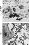
Electron microscopy of H7N7 viruses budding from 293T cells. 293T cells were transfected with plasmids encoding the FC-CC NA or FC virus. At 12 h after transfection, cells were fixed and virus budding was assessed using electron microscopy.

References

    1. Banks, J., E. S. Speidel, E. Moore, L. Plowright, A. Piccirillo, I. Capua, P. Cordioli, A. Fioretti, and D. J. Alexander. 2001. Changes in the haemagglutinin and the neuraminidase genes prior to the emergence of highly pathogenic H7N1 avian influenza viruses in Italy. Arch. Virol. 146:963-973. - PubMed
    1. Basler, C. F., and P. V. Aguilar. 2008. Progress in identifying virulence determinants of the 1918 H1N1 and the Southeast Asian H5N1 influenza A viruses. Antiviral Res. 79:166-178. - PMC - PubMed
    1. Beare, A. S., and R. G. Webster. 1991. Replication of avian influenza viruses in humans. Arch. Virol. 119:37-42. - PubMed
    1. Belser, J. A., O. Blixt, L. M. Chen, C. Pappas, T. R. Maines, N. Van Hoeven, R. Donis, J. Busch, R. McBride, J. C. Paulson, J. M. Katz, and T. M. Tumpey. 2008. Contemporary North American influenza H7 viruses possess human receptor specificity: implications for virus transmissibility. Proc. Natl. Acad. Sci. U. S. A. 105:7558-7563. - PMC - PubMed
    1. Belser, J. A., X. Lu, T. R. Maines, C. Smith, Y. Li, R. O. Donis, J. M. Katz, and T. M. Tumpey. 2007. Pathogenesis of avian influenza (H7) virus infection in mice and ferrets: enhanced virulence of Eurasian H7N7 viruses isolated from humans. J. Virol. 81:11139-11147. - PMC - PubMed

Publication types