Comparative Study
. 2010 Feb;84(4):2013-26.
doi: 10.1128/JVI.02081-09. Epub 2009 Nov 25.

Simian rotaviruses possess divergent gene constellations that originated from interspecies transmission and reassortment

Affiliations
Comparative Study

Simian rotaviruses possess divergent gene constellations that originated from interspecies transmission and reassortment

Jelle Matthijnssens et al. J Virol. 2010 Feb.

Abstract

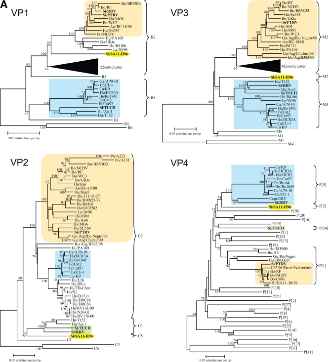
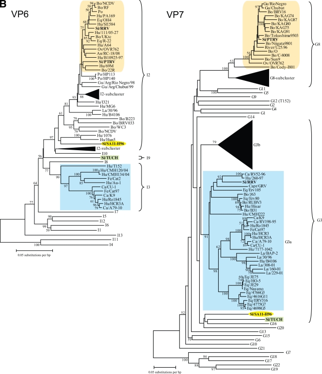
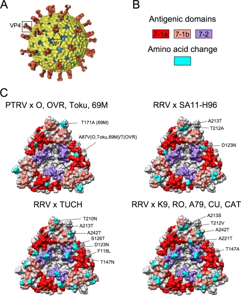
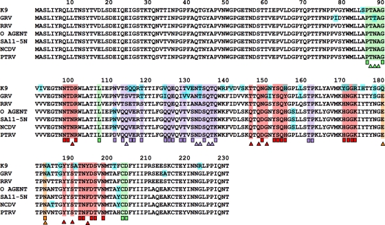
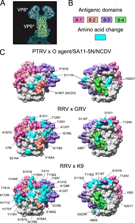
Although few simian rotaviruses (RVs) have been isolated, such strains have been important for basic research and vaccine development. To explore the origins of simian RVs, the complete genome sequences of strains PTRV (G8P[1]), RRV (G3P[3]), and TUCH (G3P[24]) were determined. These data allowed the genotype constellations of each virus to be determined and the phylogenetic relationships of the simian strains with each other and with nonsimian RVs to be elucidated. The results indicate that PTRV was likely transmitted from a bovine or other ruminant into pig-tailed macaques (its host of origin), since its genes have genotypes and encode outer-capsid proteins similar to those of bovine RVs. In contrast, most of the genes of rhesus-macaque strains, RRV and TUCH, have genotypes more typical of canine-feline RVs. However, the sequences of the canine and/or feline (canine/feline)-like genes of RRV and TUCH are only distantly related to those of modern canine/feline RVs, indicating that any potential transmission of a progenitor of these viruses from a canine/feline host to a simian host was not recent. The remaining genes of RRV and TUCH appear to have originated through reassortment with bovine, human, or other RV strains. Finally, comparison of PTRV, RRV, and TUCH genes with those of the vervet-monkey RV SA11-H96 (G3P[2]) indicates that SA11-H96 shares little genetic similarity to other simian strains and likely has evolved independently. Collectively, our data indicate that simian RVs are of diverse ancestry with genome constellations that originated largely by interspecies transmission and reassortment with nonhuman animal RVs.


Figures

FIG. 1.
FIG. 1.
Phylogenetic neighbor-joining trees based on the open reading frame sequences of the RV genes for VP1, VP2, VP3, and VP4 (A); VP6 and VP7 (B); and NSP1, NSP2, NSP3, NSP4, and NSP5 (C). Bootstrap values (500 replicates) above 50 are shown. Sequences used in building the trees were downloaded from GenBank; sequence alignments will be provided on request. Only certain genotypes are shown in detail; others are represented only by their genotype number. The annotation defines species of origin/strain name. Simian RV strains are in boldface. The phylogenetic cluster comprising simian and feline/canine/human strains is boxed in pale blue. The cluster comprising PTRV and the Bovidae/artiodactyl RV strains is boxed in pale yellow. Any simian RV not already boxed, is highlighted in green. The SA11-H96 strain is surrounded by a yellow oval.
FIG. 1.
FIG. 1.
Phylogenetic neighbor-joining trees based on the open reading frame sequences of the RV genes for VP1, VP2, VP3, and VP4 (A); VP6 and VP7 (B); and NSP1, NSP2, NSP3, NSP4, and NSP5 (C). Bootstrap values (500 replicates) above 50 are shown. Sequences used in building the trees were downloaded from GenBank; sequence alignments will be provided on request. Only certain genotypes are shown in detail; others are represented only by their genotype number. The annotation defines species of origin/strain name. Simian RV strains are in boldface. The phylogenetic cluster comprising simian and feline/canine/human strains is boxed in pale blue. The cluster comprising PTRV and the Bovidae/artiodactyl RV strains is boxed in pale yellow. Any simian RV not already boxed, is highlighted in green. The SA11-H96 strain is surrounded by a yellow oval.
FIG. 2.
FIG. 2.
Amino acid alignment of the VP7 protein of simian and related RV strains. Antigenic regions A to F are identified by heavy lines above sequences. Antigenic domains 7-1a (red), 7-1b (orange), and 7-2 (purple) are highlighted by color shading. Assignment of specific residues to antigenic domains was based on sequencing of neutralization escape mutants (triangles) and analysis of G-type specific sequence variations (squares) (1, 24). Surface-exposed residues identified in Fig. 3 that differ between certain virus strains are colored cyan.
FIG. 3.
FIG. 3.
Structure-based analysis of the antigenic domains of the VP7 trimer of simian RVs. (A) Cryoelectron microscopy image reconstruction of the SA11 RV virion, taken with permission from Pesavento et al. (34). The multimeric VP4 attachment protein and VP7 trimer are identified. (B) Color code for the VP7 antigenic domains and residues differing among VP7 proteins in panel C. Sequence alignments identifying antigenic domains and amino acid differences are shown in Fig. 2. (C) Surface renderings of the RRV VP7 trimer (PDB 3FMG) from the perspective of the virion exterior, with residues color-coded to reveal antigenic domains. Colored in cyan are the approximate positions of the surface-exposed residues of the PTRV (G8) VP7 trimer that differ from those of the G8 VP7 trimers of O-agent (O), OVR762 (OVR), Tokushima 9503 (Toku), and 69M and the surface-exposed residues of the RRV (G3) VP7 trimer that differ from those of the G3 VP7 trimers of SA11-H96, TUCH, K9, RO1845 (RO), A79-10 (A79), CU-1 (CU), and Cat97 (CAT). Only residues in one of the trimers have been labeled. VP7 GenBank accession numbers: O-agent, DQ838596; OVR762, EF554153; 69M, EF672560; K9, EU708928; RO1845, EU708895; A79-10, EU708939; CU-1, EU708917; Cat97, EU708950.
FIG. 4.
FIG. 4.
Amino acid alignment of the VP8* protein of simian and related RV strains. Antigenic domains 8-1 (red), 8-2 (orange), 8-3 (purple), and 8-4 (green) are highlighted by color shading. Assignment of specific residues to antigenic domains was based on sequencing of neutralization escape mutants (triangles) and analysis of P-type specific sequence variations (squares) (24). Surface-exposed residues identified in Fig. 5 that differ between certain virus strains are colored cyan.
FIG. 5.
FIG. 5.
Comparative analyses of the VP8* core of the RV VP4 protein. (A) Alpha-carbon traces of RRV VP8*-core monomers (white) and a VP5*-core dimer (yellow) core fit into a density map of a VP4 dimer as determined by cryoelectron-microscopy image reconstruction. The image was taken with permission from Monnier et al. (26). (B) Color code for the VP8* antigenic domains and residues differing among VP8* proteins in panel C. Sequence alignments identifying antigenic domains and amino acid differences are shown in Fig. 4. (C) Surface renderings of front (left) and back (right) orientations of the RRV VP8*-core monomer (PDB 1KQR) (4), color coded to reveal antigenic domains. The front orientation approximates that of the leftward VP8* globular head in panel A. The sialic acid binding site of the front orientation is defined by the yellow elemental stick model of a sialoside. Colored in cyan are the approximate positions of the surface-exposed residues of PTRV (P[1]) VP8* that differ from those of the P[1] VP8* proteins of O-agent, SA11-5N, and NCDV, and the surface-exposed residues of RRV (P[3]) that differ from those of the P[3] VP8* proteins of GRV and K9. Residues that differ have been labeled. VP4 GenBank accession numbers: O-agent, DQ838596; SA11-5N, DQ838602; NCDV, AB119636; GRV, AB055967; K9, EU708926.

Similar articles

Cited by

References

    1. Aoki, S. T., E. C. Settembre, S. D. Trask, H. B. Greenberg, S. C. Harrison, and P. R. Dormitzer. 2009. Structure of rotavirus outer-layer protein VP7 bound with a neutralizing Fab. Science 324:1444-1447. - PMC - PubMed
    1. Banyai, K., V. Martella, P. Molnar, I. Mihaly, M. Van Ranst, and J. Matthijnssens. 2009. Genetic heterogeneity in human G6P[14] rotavirus strains detected in Hungary suggests independent zoonotic origin. J. Infect. 59:213-215. - PubMed
    1. Centers for Disease Control and Prevention. 1999. Withdrawal of rotavirus vaccine recommendation. MMWR Morb. Mortal. Wkly. Rep. 48:1007. - PubMed
    1. Dormitzer, P. R., Z. Y. Sun, G. Wagner, and S. C. Harrison. 2002. The rhesus rotavirus VP4 sialic acid binding domain has a galectin fold with a novel carbohydrate binding site. EMBO J. 21:885-897. - PMC - PubMed
    1. Estes, M., and A. Kapikian. 2007. Rotaviruses, p. 1917-1974. In D. M. Knipe, P. M. Howley, D. E. Griffin, R. A. Lamb, M. A. Martin, B. Roizman, and S. E. Straus (ed.), Fields virology, 5th ed. Kluwer Health/Lippincott/The Williams & Wilkins Co., Philadelphia, PA.

Publication types

MeSH terms

Associated data

LinkOut - more resources