. 2010 Feb;105(2):221-31.
doi: 10.1093/aob/mcp276. Epub 2009 Nov 19.

The progamic phase of an early-divergent angiosperm, Annona cherimola (Annonaceae)

Affiliations

The progamic phase of an early-divergent angiosperm, Annona cherimola (Annonaceae)

J Lora et al. Ann Bot. 2010 Feb.

Abstract

Background and aims: Recent studies of reproductive biology in ancient angiosperm lineages are beginning to shed light on the early evolution of flowering plants, but comparative studies are restricted by fragmented and meagre species representation in these angiosperm clades. In the present study, the progamic phase, from pollination to fertilization, is characterized in Annona cherimola, which is a member of the Annonaceae, the largest extant family among early-divergent angiosperms. Beside interest due to its phylogenetic position, this species is also an ancient crop with a clear niche for expansion in subtropical climates.

Methods: The kinetics of the reproductive process was established following controlled pollinations and sequential fixation. Gynoecium anatomy, pollen tube pathway, embryo sac and early post-fertilization events were characterized histochemically.

Key results: A plesiomorphic gynoecium with a semi-open carpel shows a continuous secretory papillar surface along the carpel margins, which run from the stigma down to the obturator in the ovary. The pollen grains germinate in the stigma and compete in the stigma-style interface to reach the narrow secretory area that lines the margins of the semi-open stylar canal and is able to host just one to three pollen tubes. The embryo sac has eight nuclei and is well provisioned with large starch grains that are used during early cellular endosperm development.

Conclusions: A plesiomorphic simple gynoecium hosts a simple pollen-pistil interaction, based on a support-control system of pollen tube growth. Support is provided through basipetal secretory activity in the cells that line the pollen tube pathway. Spatial constraints, favouring pollen tube competition, are mediated by a dramatic reduction in the secretory surface available for pollen tube growth at the stigma-style interface. This extramural pollen tube competition contrasts with the intrastylar competition predominant in more recently derived lineages of angiosperms.


Figures
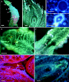
Fig. 1
Fig. 1
Gynoecium anatomy and pollen tube growth in Annona cherimola. (A) Pistil showing the stigma (stg), short style (stl) and ovary (ov) with partial postgenital fusion at the periphery of the innermost side (arrowhead). (B) Longitudinal section of the pistil showing pollen tube growth (arrowhead) through the short open stylar canal that leads to an anatropous ovule. (C) Oil cells. (D) Thick-walled sclereid cells. (E) Pollen tubes growing on the stigma towards the stigmatic furrow that leads to the stylar canal. (F) Stigma–style interface, with the stigmatic furrow leading to the narrow receptive closing margins of the stylar canal (arrowhead) and pollen tube growing through (pt). (G) Pollen tube (arrowhead) reaching the locule over the continuous papilar secretory zone. (H) Pollen tube growing through the micropyle formed by the inner integument (ii) that protrudes over a hood-shaped outer integument (oi), and reaching the nucellus (nu) 24 h after pollination. (A) Whole mount of a dissected pistil stained with aniline blue. (B, H) Aniline blue staining of a 10-μm paraffin section. (C, D) DAPI staining of a 5-μm resin section. (E, F) Aniline blue staining of squashed preparation. (G) Mixed staining of a 10-μm paraffin section. Scale bars: (A) 200 µm; (B) 200 µm; (C) 20 µm; (D) 20 µm; (E) 20 µm; (F) 100 µm; (G) 20 µm; (H) 20 µm.
Fig. 2
Fig. 2
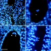
Embryo sac in Annona cherimola. (A) Ovule showing the micropyle (asterisk) formed by the inner integument (ii), the shorter outer integument (oi) and the embryo sac with two synergids (sy). (B) Egg cell (ec). (C) Two of the three antipodal cells (arrowheads). (D) Two polar nuclei (pn). DAPI staining of 5-μm resin sections. Scale bars = 20 µm.
Fig. 3
Fig. 3
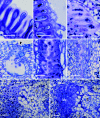
Embryo sac and endosperm development in Annona cherimola. (A) Embryo sac of flower in preanthesis with starch grains (sg) around the two polar nuclei and showing the three antipodal cells (arrowhead). The difference between the standard starch grains (sg) located in the sporophytic tissues of the ovule and the big starch grains in the female gametophyte is apparent. (B) Egg cell with a large vacuole at the base of the cell. (C) Two polar nuclei surrounded by large starch grains. (D) Two synergid cells with nucleus and cytoplasm at the micropylar end and a large vacuole at the top of the cell. (E) Filiform apparatus (fa) of a synergid cell. (F) Starch grains (sg) accumulating in the chalazal pole of the embryo sac in a fertilized ovule four days after pollination. (G) Cellular endosperm 8 d after pollination with starch (sg) accumulated at the cell of the chalazal end. (H) Zygote first division 8 d after pollination. PAS (A, F, H) and PAS and toluidine blue (B–E, G) staining of a 2-μm resin section. Scale bars: (A) 20 µm; (B) 10 µm; (C) 10 µm; (D) 10 µm; (E) 10 µm; (F) 20 µm; (G) 20 µm; (H) 10 µm.
Fig. 4
Fig. 4
Secretion along the pollen tube pathway in Annona cherimola. Longitudinal section of secretory papillae of the (A) stigma, (B) style and (C) obturator in preanthesis with the same cytohistological features although with differences in maturation and starch content: while at the stigma (A) starch has already disappeared, in the style (B) starch is still present and a rich secretion is apparent; and the obturator (C) is still full of starch. (D) Transverse section of the style showing a semi-open stylar canal (arrowhead) lined only in the outermost side with papillar cells (pc) with secretion. Stylar longitudinal section in the outermost papillar secretory zone at preanthesis (E), showing starch grains. Same area, 1 d after pollination (F), shows less starch in the cells and secretion. Transverse section of obturator (ob) cells at preanthesis (G) with starch; (ii: inner integument) and 1 d after pollination (H) with little starch and abundant secretion (ii: inner integument; oi: outer integument). PAS and toluidine blue staining of 2-μm resin sections. Scale bars = 20 µm.
Fig. 5
Fig. 5
Callose layering along the pollen tube pathway and during early endosperm development in Annona cherimola. Callose 1 d after pollination in papillar cells (arrowhead) of style (A) and obturator (arrowhead) of a pollinated (B) and an unpollinated (C) flower. (D) Callose in the nucellus micropylar pole 3 d after anthesis. (E) Deposition of callose in vascular bundles of the ovule (arrowhead) of unpollinated flowers 1 d after pollination. Callose in the cell plates (arrowhead) forming the walls of the cellular endosperm, 4 d after pollination (F) and 8 d after pollination (G). Aniline blue staining of 10-μm paraffin sections. Scale bars = 20 µm.
Fig. 6
Fig. 6
Mean pistil weight after anthesis in pollinated and unpollinated flowers of Annona cherimola. Bars indicate s.d.

Similar articles

Cited by

References

    1. APG II. An update of the Angiosperm Phylogeny Group classification for the orders and families of flowering plants: APG II. Botanical Journal of the Linnean Society. 2003;141:399–436.
    1. Arbeloa A, Herrero M. The significance of the obturator in the control of pollen tube entry into the ovary in peach (Prunus persica) Annals of Botany. 1987;60:681–685.
    1. Arbeloa A, Herrero M. Development of the ovular structures in peach [Prunus-persica (L.) Batsch] New Phytologist. 1991;118:527–533.
    1. Bernhardt P, Sage T, Weston P, et al. The pollination of Trimenia moorei (Trimeniaceae): floral volatiles, insect/wind pollen vectors and stigmatic self-incompatibility in a basal angiosperm. Annals of Botany. 2003;92:445–458. - PMC - PubMed
    1. Bhandari NN, Venkatar R. Embryology of Drimys winteri. Journal of the Arnold Arboretum. 1968;49:509–525.

Publication types

MeSH terms