Review
. 2010 Feb;298(2):H295-305.
doi: 10.1152/ajpheart.00884.2009. Epub 2009 Nov 25.

The vasa vasorum in diseased and nondiseased arteries

Affiliations
Review

The vasa vasorum in diseased and nondiseased arteries

Mary Jo Mulligan-Kehoe. Am J Physiol Heart Circ Physiol. 2010 Feb.

Abstract

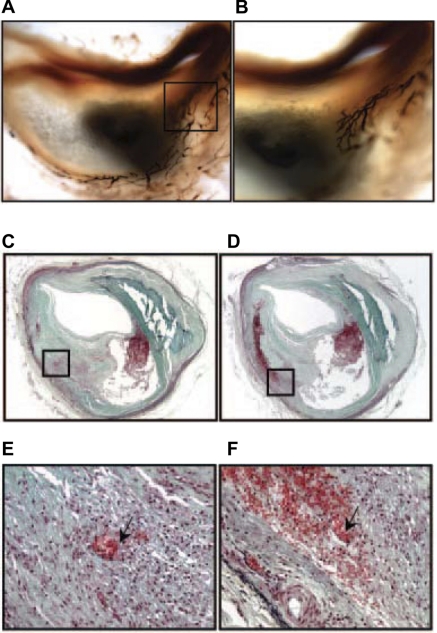
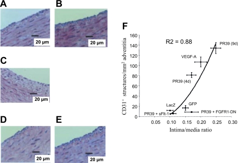
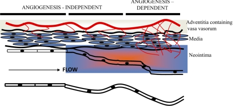
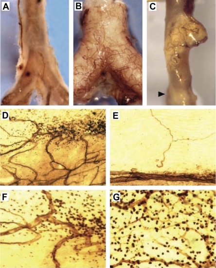
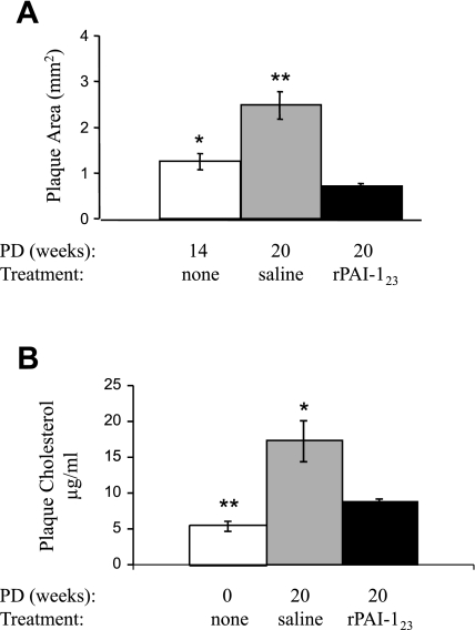
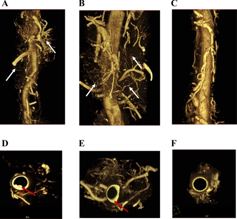
The vasa vasorum form a network of microvasculature that originate primarily in the adventitial layer of large arteries. These vessels supply oxygen and nutrients to the outer layers of the arterial wall. The expansion of the vasa vasorum to the second order is associated with neovascularization related to progression of atherosclerosis. Immunohistological analysis of human plaques from autopsied aortas have defined plaque progression and show a significant correlation with vasa vasorum neovascularization. Recent technological advances in microcomputed tomography have enabled investigation of vasa vasorum structure and function in nondiseased large arteries from pigs and dogs. Smaller mammals, particularly mice with genetic modifications that enable disease development, have been used extensively to study the vasa vasorum in diseased vessels. Despite the fact that most mouse models that are used to study atherosclerosis are unable to develop plaque to the extent found in humans, studies in both humans and mice underscore the importance of angiogenic vasa vasorum in progression of atherosclerosis. Those who have examined the vasa vasorum in occluded vessels of nondiseased pigs and dogs find that inhibition of the vasa vasorum makes the animals atheroprone. Atherosclerosis is a multifactorial disease. There is increasing evidence that factors, produced in response to changes in the arterial wall, collaborate with the vasa vasorum to enhance the disease process.


Figures

Fig. 1.
Fig. 1.
Intraplaque vasa vasorum in human coronary artery. The vasa vasorum identified with endothelial marker Ulex europaeus I is shown in the adventitial and extending through the media and into the intima (magnification ×20) (A) and the border area of the necrotic core (×40) (B). Note the multiple branches of the vasa vasorum in B. Recent hemorrhage in human thin-cap vulnerable plaque is shown in C–E. C: ×20 magnification of intraplaque hemorrhage is noted by black box; E: ×200 amplification of C to illustrate spillage of erythrocytes from vasa vasorum; D: ×20 magnification of serial section of lesion shown in C; F: ×200 amplification of D to illustrate the pool of extravasated erythrocytes surrounding microvessels (arrow). Courtesy of R. Virmani et al., Arterioscler Thromb Vasc Biol 2005 (84).
Fig. 2.
Fig. 2.
Vascular endothelial growth factor (VEGF) and fibroblast growth factor receptor (FGFR1) inhibitors block proangiogenic molecule (PR39)-induced intimal thickening and adventitial angiogenesis. Adventitia of collared carotid artery transduced with adenovirus (Ad)-expressing dominant negative (DN) FGFR (Ad-FGFR1-DN) was stained with hematoxylin and eosin 9 days posttransduction. A: VEGF inhibitor sFlt-1 (A) and FGFR1 inhibitor Ad-FGFR1-DN (B) transfer alone had no effect on neointima formation, whereas Ad-PR39 (C) increased intimal thickening after 9 days. Intimal thickening induced by Ad-PR39 was inhibited by cotransfection with sFlt-1 (D) or Ad-FGFR1-DN (E) to levels comparable to control gene transfection. F: adventitial angiogenesis is significantly correlated with degree of intimal thickening in collared arteries after treatment with angiogenesis stimulators and/or inhibitors. GFP, green fluorescent protein. Courtesy of R. Khurana et al., Circulation 2004 (41).
Fig. 3.
Fig. 3.
Adventitial angiogenesis in intimal thickening. Distinct phases of intimal thickening occur regardless of endothelial integrity. Adventitial angiogenesis contributes to intimal hyperplasia in a later phase that occurs after an injury-induced and angiogenesis-independent early phase. Courtesy of R. Khurana et al., Circulation 2005 (40).
Fig. 4.
Fig. 4.
Vasa vasorum associated with inflammatory cells in atherosclerotic plaque. CD31-probed whole-mounted aortas from either C57BL6/J (A) or ApoE−/− (B and C) mice have varied levels of vasa vasorm network associated with plaque. The normal C57BL6/J mouse in A does not have many vasa vasorum, whereas the atherogenic ApoE−/− mouse in B has extensive vasa vasorum networks associated with plaque (opaque). The vasa vasorum are not uniformly associated with advanced atheroma, as shown in C and D. E: vasa vasorum are absent in areas without plaque. F: lesion in D at ×400 magnification shows CD31+ leukocytes accumulated around the vasa vasorum. G: intense mononuclear staining associated with vasa vasorum. Courtesy of K. S. Moulton et al., Proc Natl Acad Sci USA 2003 (59).
Fig. 5.
Fig. 5.
rPAI-123 promotes regression of plaque area and plaque cholesterol in the descending aorta of atherogenic mice. Twelve-week-old female low-density lipoprotein receptor-deficient (LDLR−/−) ApoB48-deficient mice were fed Paigen's diet without cholate (PD) for varied time periods and received variable treatment in the final 6 wk of diet. A: plaque area was measured in en face preparations of descending aortas stained with Sudan IV. Plaque area relative to total area of the descending aorta was calculated in mice receiving PD for 14 wk (n = 14), PD for 20 wk [saline weeks 14–20 (n = 16)], and PD for 20 wk [rPAI-123 weeks 14–20 (n = 21)]. B: plaque cholesterol measured in the descending aorta from fasted atherogenic mice. White bars: chow diet in age-matched female mice (n = 6); gray bars, PD for 20 wk [saline weeks 14–20 (n = 6)]; black bars, PD for 20 wk [rPAI-123 weeks 14–20 (n = 6)]. Data are shown as means ± SE, and P values were determined by ANOVA. *P < 0.05 vs. rPAI-123. **P < 0.001 vs. rPAI-123. Courtesy of M. Drinane et al., Circ Res 2009 (17).
Fig. 6.
Fig. 6.
Microcomputed tomography images of second-order vasa vasorum. LDLR−/− ApoB48-deficient mice fed PD without cholate for either 14 or 20 wk were infused with Microfil. Descending aortas were scanned ex vivo at 6.5 μm, and three-dimensional volumetric images of vasa vasorum were reconstructed from mice receiving PD for 14 wk [no treatment (n = 3); A]; PD for 20 wk [saline weeks 14–20 (n = 3); B]; and PD for 20 wk [rPAI-123 weeks 14–20 (n = 3); C]. Reconstructed images were rotated to obtain luminal images from mice receiving PD for 14 wk (no treatment; D), PD for 20 wk (saline weeks 14–20; E), and PD for 20 wk (rPAI-123 weeks 14–20; F). White arrows, second-order vasa vasorum; red arrows, plaque. Courtesy of M. Drinane et al., Circ Res 2009 (17).

Similar articles

Cited by

References

    1. Barger AC, Beeuwkes R, 3rd, Lainey LL, Silverman KJ. Hypothesis: vasa vasorum and neovascularization of human coronary arteries. A possible role in the pathophysiology of atherosclerosis. N Engl J Med 310: 175–177, 1984 - PubMed
    1. Barker SG, Talbert A, Cottam S, Baskerville PA, Martin JF. Arterial intimal hyperplasia after occlusion of the adventitial vasa vasorum in the pig. Arterioscler Thromb 13: 70–77, 1993 - PubMed
    1. Cao R, Brakenhielm E, Wahlestedt C, Thyberg J, Cao Y. Leptin induces vascular permeability and synergistically stimulates angiogenesis with FGF-2 and VEGF. Proc Natl Acad Sci USA 98: 6390–6395, 2001 - PMC - PubMed
    1. Celletti FL, Hilfiker PR, Ghafouri P, Dake MD. Effect of human recombinant vascular endothelial growth factor165 on progression of atherosclerotic plaque. J Am Coll Cardiol 37: 2126–2130, 2001 - PubMed
    1. Celletti FL, Waugh JM, Amabile PG, Brendolan A, Hilfiker PR, Dake MD. Vascular endothelial growth factor enhances atherosclerotic plaque progression. Nat Med 7: 425–429, 2001 - PubMed

Publication types

MeSH terms

LinkOut - more resources