. 2010 Mar;332(3):1054-63.
doi: 10.1124/jpet.109.159244. Epub 2009 Nov 25.

Regulation of sigma-1 receptors and endoplasmic reticulum chaperones in the brain of methamphetamine self-administering rats

Affiliations

Regulation of sigma-1 receptors and endoplasmic reticulum chaperones in the brain of methamphetamine self-administering rats

Teruo Hayashi et al. J Pharmacol Exp Ther. 2010 Mar.

Abstract

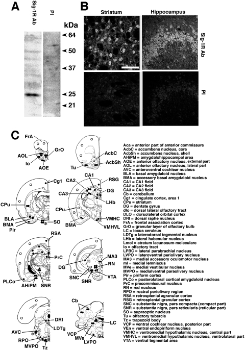
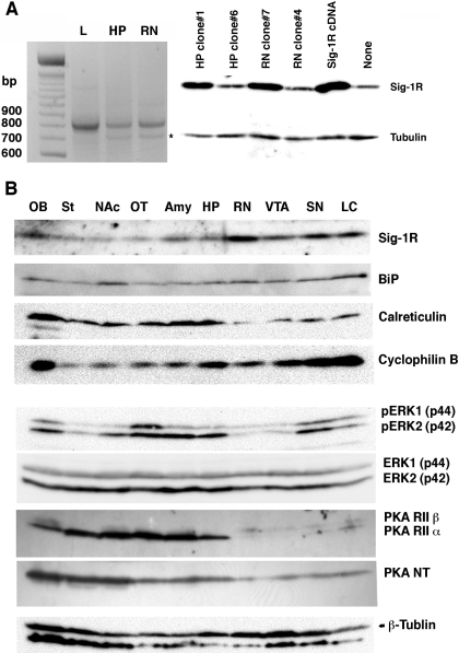
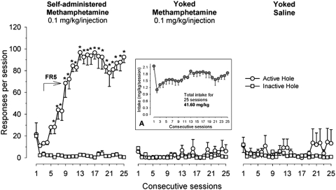
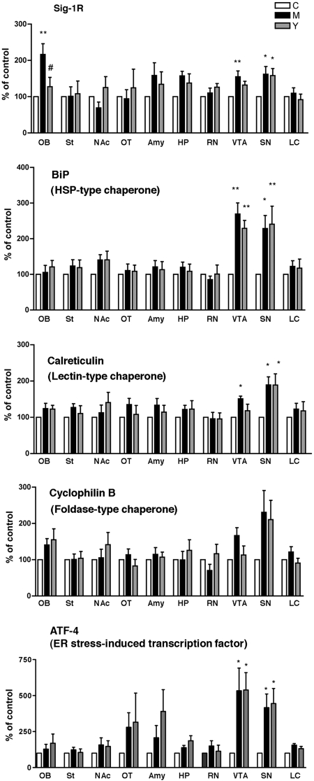
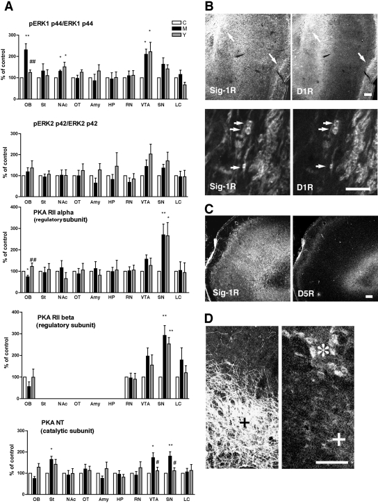
sigma-1 Receptors are endoplasmic reticulum (ER) chaperones that are implicated in the neuroplasticity associated with psychostimulant abuse. We immunocytochemically examined the distribution of sigma-1 receptors in the brain of drug-naive rats and then examined the dynamics of sigma-1 receptors and other ER chaperones in specific brain subregions of rats that self-administered methamphetamine, received methamphetamine passively, or received only saline injections. sigma-1 Receptors were found to be expressed in moderate to high levels in the olfactory bulb, striatum, nucleus accumbens shell, olfactory tubercle, amygdala, hippocampus, red nucleus, ventral tegmental area, substantia nigra, and locus ceruleus. Methamphetamine, whether self-administered or passively received, significantly elevated ER chaperones including the sigma-1 receptor, BiP, and calreticulin in the ventral tegmental area and substantia nigra. In the olfactory bulb, however, only the sigma-1 receptor chaperone was increased, and this increase occurred only in rats that actively self-administered methamphetamine. Consistent with an increase in sigma-1 receptors, extracellular signal-regulated kinase was found to be activated and protein kinase A attenuated in the olfactory bulb of methamphetamine self-administering rats. sigma-1 Receptors in the olfactory bulb were found to be colocalized with dopamine D1 receptors. These results indicate that methamphetamine induces ER stress in the ventral tegmental area and substantia nigra in rats whether the drug is received actively or passively. However, the changes seen only in rats that actively self-administered methamphetamine suggest that D1 and sigma-1 receptors in the olfactory bulb might play an important role in the motivational conditioning/learning aspects of methamphetamine self-administration in the rat.


Figures

Fig. 1.
Fig. 1.
Immunospecificity of the anti-σ-1 receptor (Sig-1R) antibody. A, Western blotting of rat brain protein lysates. Twenty micrograms of protein lysates from rat cortex was separated by 13% SDS-polyacrylamide gel electrophoresis and transblotted to a polyvinylidene difluoride membrane. Proteins were probed by either anti-Sig-1R antibody (Sig-1R Ab) or preimmune serum (PI) at a 1:750 dilution. The specific band with the 25-kDa molecular mass was detected. B, Sig-1R immunoreactivity in the rat striatum (left) or the hippocampal CA3 region (right). Top shows immunoreactivity derived from an anti-Sig-1R Ab (1:50). Bottom is from preimmune serum (1:50). Scale = 100 μm. C, schematic representation of the distribution of Sig-1R immunoreactivity in coronal sections of the rat brain. Intensities of immunoreactivities are ranked by the number of closed circles. The open circles indicate moderate expression. ∗, expression levels in pregnant rats.
Fig. 2.
Fig. 2.
σ-1 Receptor (Sig-1R) immunoreactivity in forebrain and hippocampus. A, AOL (∗) and anterior olfactory nucleus, external part (∗∗) of olfactory bulb. B, granular layer of olfactory bulb. C, olfactory tubercle. D, frontal cortex. E, layers III through IV of the parietal cortex. F, postisometric relaxation of the dorsotemporal cortex. G, nucleus accumbens shell (∗). H, dorsofrontal striatum (∗). I, cingulate cortex and corpus callosum. Note immunoreactivities along the axonal tracts (arrows). J, dentate gyrus of hippocampus. K, hippocampus CA2 (∗) and CA3 (∗∗) regions. All images are in the same scale (bar = 300 μm).
Fig. 3.
Fig. 3.
σ-1 Receptor (Sig-1R) immunoreactivity in midbrain, pons, and cerebellum. A, amygdala (∗). B, substantia nigra reticulata. C, substantia nigra compacta (∗). D, red nucleus (∗), VTA (∗∗). E, large-sized neurons in the red nucleus positive to Sig-1R. F, cochlear nucleus of a pregnant rat. G and H, trapezoid nucleus (∗) and paraolivary nucleus. ∗∗, medialventral paraolivery nucleus; #, laterolventral periolivery nucleus. I, cerebellum. J–L, locus ceruleus. Arrows, highly clustered Sig-1R at cytoplasmic structures. Scale = 50 μm in E and K, 20 μm in L. Others are in the same scale to A (bar = 300 μm).
Fig. 4.
Fig. 4.
Brain expression and distribution of σ-1 receptors (Sig-1Rs), ER chaperones, PKA, and ERK. A, expression of Sig-1R transcripts in the rat brain. Left, the result of RT-PCR using total RNAs from the liver (L), hippocampus (HP), and red nucleus (RN). One microgram of rat liver total RNA or 2 μg of rat brain total RNA was applied to RT-PCR. ∗, the 5-methyltetrahydrofolate-homocysteine methyltransferase gene nonspecifically amplified by the Sig-1R primers. All the PCR products were identified by gene sequencing. Western blotting of Sig-1Rs (right) verified the cloned HP or RN cDNA expressing Sig-1Rs in the mammalian cells. Total cell lysates (15 μg/lane) were prepared from CHO cells transfected with PCR products cloned in the pcDNA3.1 vector. HP clone 6 and RN clone 4 serve as negative controls containing the nonfunctional gene. Sig-1R cDNA is a positive vector containing the Sig-1R cDNA previously cloned (Hayashi and Su, 2001). B, brain distribution of ER chaperones and kinases. See Materials and Methods for details.
Fig. 5.
Fig. 5.
Yoked methamphetamine self-administration. The mean number (±S.E.M.) of responses in the active and inactive holes for rats that were allowed to acquire self-administration of methamphetamine at a dose of 0.1 mg/kg/injection (n = 10) and their yoked controls that received yoked infusions of methamphetamine (n = 9) or saline (n = 9) during each of the daily 2-h sessions. The arrow indicates the period when methamphetamine self-administration was maintained under the final five-response fixed-ratio schedule of reinforcement. Asterisks (∗) denote significant differences p < 0.01 between active and inactive nose pokes. Inset A, the values (mean ± S.E.M.) represent the total amount of actively self-administered (first group) or passively delivered (yoked methamphetamine group) methamphetamine during each of 25 experimental sessions.
Fig. 6.
Fig. 6.
Effect of methamphetamine self-administration on expression levels of ER chaperones. Protein levels of each molecular chaperone were measured by Western blotting. Data are shown as percentage of animals receiving saline (control). ∗∗, p < 0.01, ∗, p < 0.05 compared with control (n = 6/group). Data analyzed by one-way ANOVA followed by Tukey's multiple comparison test. See Materials and Methods for details in the sample preparation. C, yoked saline control; M, methamphetamine self-administration; Y, yoked methamphetamine control. OB, olfactory bulb; St, striatum; NAc, nucleus accumbens; OT, olfactory tubercle; Amy, basolateral amygdala; HP, dorsolateral hippocampus; RN, red nucleus; SN, substantia nigra (compacta and reticulata); LC, locus ceruleus.
Fig. 7.
Fig. 7.
Effect of methamphetamine self-administration on expression levels of PKA or phosphorylation of ERK. Protein levels or phosphorylation were measured by Western blotting. Data are shown as percentage of animals receiving saline (control). ∗∗, p < 0.01, p < 0.05 compared with control (n = 6/group). Data analyzed by one-way ANOVA followed by Tukey's multiple comparison test. See Materials and Methods for details of the sample preparation. C, yoked saline control; M, methamphetamine self-administration; Y, yoked methamphetamine control. OB, olfactory bulb; St, striatum; NAc, nucleus accumbens; OT, olfactory tubercle; Amy, basolateral amygdala; HP, dorsolateral hippocampus; RN, red nucleus; SN, substantia nigra (compacta and reticulata); LC, locus ceruleus. B, localization of σ-1 receptors (Sig-1Rs) with D1R in the olfactory bulb. Arrows indicate the potential colocalization between Sig-1Rs and D1R seen at lower magnification of AOL (left) and anterior olfactory nucleus, external part (right) regions. Bottom two panels are at the higher magnification of the AOL region. Note the clear localization of Sig-1Rs in cell bodies of D1R-postive neurons. C, no colocalization of Sig-1Rs with dopamine D5 receptors (D5R) in the olfactory bulb. D, expression of Sig-1Rs in tyrosine hydroxylase-positive VTA neurons (+). ∗, large neurons in the red nucleus positive to Sig-1R immunoreactivity. Scale = 300 μm (B, top, and C), and 100 μm (B, bottom, and D).

References

    1. Alonso G, Phan V, Guillemain I, Saunier M, Legrand A, Anoal M, Maurice T. (2000) Immunocytochemical localization of the sigma(1) receptor in the adult rat central nervous system. Neuroscience 97:155–170 - PubMed
    1. Cecchi GA, Petreanu LT, Alvarez-Buylla A, Magnasco MO. (2001) Unsupervised learning and adaptation in a model of adult neurogenesis. J Comput Neurosci 11:175–182 - PubMed
    1. Cormaci G, Mori T, Hayashi T, Su TP. (2007) Protein kinase A activation down-regulates, whereas extracellular signal-regulated kinase activation up-regulates sigma-1 receptors in B-104 cells: implication for neuroplasticity. J Pharmacol Exp Ther 320:202–210 - PubMed
    1. Davila NG, Blakemore LJ, Trombley PQ. (2003) Dopamine modulates synaptic transmission between rat olfactory bulb neurons in culture. J Neurophysiol 90:395–404 - PubMed
    1. Deng X, Ladenheim B, Jayanthi S, Cadet JL. (2007) Methamphetamine administration causes death of dopaminergic neurons in the mouse olfactory bulb. Biol Psychiatry 61:1235–1243 - PubMed

Publication types

MeSH terms

Substances