. 2010 Feb 5;285(6):4038-4048.
doi: 10.1074/jbc.M109.078345. Epub 2009 Nov 23.

c-Jun regulates shear- and injury-inducible Egr-1 expression, vein graft stenosis after autologous end-to-side transplantation in rabbits, and intimal hyperplasia in human saphenous veins

Affiliations

c-Jun regulates shear- and injury-inducible Egr-1 expression, vein graft stenosis after autologous end-to-side transplantation in rabbits, and intimal hyperplasia in human saphenous veins

Jun Ni et al. J Biol Chem. .

Erratum in

  • J Biol Chem. 2013 Nov 1;288(44):31918

Retraction in

Abstract

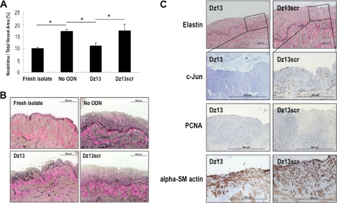
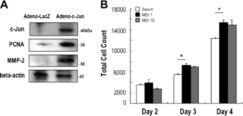
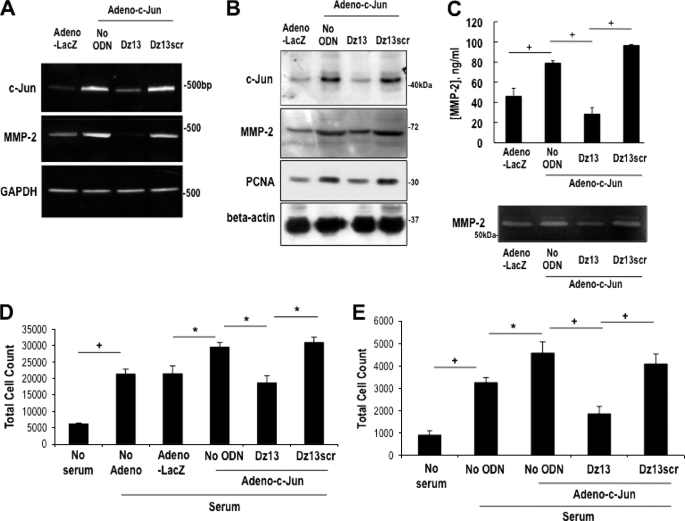
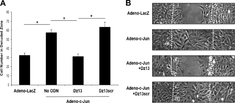
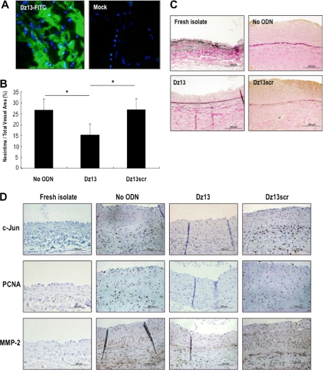
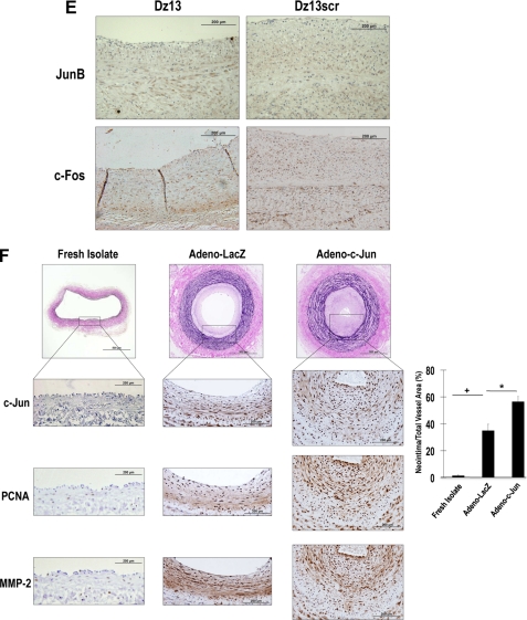
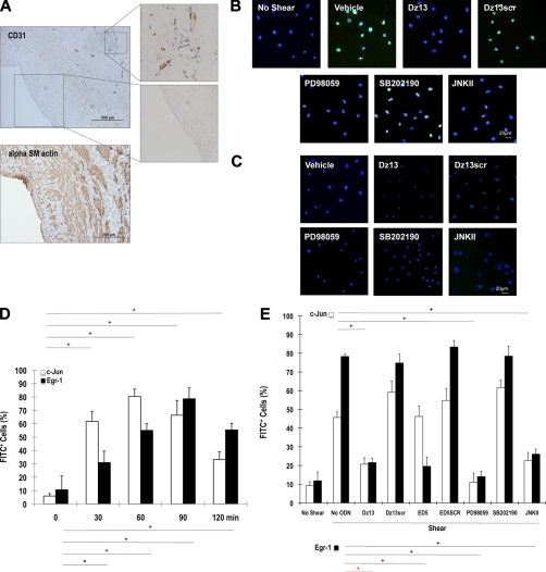
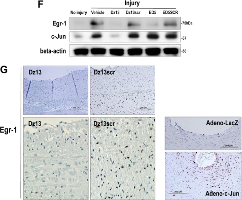
Coronary artery bypass graft failure represents an unsolved problem in interventional cardiology and heart surgery. Late occlusion of autologous saphenous vein bypass grafts is a consequence of neointima formation underpinned by smooth muscle cell (SMC) migration and proliferation. Poor long term patency and the lack of pharmacologic agents that prevent graft failure necessitate effective alternative therapies. Our objective here was to evaluate the effect of targeted inhibition of the bZIP transcription factor c-Jun on intimal hyperplasia in human saphenous veins and vein graft stenosis after autologous end-to-side transplantation. DNAzymes targeting c-Jun attenuated intimal hyperplasia in human saphenous vein explants. Adenovirus-forced c-Jun expression stimulated SMC proliferation, proliferating cell nuclear antigen, and MMP-2 expression. c-Jun DNAzymes abrogated Adeno-c-Jun-inducible SMC growth and wound repair and reduced intimal thickening in jugular veins of New Zealand white rabbits 4 weeks after autologous end-to-side transplantation to carotid arteries. Conversely, in a DNAzyme-free setting, Adeno-c-Jun potentiated neointima formation in the veins compared with Adeno-LacZ. Inducible c-Jun expression is ERK1/2- and JNK-dependent but p38-independent. Injury- and shear-inducible c-Jun controls early growth response-1. These data demonstrate that strategies targeting c-Jun may be useful for the prevention of vein graft stenosis. Control of one important shear-responsive transcription factor by another indicates the existence of transcriptional amplification mechanisms that magnify the vascular response to cell injury or stress through inducible transcriptional networks.


Figures

FIGURE 1.
FIGURE 1.
c-Jun DNAzymes attenuate intimal hyperplasia in human saphenous vein grafts. A, c-Jun DNAzyme Dz13, but not the scrambled arm counterpart (Dz13scr), attenuates neointima formation in human saphenous veins within 2 weeks. Vein explants in medium containing 20% serum were incubated with Dz13 or Dz13scr (0.4 μmol/liter), or vehicle alone, and after 14 days veins were harvested, fixed, cross-sectioned, and stained with Miller's elastin and van Gieson staining. Intimal thickening is expressed as the neointimal area as a percentage of total vessel area. B, representative cross-sections of DNAzyme, vehicle treated at 14 days, or freshly isolated saphenous veins. Magnification is 200×. C, immunohistochemical analysis for c-Jun, PCNA, and α-SM actin after 14 days. Magnification is 400×.
FIGURE 2.
FIGURE 2.
Adenovirus-c-Jun stimulates SMC proliferation. A, expression of c-Jun, PCNA, and MMP-2 in rat SMCs after transduction with adenovirus-LacZ or adenovirus c-Jun. Twenty-four hours after transduction, total cell extracts were prepared in radioimmune precipitation assay buffer prior to Western blot analysis with appropriate antibodies. B, rat SMCs in 96-well plates (1000 cells seeded per well) were transduced with adenovirus and after 24 h incubated in serum-free media for a further 24 h. The cells were then incubated in medium containing 5% FBS for up to 2 days subsequently. Days 2, 3, and 4 refer to the period since adenovirus transduction. The data are representative of at least two experiments performed in triplicate.
FIGURE 3.
FIGURE 3.
Dz13 suppresses adenovirus c-Jun-inducible SMC proliferation. A, RT-PCR, and B, Western blot analysis 24 h following adenovirus-LacZ or adenovirus-c-Jun transduction and transfection with 0.4 μmol/liter Dz13 or Dz13scr. C, adenovirus-inducible MMP-2 protein and proteolytic activity in the supernatant was determined by ELISA and zymography, respectively. D, rat SMCs (5000 cells seeded per well), and E, rabbit SMCs (1000 cells seeded per well) in 96-well plates for 24 h were transduced with adenovirus. The cells were incubated in serum-free media for 24 h after transduction, with or without transfection with 0.4 μmol/liter Dz13 or Dz13scr. After 72 h in medium containing 5% FBS, the cells were trypsinized and suspension was quantitated in a Coulter counter. The data are representative of at least two independent experiments performed in triplicate.
FIGURE 4.
FIGURE 4.
Dz13 suppresses adenovirus c-Jun-inducible SMC wound repair. A, rat SMCs grown in 6-well plates were transduced with adenovirus-c-Jun or adenovirus-LacZ and transfected with 0.4 μmol/liter Dz13 or Dz13scr in serum-free medium. The cells were scraped with a sterile P1000 pipette tip and left for 48 h prior to photomicroscopy. B, representative images at 200×. Dashed lines denote the wound edge. The data are representative of at least two independent experiments performed in triplicate.
FIGURE 5.
FIGURE 5.
Dz13 inhibits vein graft stenosis after autologous end-to-side transplantation in rabbits. A, jugular veins were extracted and transfected with FITC-labeled Dz13. Tissue was frozen in OCT, cross-sectioned, DAPI-stained, and viewed under fluorescence microscopy. B, veins were treated intraoperatively with 500 μg of DNAzyme or vehicle, and sutured end-to-side into the carotid arteries of the same rabbits. After 28 days, vein grafts were harvested, fixed, and embedded in paraffin. Sections were stained by modified Miller's elastic and van Gieson staining and photographed under 200× magnification. Intimal thickening is expressed as the neointimal area as a percentage of total vessel area. C, representative cross-sections of grafts at 28 days or of freshly isolated veins. D, immunohistochemical analysis for c-Jun, PCNA, and MMP-2 in the respective groups (magnification, 400×). E, immunohistochemical analysis for JunB and c-Fos in Dz13- and Dz13scr-treated veins (magnification, 400×). F, jugular vein segments were transduced ex vivo with adenovirus-c-Jun (1010 pfu/ml) over 30 min, prior to end-to-side transplantation into carotid arteries, and assessment of intimal thickening after 28 days. Immunohistochemical analysis was performed for c-Jun, PCNA, and MMP-2 (magnification, 400×).
FIGURE 5.
FIGURE 5.
Dz13 inhibits vein graft stenosis after autologous end-to-side transplantation in rabbits. A, jugular veins were extracted and transfected with FITC-labeled Dz13. Tissue was frozen in OCT, cross-sectioned, DAPI-stained, and viewed under fluorescence microscopy. B, veins were treated intraoperatively with 500 μg of DNAzyme or vehicle, and sutured end-to-side into the carotid arteries of the same rabbits. After 28 days, vein grafts were harvested, fixed, and embedded in paraffin. Sections were stained by modified Miller's elastic and van Gieson staining and photographed under 200× magnification. Intimal thickening is expressed as the neointimal area as a percentage of total vessel area. C, representative cross-sections of grafts at 28 days or of freshly isolated veins. D, immunohistochemical analysis for c-Jun, PCNA, and MMP-2 in the respective groups (magnification, 400×). E, immunohistochemical analysis for JunB and c-Fos in Dz13- and Dz13scr-treated veins (magnification, 400×). F, jugular vein segments were transduced ex vivo with adenovirus-c-Jun (1010 pfu/ml) over 30 min, prior to end-to-side transplantation into carotid arteries, and assessment of intimal thickening after 28 days. Immunohistochemical analysis was performed for c-Jun, PCNA, and MMP-2 (magnification, 400×).
FIGURE 6.
FIGURE 6.
c-Jun is induced in SMCs by fluid shear stress in an ERK1/2- and JNK-dependent and p38-independent manner and controls induction of the transcription factor Egr-1. A, CD31 α-SM actin+ cells reside at the inner surface of human saphenous vein conduits making contact with flowing blood (magnification, 200×). B, growth-quiescent SMCs, transfected with Dz13, Dz13scr, or incubated with PD98059, SB202190, or JNKII, were exposed to 10 dynes/cm2 fluid shear stress for 1 h and fixed, and immunocytochemical analysis was performed. c-Jun expression (FITC+) was visualized under fluorescence microscopy. Blue, DAPI-Blue staining indicates nuclei. C, SMCs were treated as in B, except that the cells remained under static conditions for the 1 h. D, SMCs were exposed to shear for the indicated times, and immunocytochemical analysis was performed for c-Jun or Egr-1 on fixed cells. FITC+ staining was expressed as a proportion of FITC+- and DAPI-Blue-stained nuclei in five random fields of view. E, effect of Dz13, Dz13scr, ED5, ED5 SCR, PD98059, SB202190, or JNKII on shear-inducible c-Jun and Egr-1 expression after 90-min exposure to fluid shear stress. FITC+ staining was expressed as a proportion of FITC+- and DAPI-Blue-stained nuclei in five random fields of view. F, Western blot analysis for Egr-1 or c-Jun using total cell extracts of SMCs (pretreated with Dz13, Dz13scr, ED5, or ED5SCR), injured by scraping and left for 1 h prior to harvest. In G: Left/upper panels, immunohistochemical analysis for Egr-1 in rabbit venoarterial autologous bypass transplants 28 days after the veins were treated ex vivo with Dz13, Dz13scr, and anastomosis. Right/lower panels, Egr-1 staining on vein transplants 28 days after the veins were transduced with Adeno-c-Jun or Adeno-LacZ.
FIGURE 6.
FIGURE 6.
c-Jun is induced in SMCs by fluid shear stress in an ERK1/2- and JNK-dependent and p38-independent manner and controls induction of the transcription factor Egr-1. A, CD31 α-SM actin+ cells reside at the inner surface of human saphenous vein conduits making contact with flowing blood (magnification, 200×). B, growth-quiescent SMCs, transfected with Dz13, Dz13scr, or incubated with PD98059, SB202190, or JNKII, were exposed to 10 dynes/cm2 fluid shear stress for 1 h and fixed, and immunocytochemical analysis was performed. c-Jun expression (FITC+) was visualized under fluorescence microscopy. Blue, DAPI-Blue staining indicates nuclei. C, SMCs were treated as in B, except that the cells remained under static conditions for the 1 h. D, SMCs were exposed to shear for the indicated times, and immunocytochemical analysis was performed for c-Jun or Egr-1 on fixed cells. FITC+ staining was expressed as a proportion of FITC+- and DAPI-Blue-stained nuclei in five random fields of view. E, effect of Dz13, Dz13scr, ED5, ED5 SCR, PD98059, SB202190, or JNKII on shear-inducible c-Jun and Egr-1 expression after 90-min exposure to fluid shear stress. FITC+ staining was expressed as a proportion of FITC+- and DAPI-Blue-stained nuclei in five random fields of view. F, Western blot analysis for Egr-1 or c-Jun using total cell extracts of SMCs (pretreated with Dz13, Dz13scr, ED5, or ED5SCR), injured by scraping and left for 1 h prior to harvest. In G: Left/upper panels, immunohistochemical analysis for Egr-1 in rabbit venoarterial autologous bypass transplants 28 days after the veins were treated ex vivo with Dz13, Dz13scr, and anastomosis. Right/lower panels, Egr-1 staining on vein transplants 28 days after the veins were transduced with Adeno-c-Jun or Adeno-LacZ.

Similar articles

Cited by

References

    1. Wang X. L., Tam C., McCredie R. M., Wilcken D. E. (1994) Circulation 89, 1974–1981 - PubMed
    1. Goldman S., Zadina K., Moritz T., Ovitt T., Sethi G., Copeland J. G., Thottapurathu L., Krasnicka B., Ellis N., Anderson R. J., Henderson W. (2004) J. Am. Coll. Cardiol. 44, 2149–2156 - PubMed
    1. Fitzgibbon G. M., Kafka H. P., Leach A. J., Keon W. J., Hooper G. D., Burton J. R. (1996) J. Am. Coll. Cardiol. 28, 616–626 - PubMed
    1. The Post Coronary Artery Bypass Graft Trial Investigators (1997) N. Engl. J. Med. 336, 153–162 - PubMed
    1. Topol E. J., Serruys P. W. (1998) Circulation 98, 1802–1820 - PubMed

Publication types

MeSH terms

Substances