. 2010 Jan 15;285(3):1716-25.
doi: 10.1074/jbc.M109.021576. Epub 2009 Nov 23.

Plasmodium falciparum BAEBL binds to heparan sulfate proteoglycans on the human erythrocyte surface

Affiliations

Plasmodium falciparum BAEBL binds to heparan sulfate proteoglycans on the human erythrocyte surface

Kyousuke Kobayashi et al. J Biol Chem. .

Abstract

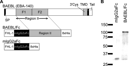
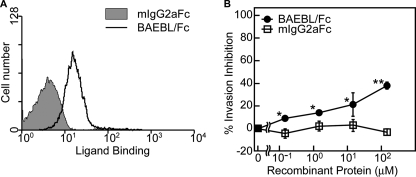
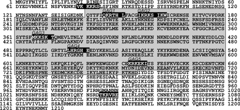
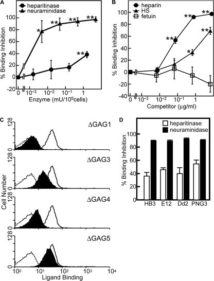
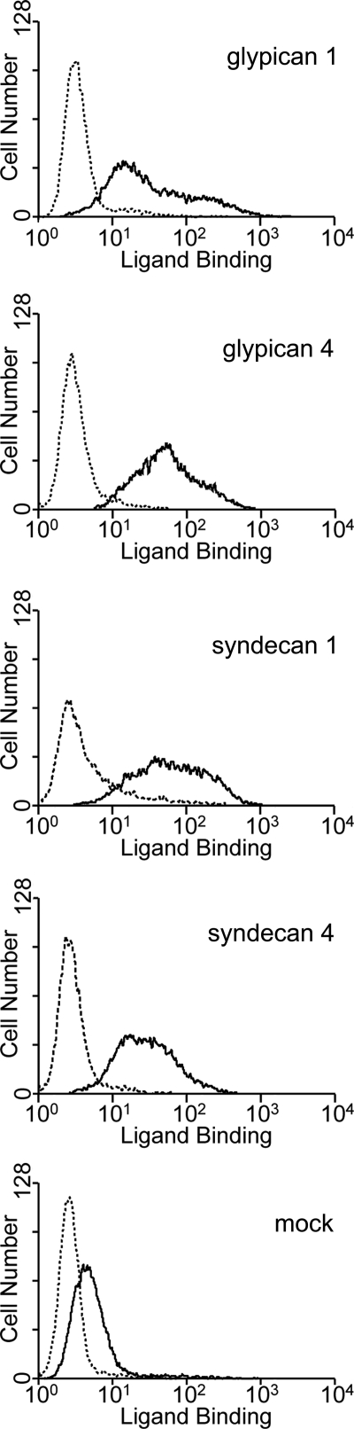
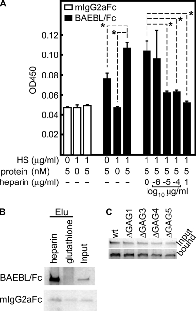
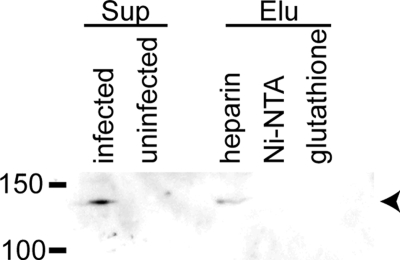
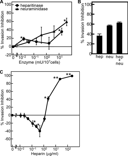
Erythrocyte invasion is critical to the pathogenesis and survival of the malarial parasite, Plasmodium falciparum. This process is partly mediated by proteins that belong to the Duffy binding-like family, which are expressed on the merozoite surface. One of these proteins, BAEBL (also known as EBA-140), is thought to bind to glycophorin C in a sialic acid-dependent manner. In this report, by the binding assay between recombinant BAEBL protein and enzyme-treated erythrocytes, we show that the binding of BAEBL to erythrocytes is mediated primarily by sialic acid and partially through heparan sulfate (HS). Because BAEBL binds to several kinds of HS proteoglycans or purified HS, the BAEBL-HS binding was found to be independent of the HS proteoglycan peptide backbone and the presence of sialic acid moieties. Furthermore, both the sialic acid- and HS-dependent binding were disrupted by the addition of soluble heparin. This inhibition may be the result of binding between BAEBL and heparin. Invasion assays demonstrated that HS-dependent binding was related to the efficiency of merozoite invasion. These results suggest that HS functions as a factor that promotes the binding of BAEBL and merozoite invasion. Moreover, these findings may explain the invasion inhibition mechanisms observed following the addition of heparin and other sulfated glycoconjugates.


Figures

FIGURE 1.
FIGURE 1.
Recombinant region II of BAEBL protein expressed as Fc fusion protein. A, schematic representations of P. falciparum BAEBL and the recombinant fusion proteins. BAEBL is composed of a signal peptide (SP), a region II, a 3′ cysteine-rich region (3Cys), a transmembrane domain (TMD), and a cytoplasmic tail (Tail). The region II of BAEBL was cloned into the pBSV-Fc-His8 vector, which contains a secretion signal sequence (FHL-1), the Fc region of mouse IgG2a (mIgG2aFc), and an octahistidine C-terminal tag (8xHis). B, recombinant proteins expressed using the baculovirus expression system and purified on Ni2+-NTA-agarose. Each protein (25 ng) was separated by 5–20% gradient SDS-PAGE and subjected to silver staining. The molecular masses (kDa) are indicated on the left.
FIGURE 2.
FIGURE 2.
BAEBL/Fc binds to erythrocytes and inhibits the merozoite invasion. A, binding of recombinant proteins to erythrocytes. The cells were incubated with each of the recombinant proteins. Cell surface binding was detected using the FACSCalibur system. B, merozoite invasion inhibition by recombinant proteins. Erythrocytes were preincubated with buffer or a 0.15, 1.5, 15, or 150 nm concentration of each recombinant protein at 37 °C for 1 h and then mixed with iRBC. Parasitemia was assessed at 20 h postinoculation, as described under “Experimental Procedures.” Results are shown as the means of three independent experiments, and the error bars represent S.E. values. Asterisks and double asterisks indicate significant differences (p < 0.05 and p < 0.001, respectively), as determined by t test.
FIGURE 3.
FIGURE 3.
The amino acid sequence of P. falciparum BAEBL. The locations of potential GAG-binding motifs are shown in white letters on a black background. The region II expressed as the fusion protein in the present study is shaded in gray. These sequences are available from GenBankTM (accession number AAK49521).
FIGURE 4.
FIGURE 4.
Recombinant BAEBL/Fc binds to erythrocytes via HS and sialic acid. A, binding inhibition of BAEBL/Fc to enzyme-treated erythrocytes. Erythrocytes (105 cells) were pretreated with buffer; 0.0016, 0.016, 0.16, or 1.6 milliunits of heparitinase; or 0.004, 0.04, 0.4, or 4 milliunits of neuraminidase and then incubated with BAEBL/Fc or mIgG2aFc. B, binding inhibition of BAEBL/Fc preincubated with soluble heparin. The recombinant protein was mixed with buffer, heparin (at a final concentration of 0.0077, 0.077, 0.77 or 7.7 μg/ml), HS, or fetuin (at a final concentration of 0.0067, 0.067, 0.67, or 6.7 mg/ml). The mixtures were incubated at 4 °C for 1 h and then added to erythrocytes. C, the binding of motif-deleted BAEBLs to erythrocytes. Erythrocytes (105 cells) were incubated with 24 nm mIgG2aFc (dashed line), BAEBL/Fc (solid line), BAEBL-ΔGAG1/Fc, BAEBL-ΔGAG3/Fc, BAEBL-ΔGAG4/Fc, or BAEBL-ΔGAG5/Fc. The bindings of motif-deleted BAEBLs are shaded in black. D, binding inhibition of BAEBL/Fc derived from four types of BAEBL variants to enzyme-treated erythrocytes. BAEBL/Fc from HB3 (VSKK), E12 (ISKK), Dd2/Nm (VSTK), and PNG3 (INRE) were incubated with erythrocytes pretreated with buffer, 1.6 milliunits of heparitinase, or 4 milliunits of neuraminidase. Bound protein was quantified, and percentage of binding inhibition was calculated as described in “Experimental Procedures.” Results are shown as the means of three independent experiments. Error bars, S.E. Asterisks and double asterisks indicate significant differences (p < 0.05 and p < 0.001, respectively) as determined by t test.
FIGURE 5.
FIGURE 5.
Binding of BAEBL to four types of HSPGs expressed on Jurkat cells. Jurkat-EcoVRc cells transduced with a retrovirus vector from an empty plasmid (mock) or the vectors that carry the cDNA of glypican 1, glypican 4, syndecan 1, or syndecan 4 were incubated with BAEBL/Fc (solid line) or mIgG2aFc (dashed line). Bound protein was detected as described under “Experimental Procedures.”
FIGURE 6.
FIGURE 6.
Recombinant BAEBL/Fc binds to purified HS and heparin. A, ELISA-based binding assays of BAEBL to HS. Purified HS was coated onto the wells of an ELISA plate and tested for binding efficiency to the recombinant proteins. The bound protein was detected with a horseradish peroxidase-conjugated anti-mouse IgG F(ab′) fragment. Binding inhibition was evaluated by the addition of buffer or 10−6, 10−5, 10−4, or 1 μg/ml soluble heparin to the HS-coated wells immediately before the addition of equal amounts of BAEBL. The optical densities at 450 nm (OD450) are shown as the means of three independent experiments. Error bars, S.E. The asterisks indicate a significant difference (p < 0.01) as determined by t test. B, heparin bead pull-down assays for recombinant proteins. A recombinant protein (500 ng) was incubated with one of two bead types (heparin-agarose and glutathione-Sepharose) for 3.5 h at 4 °C. Eluates (Elu) from the washed beads and 50 ng of untreated protein (Input) were separated by 5–20% gradient SDS-PAGE and subjected to silver staining. C, BAEBL/Fc (wild type (wt)) and four kinds of motif-deleted BAEBL/Fc (ΔGAG1–ΔGAG5) were incubated with heparin-agarose beads. After they were washed, the bound protein and 25 ng of untreated protein (input) were analyzed as described above.
FIGURE 7.
FIGURE 7.
BAEBL secreted from P. falciparum also binds to heparin. Culture supernatants of the P. falciparum HB3 clone were incubated with heparin-agarose, Ni2+-NTA-agarose or glutathione-Sepharose for 3.5 h at 4 °C. The culture supernatants (Sup) of P. falciparum-infected and uninfected erythrocytes and the eluates (Elu) from the washed beads were separated on 8% SDS-PAGE and transferred to a polyvinylidene difluoride membrane. The specific band (arrowhead) for BAEBL was detected using a rabbit anti-BAEBL polyclonal antibody. The molecular masses (kDa) are indicated on the left.
FIGURE 8.
FIGURE 8.
HS-dependent binding is involved in merozoite invasion. A, inhibition of merozoite invasion of enzyme-treated erythrocytes. Erythrocytes (107 cells) were treated with buffer, with 0.016, 0.16, 1.6, or 16 milliunits (mU) of heparitinase, or with 0.04, 0.4, 4, or 40 milliunits of neuraminidase at 37 °C for 2 h, followed by the invasion assay. B, inhibition of merozoite invasion of erythrocytes treated with heparitinase (16 milliunits) and neuraminidase (40 milliunits) simultaneously. In parallel, the treatment with either enzyme was performed. C, inhibition of merozoite invasion by the addition of soluble heparin. Erythrocytes were mixed with iRBC (2 × 10⁵ cells), immediately before the addition of buffer or heparin at a final concentration of 0.01, 0.02, 0.05, 0.12, 0.23, 0.47, 1.17, 2.33, 23.3, or 233 μg/ml. In all experiments, parasitemia was assessed at 20 h post-inoculation, and percentage of invasion inhibition was calculated, as described under “Experimental Procedures.” Results are shown as the means of three independent experiments. Error bars, S.E. Asterisks and double asterisks indicate significant differences (p < 0.05 and p < 0.001, respectively) as determined by t test.

Similar articles

Cited by

References

    1. Dvorak J. A., Miller L. H., Whitehouse W. C., Shiroishi T. (1975) Science 187, 748–750 - PubMed
    1. Adams J. H., Blair P. L., Kaneko O., Peterson D. S. (2001) Trends Parasitol. 17, 297–299 - PubMed
    1. Sim B. K., Chitnis C. E., Wasniowska K., Hadley T. J., Miller L. H. (1994) Science 264, 1941–1944 - PubMed
    1. Dolan S. A., Miller L. H., Wellems T. E. (1990) J. Clin. Invest. 86, 618–624 - PMC - PubMed
    1. Pasvol G. (2003) Trends Parasitol. 19, 430–432 - PubMed

Publication types

MeSH terms

LinkOut - more resources