. 2010 Jan 22;285(4):2580-90.
doi: 10.1074/jbc.M109.068726. Epub 2009 Nov 23.

Location of 3-hydroxyproline residues in collagen types I, II, III, and V/XI implies a role in fibril supramolecular assembly

Affiliations

Location of 3-hydroxyproline residues in collagen types I, II, III, and V/XI implies a role in fibril supramolecular assembly

Mary Ann Weis et al. J Biol Chem. .

Abstract

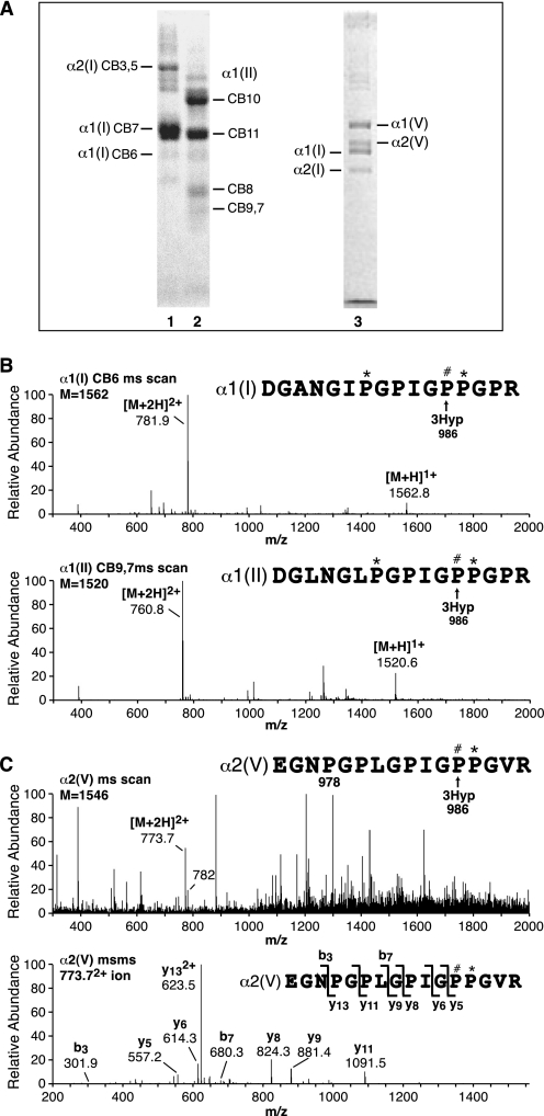
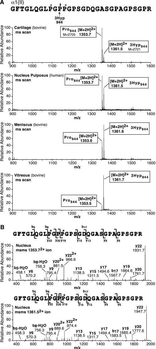
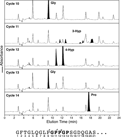
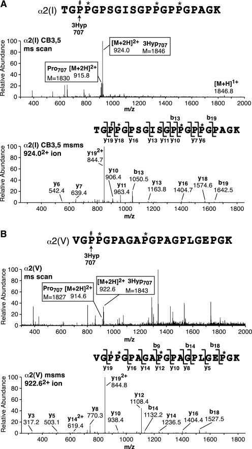
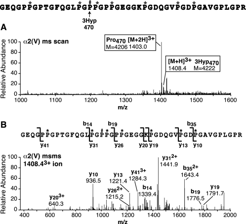
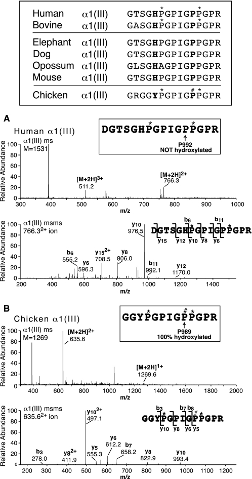
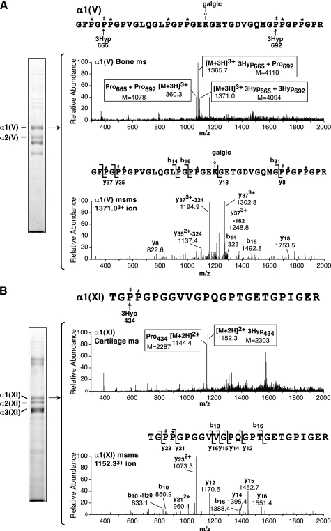
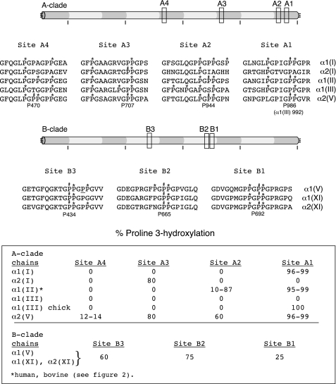
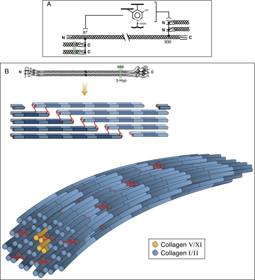
Collagen triple helices are stabilized by 4-hydroxyproline residues. No function is known for the much less common 3-hydroxyproline (3Hyp), although genetic defects inhibiting its formation cause recessive osteogenesis imperfecta. To help understand the pathogenesis, we used mass spectrometry to identify the sites and local sequence motifs of 3Hyp residues in fibril-forming collagens from normal human and bovine tissues. The results confirm a single, essentially fully occupied 3Hyp site (A1) at Pro(986) in A-clade chains alpha1(I), alpha1(II), and alpha2(V). Two partially modified sites (A2 and A3) were found at Pro(944) in alpha1(II) and alpha2(V) and Pro(707) in alpha2(I) and alpha2(V), which differed from A1 in sequence motif. Significantly, the distance between sites 2 and 3, 237 residues, is close to the collagen D-period (234 residues). A search for additional D-periodic 3Hyp sites revealed a fourth site (A4) at Pro(470) in alpha2(V), 237 residues N-terminal to site 3. In contrast, human and bovine type III collagen contained no 3Hyp at any site, despite a candidate proline residue and recognizable A1 sequence motif. A conserved histidine in mammalian alpha1(III) at A1 may have prevented 3-hydroxylation because this site in chicken type III was fully hydroxylated, and tyrosine replaced histidine. All three B-clade type V/XI collagen chains revealed the same three sites of 3Hyp but at different loci and sequence contexts from those in A-clade collagen chains. Two of these B-clade sites were spaced apart by 231 residues. From these and other observations we propose a fundamental role for 3Hyp residues in the ordered self-assembly of collagen supramolecular structures.


Figures

FIGURE 1.
FIGURE 1.
Tandem mass spectra of tryptic peptides containing Pro986 (site A1), the single, fully occupied 3Hyp site in collagen chains α1(I), α1(II), and α2(V). A, peptides were prepared by in-gel trypsin digestion after SDS-PAGE of CNBr digests of human bone collagen (lane 1), human articular cartilage collagen (lane 2), and pepsin-solubilized human bone collagen, 1.2 m NaCl precipitate (lane 3). B, full scan mass spectra from the tryptic peptide LC-MS profiles of α1(I)CB6 and α1(II)CB9,7 across the elution window of the post-translational variants of the tryptic peptide containing Pro986. Any unhydroxylated peptides would be included so this provides a measure of hydroxylation of Pro986. This site is fully 3-hydroxylated in type I collagen of bone and type II collagen of cartilage. C, full scan mass spectrum from the LC-MS profile of the α2(V) chain over the elution window of the tryptic peptide containing Pro986 (upper) and MS/MS analysis of ion 773.72+ (lower). The y5 fragment ion establishes the added 16 Da on Pro986. The 773.72+ peptide ion lacks the 4-hydroxylation at Pro978, whereas the 7822+ ion has both 3Hyp at Pro986 and 4Hyp at Pro978; α2(V) is consistently under-hydroxylated at Pro978. (The 7822+ ion is not a contaminant from α1(I).) P#, 3Hyp; P*, 4Hyp.
FIGURE 2.
FIGURE 2.
Tandem mass spectra of an α1(II) tryptic peptide revealing a secondary, variably hydroxylated 3Hyp site at Pro944 (site A2). A, upper four panels are full scan mass spectra from LC-MS profiles of in-gel trypsin digests of α1(II)CB9,7 from cartilage (bovine), nucleus pulposus (human), meniscus (bovine), and vitreous (bovine) over the elution window of hydroxylated and prolyl versions of the Pro944-containing peptide. The relative abundance of the ions shown provides an index of the degree of hydroxylation at Pro944. B, the bottom two panels show MS/MS spectral analyses of the prolyl and suspected 3-hydroxyprolyl versions of the peptide, from which the y ion ladder establishes the position of the added 16 Da on Pro944. Hydroxylation ranged from 10% for hyaline cartilage to 87% for vitreous type II collagen with intermediate values for intervertebral disc and meniscus collagens. See Table 1 for a guide to how fragment ions establish the sequence and position of the 3Hyp residue. P#, 3Hyp; P*, 4Hyp.
FIGURE 3.
FIGURE 3.
Edman N-terminal sequence analysis establishes that the modified residue at Pro944 (A2 site) is 3-hydroxyproline. The tryptic peptide containing Pro944 from α1(II) of sequence GFTGLQGLP*GP#P*GPSGDQGASGPAGPSGPR was prepared from human nucleus pulposus (a rich source of the putative 3Hyp version). Sequential phenylthiohydantoin-derivative HPLC profiles are shown for sequencer steps 10–14. At cycle 11 a profile very similar to that reported for a 3Hyp residue in α1(IV) collagen (19) is evident, quite distinct from that of proline (cycle 14) or 4Hyp (cycle 12).
FIGURE 4.
FIGURE 4.
Tandem mass spectra identifying a third molecular site of 3Hyp at Pro707 in α2(I) and α2(V) chains (site A3). A and B show pairs of spectra from LC-MS profiles of tryptic peptides from α2(I)CB3,5 and α2(V), respectively, prepared as in Fig. 1A. In each the upper panel is the full scan mass spectrum over the LC elution window of the tryptic peptide of sequence shown, and the top panel is an MS/MS analysis of the prolyl 3-hydroxylated version. The ratio of prolyl to 3-hydroxyprolyl forms in the collagen chains can be estimated by the ion ratios 9162+/9242+ and 9152+/9232+ for α2(I) and α2(V), respectively, at this Pro707 site. P#, 3Hyp; P*, 4Hyp.
FIGURE 5.
FIGURE 5.
Tandem mass spectra identifying a fourth molecular site of 3Hyp at Pro470 in α2(V) (site A4). The spectra were derived from an LC-MS profile of tryptic peptides prepared from the α2(V) chain from bone resolved by SDS-PAGE as shown in Figs. 1A and 7A. A is the full scan mass spectrum over the LC elution window of the peptide of sequence shown. B is an MS/MS analysis of the prolyl 3-hydroxylated version that establishes the position of the extra 16Da on Pro470. P#, 3Hyp; P*, 4Hyp.
FIGURE 6.
FIGURE 6.
Comparative protein sequences at the candidate A1 3Hyp site (Pro992) in the collagen α1(III) chain and tandem mass spectra of the corresponding tryptic peptides from human and chicken α1(III). The top panel compares the homologous sequences for a diverse range of mammalian species with chicken (Ensemble genomic data base). For human, bovine, and chicken the 4Hyp (*) and 3Hyp (#) modifications established by MS are shown. The upper pair (A) and lower pair (B) of spectra are from LC-MS profiles of tryptic peptides from human and chicken α1(III), respectively. In each, the upper spectrum is the full scan mass spectrum over an LC elution window that would combine all post-translational forms of the peptide shown, and the lower spectrum is an MS/MS analysis of the single peptide so found. The 755 ion is from an unrelated α1(III) tryptic peptide. From human and bovine (not shown) α1(III), Pro992 was not hydroxylated but from chicken the homologous Pro989 was 100% hydroxylated. P#, 3Hyp; P*, 4Hyp.
FIGURE 7.
FIGURE 7.
Tandem mass spectral identification of 3Hyp sites in α1(V) and α1(XI) collagen chains (sites B1, B2, and B3). The peptides were prepared by in-gel trypsin digestion after SDS-PAGE of collagen types V and XI prepared by pepsin digestion and salt precipitation from human bone and articular cartilage, respectively. A, full scan mass spectrum from the tryptic peptide LC-MS profile of α1(V) across the elution window of the peptide of sequence shown (upper spectrum). An MS/MS spectrum of the 13713+ ion confirms the presence of two extra 16-Da units as hydroxyls on Pro665 and Pro692 (lower spectrum). The fragment ions resulting from neutral hexose losses (−162 and −324) are also indicated. Similarly, MS/MS spectra showed that 13663+ had one extra 16 Da on Pro665 and Pro1360 3+ was mostly the sequence with six 4Hyp and no 3Hyp residues (not shown). B, full scan mass spectrum from the tryptic peptide LC-MS profile of α1(XI) across the elution window of the peptide of sequence shown (upper spectrum). An MS/MS spectrum of the 11522+ ion establishes an extra 16 Da as a hydroxyl on Pro434 (lower spectrum). Similarly, the MS/MS spectrum of 11442+ showed that it lacked the 16Da on Pro434 (not shown). P#, 3Hyp; P*, 4Hyp; galglc, glucosylgalactosyl.
FIGURE 8.
FIGURE 8.
Summary of human sequences and molecular locations of 3Hyp residues identified in the A-clade and B-clade collagen chains. Relative positions along the triple helix are indicated by boxes and identified by A1-A4 for A-clade sites and B1-B3 for B-clade sites. Homologous stretches of human sequence are shown for all A-clade chain types and all B-clade chain types. A D-periodic spacing is evident between Pro470 and Pro707, between Pro707 and Pro944 in the A-clade triple helix, and between Pro434 and Pro665 in the B-clade triple helix. The underlined P for proline at B3 in α1(V) from bovine meniscus was also ∼50% hydroxylated. P#, 3Hyp; P*, 4Hyp.
FIGURE 9.
FIGURE 9.
Speculated concept of fibril molecular packing in which the subunits are molecular dimers in register and staggered axially by d-periods. A shows the placement of complex intermolecular cross-links in skeletal tissue collagens. B illustrates a molecular model of fibril packing. Such an arrangement could result from the influence of inter-triple-helical hydrogen bonding between 3Hyp-containing loci. The location of site A1 at Pro986 and potential for inter-helical bonding is illustrated. The remaining 3Hyp sites could similarly facilitate D-staggered helical association during the macromolecular assembly process.

Similar articles

Cited by

References

    1. Berg R. A., Prockop D. J. (1973) Biochem. Biophys. Res. Commun. 52, 115–120 - PubMed
    1. Ogle J. D., Arlinghaus R. B., Logan M. A. (1962) J. Biol. Chem. 237, 3667–3673 - PubMed
    1. Gryder R. M., Lamon M., Adams E. (1975) J. Biol. Chem. 250, 2470–2474 - PubMed
    1. Vranka J. A., Sakai L. Y., Bächinger H. P. (2004) J. Biol. Chem. 279, 23615–23621 - PubMed
    1. Tiainen P., Pasanen A., Sormunen R., Myllyharju J. (2008) J. Biol. Chem. 283, 19432–19439 - PubMed

Publication types

MeSH terms

LinkOut - more resources