. 2010 Jan 22;285(4):2610-21.
doi: 10.1074/jbc.M109.071159. Epub 2009 Nov 23.

Cordycepin inhibits protein synthesis and cell adhesion through effects on signal transduction

Affiliations

Cordycepin inhibits protein synthesis and cell adhesion through effects on signal transduction

Ying Ying Wong et al. J Biol Chem. .

Abstract

3'-Deoxyadenosine, also known as cordycepin, is a known polyadenylation inhibitor with a large spectrum of biological activities, including anti-proliferative, pro-apoptotic and anti-inflammatory effects. In this study we confirm that cordycepin reduces the length of poly(A) tails, with some mRNAs being much more sensitive than others. The low doses of cordycepin that cause poly(A) changes also reduce the proliferation of NIH3T3 fibroblasts. At higher doses of the drug we observed inhibition of cell attachment and a reduction of focal adhesions. Furthermore, we observed a strong inhibition of total protein synthesis that correlates with an inhibition of mammalian target of rapamycin (mTOR) signaling, as observed by reductions in Akt kinase and 4E-binding protein (4EBP) phosphorylation. In 4EBP knock-out cells, the effect of cordycepin on translation is strongly reduced, confirming the role of this modification. In addition, the AMP-activated kinase (AMPK) was shown to be activated. Inhibition of AMPK prevented translation repression by cordycepin and abolished 4EBP1 dephosphorylation, indicating that the effect of cordycepin on mTOR signaling and protein synthesis is mediated by AMPK activation. We conclude that many of the reported biological effects of cordycepin are likely to be due to its effects on mTOR and AMPK signaling.


Figures
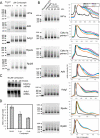
FIGURE 1.
FIGURE 1.
Cordycepin affects poly(A) tail length and cell proliferation at low concentrations. A, RNA ligation poly(A) tests on RNA from NIH3T3 cells treated with different doses of cordycepin. RNase H + Oligo(dT) indicates RNA from untreated cells digested with RNase H and oligo(dT) to remove the poly(A) tail as a control. The GenBank™ gene name abbreviations indicate which mRNAs were tested: Hif1a (hypoxia-inducible factor 1α), Cdkn1a (p21/Waf/Cip), Atf4 (activating transcription factor 4), Actg1 (gamma1 actin), Rps4X (X-linked ribosomal protein S4), and Rpl28 (ribosomal protein L28). B, Klenow priming poly(A) tests on a time course of cordycepin treatment. On the right side the panels shows the distribution of the intensity in each lane as a percentage of the maximum intensity in that lane on the vertical axis and the size of the poly(A) test products in base pairs on the horizontal axis. Black, oligo(dT) RNase A-treated sample; purple, no treatment; green, 15-min cordycepin; dark blue, 30-min cordycepin; light blue, 45-min cordycepin; and orange, 60-min cordycepin. C, Northern blot for Cdkn1a and β-actin (Act1b) on total RNA isolated from cells treated with 50 μm cordycepin for the indicated times. D, cell numbers after 72 h of treatment with cordycepin (medium refreshed daily, including drug).
FIGURE 2.
FIGURE 2.
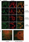
Cordycepin affects the actin cytoskeleton and dissolves focal adhesions. NIH3T3 cells were plated on coverslips the day before and treated for 3 h with cordycepin (50 or 200 μm) or cycloheximide (100 μg/ml). The cells were fixed and stained with an antibody against vinculin (red) to detect focal adhesions and phalloidin (green) to detect filamentous actin. A, microscopy of control cells treated with the indicated drugs with the stains imaged separately as well as merged. B, higher magnification merged image with the same stains for control and cordycepin-treated cells. Arrows indicate focal adhesions in the untreated cell.
FIGURE 3.
FIGURE 3.
Cordycepin inhibits cell spreading. NIH3T3 cells were detached, suspended for 1 h in serum-free medium, and allowed to re-attach to coverslips with serum for 5 h in the presence or absence of cordycepin, cycloheximide, or actinomycin D at the concentrations indicated. After fixation, cells were stained with phalloidin to visualize the actin cytoskeleton. A, images of typical control and cordycepin-treated cells. B and C, quantitation of the percentage of unspread cells (largest diameter, 25 μm or less) in cells incubated with the indicated doses of drugs.
FIGURE 4.
FIGURE 4.
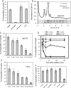
Cordycepin inhibits protein synthesis. A, protein synthesis rates of NIH3T3 cells as measured by 35S incorporation into protein, corrected for total protein concentration. The effects of treatment with 200 μm cordycepin or adenosine or with 10 μg/ml actinomycin D (AcD) are shown. B, polyribosome profiles of control cells and cells treated with cordycepin (200 μm) for 2 h. 40S and 60S indicate the dissociated free ribosomal subunit peaks, polysomes indicates ribosomes translating mRNA. The inset shows the RNA isolated from a control gradient, proving the identification of the 40 S peak. C, dose response of the effect of cordycepin on protein synthesis rates in NIH3T3 cells treated for 2 h. D, time course of the response of protein synthesis rates to three doses of cordycepin. E, dose response of the effect of cordycepin on protein synthesis rates in HeLa cells treated for 2 h. F, incorporation of radioactive methionine into protein in vitro in reticulocyte lysate supplemented with cordycepin, adenosine, cordycepin triphosphate (cordyTP), ATP, or cap analogue (m7GpppG) (all at 200 μm).
FIGURE 5.
FIGURE 5.
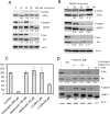
Cordycepin inhibits mTOR signaling. A, Western blots of extracts from NIH3T3 cells treated for 2 h with increasing doses of cordycepin. Blots were developed as indicated with antibodies specific for total eIF2α and its phosphorylated form (Ser51), total 4EBP1 and its phosphorylated forms (Thr37/46), Akt1 and its phosphorylated form (Ser473), and β-actin (as a loading control). Ratios between phosphorylated and total protein levels are given under the blots, with the ratio for the control set at 1. B, Western blots of extracts of cells treated for different time periods with 200 μm cordycepin. C and D, the effect of the mTORC1 inhibitor rapamycin and the phosphatidylinositol 3-kinase inhibitor LY294,002 on protein synthesis and phosphorylation of proteins. C, protein synthesis rates of NIH3T3 cells as measured by 35S incorporation into protein, corrected for total protein concentration. D, Western blots of extracts treated with inhibitors as described above.
FIGURE 6.
FIGURE 6.
Protein synthesis in murine embryonic fibroblasts lacking 4EBP expression or containing non-phosphorylatable eIF2α is resistant to cordycepin. Protein synthesis in MEFs from animals lacking expression of 4EBP1 and 4EBP2 (double knock-out, DKO) (A) and MEFs from animals with the S51A mutation in eIF2α (B), and their corresponding wild-type controls, was measured by [35S]methionine incorporation into protein. The cells were pre-treated with 0, 50, and 200 μm cordycepin for 1.5 h and then labeled for 1 h. The data are corrected for total protein concentration and are shown as the means ± S.E. of three replicates for each treatment.
FIGURE 7.
FIGURE 7.
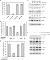
Cordycepin activates AMPK. A, adenosine blocks cordycepin-mediated repression of protein synthesis. NIH3T3 cells were treated with cordycepin and/or adenosine for 2 h, and protein synthesis rates were measured by 35S incorporation, corrected for total protein concentration. B, import and phosphorylation of cordycepin are required for inhibition of protein synthesis. Cells were treated with nitrobenzylthioinosine (NBTI, 10 μm) or iodotubericidin (ITu, 0.1 μm) for 15 min before treatment with cordycepin (200 μm) for 1 h. Protein synthesis rates were determined as before. C, cordycepin induces AMPK activation. Western blots are shown for AMPK β1 and its autophosphorylation site (Ser108), acetyl-CoA carboxylase and its AMPK phosphorylation site (Ser79) as well as for the proteins listed in the legend to Fig. 5. Treatment with cordycepin was for the indicated times and dose. D, protein synthesis in cells pretreated with Compound C or DMSO for 15 min prior to the addition of 200 μm cordycepin and incubation for 1 h. E, cells were treated as in D, and Western blotting was performed for the proteins indicated as described above.
FIGURE 8.
FIGURE 8.
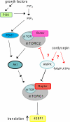
Summary of the AMPK and mTOR signaling pathways. Phosphatidylinositol 3 kinase (PI3K) is activated when growth factors bind to their receptors. Phosphatidylinositol bisphosphate (PIP2) is converted by PI3K into phosphatidylinositol trisphosphate (PIP3). This leads to activation of phosphoinositide-dependent protein kinase-1 (PDK1). PDK1 phosphorylates Akt1 to partially activate it and prime it for further activation. Full activation is dependent on an additional phosphorylation of Akt1by mTORC2, a complex that includes the kinase, mammalian target of rapamycin (mTOR) and the regulatory subunit Rictor. Akt1 phosphorylates and activates the mTORC1 complex that contains mTOR and the regulatory subunit Raptor. mTORC1 phosphorylates the translation repressor 4EBP and inactivates it. AMPK is known to inhibit mTORC1 activation at multiple levels. More detail can be found in Refs. , . We propose that cordycepin activates AMPK by an unknown mechanism and that active AMPK somehow also inhibits mTORC2 activity (interactions shown in red), leading to a double block of the mTOR signaling pathway.

Similar articles

Cited by

References

    1. Yang F. Q., Li S. P. (2008) J. Pharm. Biomed. Anal. 48, 231–235 - PubMed
    1. Paterson R. R. (2008) Phytochemistry 69, 1469–1495 - PMC - PubMed
    1. Ng T. B., Wang H. X. (2005) J. Pharm. Pharmacol. 57, 1509–1519 - PubMed
    1. Chang W., Lim S., Song H., Song B. W., Kim H. J., Cha M. J., Sung J. M., Kim T. W., Hwang K. C. (2008) Eur. J. Pharmacol. 597, 64–69 - PubMed
    1. Shi P., Huang Z., Tan X., Chen G. (2008) Methods Find. Exp. Clin. Pharmacol. 30, 347–353 - PubMed

Publication types

MeSH terms

LinkOut - more resources