

Basal cerebral metabolism may modulate the cognitive effects of Abeta in mild cognitive impairment: an example of brain reserve
- PMID: 19940172
- PMCID: PMC2810461
- DOI: 10.1523/JNEUROSCI.3669-09.2009
Basal cerebral metabolism may modulate the cognitive effects of Abeta in mild cognitive impairment: an example of brain reserve
Abstract
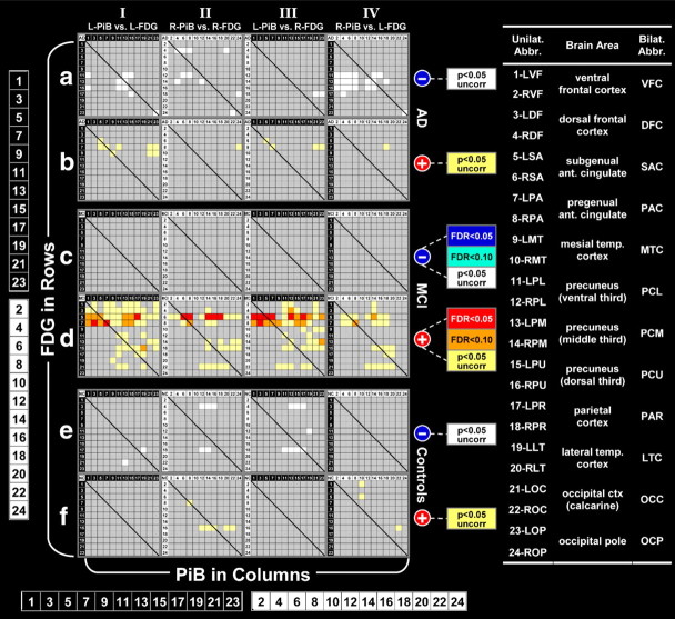
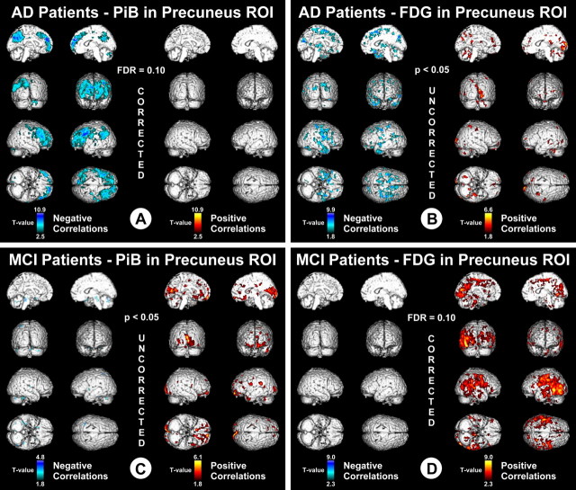
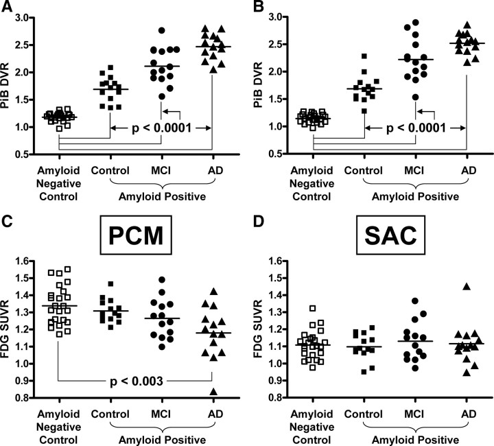
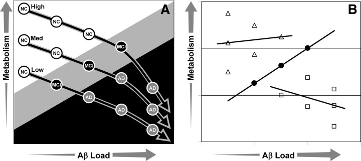
Inverse correlations between amyloid-beta (Abeta) load measured by Pittsburgh Compound-B (PiB) positron emission tomography (PET) and cerebral metabolism using [(18)F]fluoro-2-deoxy-d-glucose (FDG) in Alzheimer's disease (AD) patients, suggest local Abeta-induced metabolic insults. However, this relationship has not been well studied in mild cognitive impairment (MCI) or amyloid-positive controls. Here, we explored associations of Abeta deposition with metabolism via both region-of-interest-based and voxel-based analyses in amyloid-positive control subjects and patients with MCI or AD. Metabolism in parietal and precuneus cortices of AD patients was negatively correlated with PiB retention locally, and more distantly with PiB retention in frontal cortex. In amyloid-positive controls, no clear patterns in correlations were observed. In MCI patients, there were essentially no significant, negative correlations, but there were frequent significant positive correlations between metabolism and PiB retention. Metabolism in anterior cingulate showed positive correlations with PiB in most brain areas in MCI, and metabolism and PiB retention were positively correlated locally in precuneus/parietal cortex. However, there was no significant increase in metabolism in MCI compared to age-matched controls, negating the possibility that Abeta deposition directly caused reactive hypermetabolism. This suggests that, in MCI, higher basal metabolism could either be exacerbating Abeta deposition or increasing the level of Abeta necessary for cognitive impairment sufficient for the clinical diagnosis of AD. Only after extensive Abeta deposition has been present for longer periods of time does Abeta become the driving force for decreased metabolism in clinical AD and, only in more vulnerable brain regions such as parietal and precuneus cortices.
Figures
References
-
- Aizenstein HJ, Nebes RD, Saxton JA, Price JC, Mathis CA, Tsopelas ND, Ziolko SK, James JA, Snitz BE, Houck PR, Bi W, Cohen AD, Lopresti BJ, DeKosky ST, Halligan EM, Klunk WE. Frequent amyloid deposition without significant cognitive impairment among the elderly. Arch Neurol. 2008;65:1509–1517. - PMC - PubMed
-
- Ashburner J, Friston KJ. Unified segmentation. Neuroimage. 2005;26:839–851. - PubMed
-
- Becker JT, Boller F, Saxton J, McGonigle-Gibson KL. Normal rates of forgetting of verbal and non-verbal material in Alzheimer's disease. Cortex. 1987;23:59–72. - PubMed
-
- Bennett DA, Schneider JA, Wilson RS, Bienias JL, Arnold SE. Neurofibrillary tangles mediate the association of amyloid load with clinical Alzheimer disease and level of cognitive function. Arch Neurol. 2004;61:378–384. - PubMed
-
- Bennett DA, Schneider JA, Arvanitakis Z, Kelly JF, Aggarwal NT, Shah RC, Wilson RS. Neuropathology of older persons without cognitive impairment from two community-based studies. Neurology. 2006;66:1837–1844. - PubMed
Publication types
MeSH terms
Substances
Grants and funding
LinkOut - more resources
Full Text Sources
Other Literature Sources
Medical
Molecular Biology Databases