. 2009 Nov 25;29(47):14790-802.
doi: 10.1523/JNEUROSCI.2059-09.2009.

Proneurotrophin-3 is a neuronal apoptotic ligand: evidence for retrograde-directed cell killing

Affiliations

Proneurotrophin-3 is a neuronal apoptotic ligand: evidence for retrograde-directed cell killing

Hiroko Yano et al. J Neurosci. .

Abstract

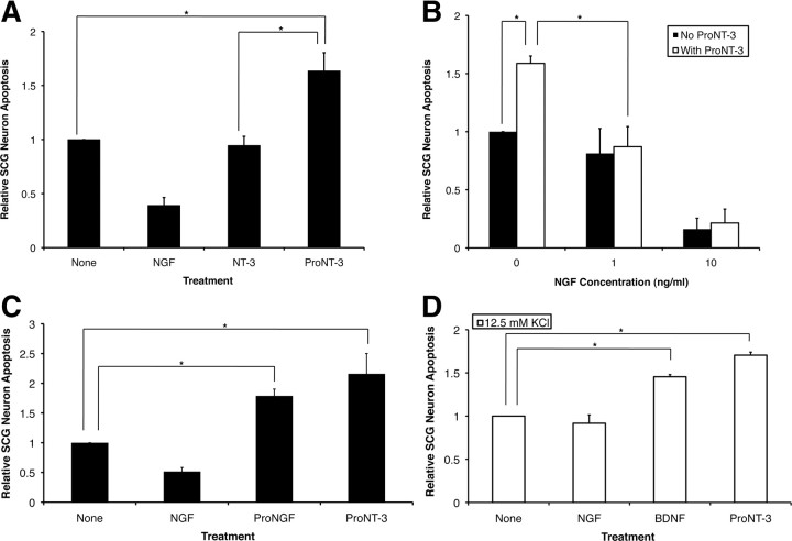
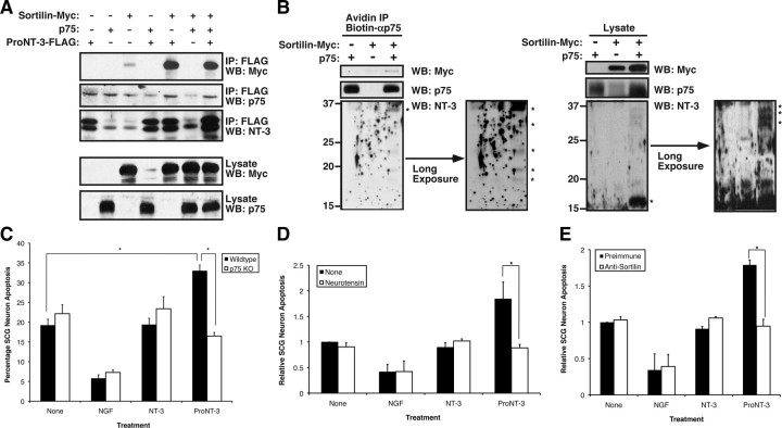
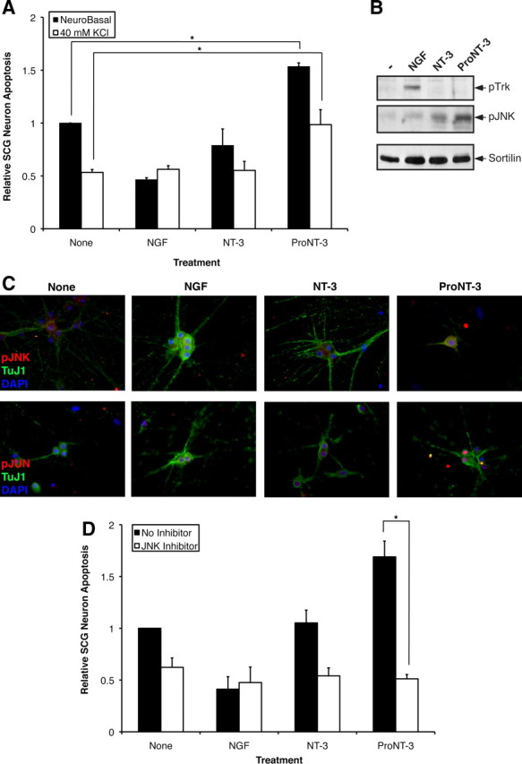
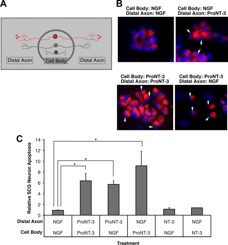
Although mature neurotrophins are well described trophic factors that elicit retrograde survival signaling, the precursor forms of neurotrophins (i.e., proneurotrophins) can function as high-affinity apoptotic ligands for selected neural populations. An outstanding question is whether target-derived proneurotrophins might affect neuronal survival/death decisions through a retrograde transport mechanism. Since neurotrophin-3 (NT-3) is highly expressed in non-neural tissues that receive peripheral innervation, we investigated the localized actions of its precursor (proNT-3) on sympathetic neurons in the present study. Pharmacological inhibition of intracellular furin proteinase activity in 293T cells resulted in proNT-3 release instead of mature NT-3, whereas membrane depolarization in cerebellar granule neurons stimulated endogenous proNT-3 secretion, suggesting that proNT-3 is an inducible bona fide ligand in the nervous system. Our data also indicate that recombinant proNT-3 induced sympathetic neuron death that is p75(NTR)- and sortilin-dependent, with hallmark features of apoptosis including JNK (c-Jun N-terminal kinase) activation and nuclear fragmentation. Using compartmentalized culture systems that segregate neuronal cell bodies from axons, proNT-3, acting within the distal axon compartment, elicited sympathetic neuron death and overrode the survival-promoting actions of NGF. Together, these results raise the intriguing possibility that dysregulation of proneurotrophin processing/release by innervated targets can be deleterious to the neurons projecting to these sites.


Figures

Figure 1.
Figure 1.
Characterization of anti-proNT-3 antiserum. A, Sf9 cells expressing full-length preproNT-3 cDNA synthesize both proNT-3 and mature NT-3 as indicated (red arrows). Total cellular lysates, along with recombinant NT-3 standard (Promega), were Western blotted with an anti-NT-3 antiserum (sc-547; Santa Cruz Biotechnology) followed by reprobing the blot with an anti-proNT-3 antiserum (see Materials and Methods) to demonstrate the specificity of the prodomain specific antiserum. The numbers on the left indicate positions of the molecular weight markers. B, Western blot of Sf9 cells expressing proNGF, proBDNF, or proNT-3 using preimmune or affinity-purified proNT-3 specific antibody as indicated. Note that the anti-proNT-3 antiserum does not recognize proNGF or proBDNF. Reprobing the blot with anti-NGF and anti-BDNF antisera confirmed the expression of the corresponding proneurotrophins in the appropriate cell lysates (data not shown). The red arrow on the right indicates position of proNT-3, and the numbers on the left indicate positions of the molecular weight markers. C, HEK 293T cells were cotransfected with plasmids encoding GFP (to identify transfected cells) and proNT-3, proBDNF, or proNGF as indicated. Forty-eight hours later, cells were fixed and indirect immunofluorescence microscopy was performed using preimmune serum or the affinity-purified anti-proNT-3 antibody. The top panels represent merged images (with DAPI) of GFP epifluorescence and preimmune or proNT-3 immunoreactivity (bottom insets) from the corresponding transfected cells.
Figure 2.
Figure 2.
Neuronal release of endogenous proNT-3. A, Tissue-specific expression of proNT-3 and mature NT-3. Detergent lysates were prepared from the indicated tissues. Except for heart and kidney in which 100 μg of the respective lysates were analyzed, equal amounts of visceral tissue lysates (50 μg) and 150 μg of CNS tissue lysates were Western blotted with an anti-NT-3 antiserum (sc-547). This was done because the range of proNT-3/NT-3 expression varies greatly between these tissues (Kaisho et al., 1994; Katoh-Semba et al., 1996); the resulting enhanced chemiluminescence signals would have exceeded the linearity of the film without the loading adjustment. Note that various non-neural and CNS tissues express mature NT-3 and proNT-3 at varying proportion and abundance. The numbers on the left indicate positions of the molecular weight markers. The brackets on the right indicate multiple proNT-3 species whose identities were confirmed by reprobing the blot with anti-proNT-3 antiserum and an independently generated anti-NT-3 antibody (data not shown). Ctx, Cortex; Cbm, cerebellum; Hip, hippocampus; Spl, spleen; Hrt, heart; Lng, lung; Kid, kidney; Liv, liver. B, To demonstrate specificity of the proNT-3 and mature NT-3 species, anti-NT-3 antibody (sc-547) was preincubated with excess recombinant NT-3 before addition to a replica tissue lysates blot but was otherwise processed in identical manner as in Figure 2A. C, Age-dependent expression of proNT-3 and mature NT-3. Cerebellar lysates from early postnatal (P3) and adult female rat were Western blotted with anti-NT-3 (sc-547). Positions of proNT-3 and mature NT-3 are indicated on the right. D, P6 cerebellar granule neurons in culture were treated with either 5 mm KCl (None) or with 25 mm KCl for 24 h in the presence of a goat anti-NT-3 antibody (sc-13380) as described in Materials and Methods. Anti-NT-3 immunoprecipitates were Western blotted with either rabbit anti-NT-3 antiserum (sc-547; left panel) or with anti-proNT-3 specific antiserum (right panel) to verify the identify of proNT-3. The position of proNT-3 is indicated on the left as are positions of nonspecific bands (NS).
Figure 3.
Figure 3.
Purification of recombinant NT-3 and furin-resistant proNT-3. A, Right panel, HEK 293T cells were transfected with control GFP encoding plasmid or with plasmids encoding FLAG-tagged wild-type NT-3 or proNT-3 in which the putative furin cleavage motif had been mutated. Replica cultures were treated, or not, with 30 μm Dec-RVRK-CMK 24 h after transfection. Conditioned media were immunoprecipitated with anti-FLAG antiserum (M2; Sigma-Aldrich) after another 48 h. Immunoprecipitates were then analyzed by Western blotting using a commercial anti-NT-3 antiserum (sc-547). Left panel, Cultured cerebellar granule neurons were treated or not with 30 μm furin inhibitor Dec-RVKR-CMK for 24 h as indicated. Conditioned media were immunoprecipitated with a goat anti-NT-3 antiserum followed by proNT-3 Western blotting as described (see Materials and Methods). Position of proNT-3 is indicated on the left as are position of nonspecific bands (NS). B, His7-tagged mature NT-3 from lysates of insect cells infected with wild-type preproNT-3-His7 encoding baculoviral vector was purified as described in Materials and Methods. C, Purification of His7-tagged proNT-3 from lysates of insect cells infected with cleavage resistant proNT-3-His7 encoding baculoviral vector. For B and C, the extent of purification was monitored by anti-NT-3 Western blot analysis of equal proportion (1/200 by volume) of each indicated fractions and by silver staining of the NT-3- and proNT-3-enriched eluates. The numbers on the left indicate positions of the molecular weight markers. Where appropriate, arrows mark the positions of proNT-3 and mature NT-3.
Figure 4.
Figure 4.
ProNT-3 does not activate TrkC. A, TrkA-deficient PC12nnr5 cells were transfected with a mammalian TrkC expression plasmid (Klein et al., 2005) followed by 500 μg/ml G418 selection to obtain TrkC-PC12nnr5 cell clones. Serum-starved TrkC-PC12nnr5 cells were treated, or not, with 100 ng/ml NGF, 25 ng/ml mature NT-3, or 25 ng/ml proNT-3 for 10 min as indicated. Detergent-extracted cellular lysates were immunoprecipitated with an anti-Trk antiserum followed by anti-phosphotyrosine Western blotting to assess TrkC activation. The blot was then reprobed with an anti-Trk antiserum to verify the presence of Trk in all the samples. In addition, whole-cell lysates were Western blotted with pERK or total ERK antibody to demonstrate ERK activation by NT-3, but not proNT-3, in TrkC-PC12nnr5 cells. B, Wild-type PC12 cells were transiently transfected with expression plasmids encoding GFP (to mark transfected cells) with or without TrkC. Forty-eight hours later, replica cultures were treated or not with 20 ng/ml NGF, and equal molar of NT-3 or proNT-3 (i.e., 20 ng/ml NT-3 or 40 ng/ml proNT-3) as indicated. The percentage of neurite-bearing populations were assessed 48 h later as described (see Materials and Methods). Data represent the results obtained from two independently conducted experiments. Error bars indicate ranges of values from two independent experiments.
Figure 5.
Figure 5.
Apoptotic actions of proNT-3. A, P1 rat SCG neurons (DIV 7), washed free of NGF, were treated with no additive (None), 10 ng/ml NGF, or equal molar of NT-3 or proNT-3 (i.e., 2 ng/ml NT-3 or 4 ng/ml proNT-3) as indicated. The percentage of apoptotic neurons were assessed as described (see Materials and Methods) and were normalized to that of apoptotic neurons without any trophic factor treatment (typically between 15 and 25% from experiment to experiment). Data represent the results obtained from six independently conducted experiments. The vertical error bars indicate SEM. B, NGF rescues proNT-3-treated neurons. P1 rat SCG neurons (DIV 7), washed free of NGF, were treated with either no additive or 4 ng/ml proNT-3 in the presence or absence of increasing concentrations of NGF as indicated. Neuronal apoptosis was assessed 48 h later. Data summarize the results of three independently conducted experiments. The vertical error bars indicate SEM. C, Comparable apoptotic effects of proNGF and proNT-3. P1 rat SCG neurons plated on collagen-coated slides were washed free of NGF on DIV 7 and were then treated with no additive (None) or equal molar concentrations of proNGF or proNT-3 (4 ng/ml) as indicated. Neuronal apoptosis was assessed as above. The data were normalized so that the percentage of dying SCG neurons caused by NGF deprivation alone equals 1. Results represent three independently conducted experiments, and the vertical error bars indicate SEM. Note that, for this particular experiment, SCG neurons were plated on collagen instead of laminin because we found that laminin interferes with the apoptotic activity of proNGF (data not shown). D, Apoptotic actions of BDNF and proNT-3. Replica cultures of rat SCG neurons (DIV 7) were washed free of NGF, and were treated, or not, with 10 ng/ml NGF, 100 ng/ml BDNF, or 4 ng/ml proNT-3 in the presence 12.5 mm KCl. Under this culture condition, neuronal survival can be maintained by KCl in the absence of NGF but high concentration of BDNF can elicit death via p75NTR activation (Bamji et al., 1998). Neuronal apoptosis was assessed for each culture condition 48 h later. The data, normalized to the number of apoptotic neurons on KCl treatment alone, represent values from three independently conducted experiments. The vertical error bars indicate SEM. Note that membrane depolarization prevented NGF-deprived neuron death (Franklin et al., 1995), but not did not affect proNT-3-induced apoptosis. For A–D, asterisks denote statistical significance between the paired samples.
Figure 6.
Figure 6.
Dual-receptor requirement for ProNT-3 action. A, Coimmunoprecipitation of proNT-3 and sortilin. HEK 293T cells were transfected with Myc-tagged sortilin, p75NTR, FLAG-tagged proNT-3 alone or in combination as indicated. Forty-eight hours later, detergent lysates were immunoprecipitation with anti-FLAG beads (Sigma-Aldrich), followed by Western blotting analysis with anti-NT-3, anti-p75NTR, or anti-Myc antisera as indicated. Note that sortilin but not p75NTR readily coimmunoprecipitated with proNT-3 and that stable complex between sortilin and proNT-3 was not enhanced by p75NTR. B, Sortilin expression is required for proNT-3 binding to p75NTR. HEK 293T cells were transiently transfected with either p75NTR, Myc-tagged sortilin, or both receptors as indicated. Forty-eight hours later, exogenous recombinant proNT-3 (for proNT-3 production in 293T cells, see Fig. 3A) was added to these cells for 1 h. Left panel, Anti-p75NTR immunoprecipitation was performed as described (see Materials and Methods), followed by Western blotting for proNT-3 (anti-NT3), sortilin (anti-Myc), and p75NTR. Right panel, The corresponding total cellular lysates were similarly Western blotted with the indicated antisera. Note that proNT-3 internalization, which occurred only in cells that coexpress both p75NTR and sortilin, also resulted in significant degradation of the molecule. The numbers on the left indicate positions of the molecular weight markers. The asterisks (*) on the left indicate full-length proNT-3 (∼37 kDa) and various partially cleaved NT-3 species (∼32–16 kDa). Note also that, in these experiments, no mature NT-3 of the expected molecular weight (i.e., 13 kDa) was detected, suggesting that endocytosed proNT-3 is not processed by the same furin-based mechanism for mature NT-3 production. C, p75NTR is required for proNT-3-induced neuronal apoptosis. Wild-type or p75NTR-null SCG neurons were cultured as described previously (Nykjaer et al., 2004; Teng et al., 2005). Replica cultures (DIV 7), washed free of NGF, were treated or not with 10 ng/ml NGF or equal molar of NT-3 or proNT-3 (i.e., 2 ng/ml NT-3 or 4 ng/ml proNT-3) as indicated. The percentage of apoptotic neurons were assessed 36 h later. The data were not further normalized because neurons for these experiments were from two different sources (i.e., wild-type and p75NTR-null mice). The vertical error bars indicate SEM (n = 5). D, Sortilin antagonist neurotensin abrogates proNT-3 induced SCG apoptosis. NGF-deprived rat SCG neuron cultures were treated with no ligand (None), 10 ng/ml NGF, or equal molar of NT-3 or proNT-3 (i.e., 2 ng/ml NT-3 or 4 ng/ml proNT-3) in the presence or absence of 20 μm neurotensin as indicated. Neuronal apoptosis under each culture condition was evaluated 48 h later, and the data were normalized to that of apoptotic neurons without any trophic factor or neurotensin treatment. Data represent the results obtained from three independently conducted experiments. The vertical error bars indicate SEM. E, Sortilin antiserum blocks apoptosis of proNT-3-treated SCG neurons. NGF-deprived rat SCG neuron cultures were treated with 10 ng/ml NGF or equal molar of NT-3 or proNT-3 (i.e., 2 ng/ml NT-3 or 4 ng/ml proNT-3) in the presence of an anti-sortilin antiserum (1:10 dilution) or the equivalent dilution of preimmune serum as a control (for characterization of the antiserum, see supplemental Fig. 1, available at www.jneurosci.org as supplemental material). Neuronal apoptosis under each culture condition was evaluated 48 h later. The data represent the results obtained from three independently conducted experiments. The vertical error bars indicate SEM. Note that sortilin antiserum specifically inhibits proNT-3-induced cell killing without additional survival-promoting effects on NGF-deprived neurons. For C–E, asterisks denote statistical significance between the paired samples.
Figure 7.
Figure 7.
A role for JNK in proNT-3-induced neuronal apoptosis. A, Replica cultures of rat SCG neurons (DIV 7) were washed free of NGF and were treated or not with 10 ng/ml NGF or equal molar NT-3 or proNT-3 (i.e., 2 ng/ml NT-3 or 4 ng/ml proNT-3) in the presence or absence of 40 mm KCl as indicated. Neuronal apoptosis was assessed for each culture condition 48 h later. The data, normalized to the number of apoptotic neurons on NGF deprivation, represent values from three independently conducted experiments. The vertical error bars indicate SEM. Note that membrane depolarization prevented NGF-deprived neuron death (Franklin et al., 1995) but not did not affect proNT-3-induced apoptosis. B, Replica cultures of rat SCG neurons (DIV 7) were washed free of NGF and were then treated with 12.5 mm KCl as described for analysis of JNK activation (Linggi et al., 2005). SCG neurons were treated or not with 50 ng/ml NGF, 25 ng/ml mature NT-3, or 25 ng/ml proNT-3 as indicated for 4 h. Cellular lysates (100 μg each) were Western blotted with the indicated antisera to assess proNT-3-induced JNK activation relative to TrkA-specific signaling. Equality of sample loading was verified by reprobing the blot with an anti-sortilin antiserum (BD Biosciences). C, Replica cultures of P1 SCG neurons (DIV 7) were washed free of NGF and maintained in 12.5 mm KCl either in presence or absence of NGF, NT-3, or proNT-3 as indicated. Forty-eight hours later, cells were fixed and stained with TuJ1 (Covance), pJNK (Cell Signaling), and DAPI as indicated. Only proNT-3-treated, apoptotic neurons exhibited pJNK immunoreactivity (top panels). Alternatively, cultures were stained with TuJ1 and pJun as corroborative evidence of JNK activation (bottom panels). D, Replica cultures of rat SCG neurons (DIV 7) were washed free of NGF and were treated or not with 10 ng/ml NGF or equal molar of NT-3 or proNT-3 (i.e., 2 ng/ml NT-3 or 4 ng/ml proNT-3) in the presence or absence of 15 μm SP600125 as indicated. Neuronal apoptosis under standard (non-depolarizing) culture condition were assessed 48 h after treatment. The data, normalized to the number of apoptotic neurons on NGF deprivation, represent values from three independently conducted experiments, and the vertical error bars indicate SEM. For A and D, asterisks denote statistical significance between the paired samples.
Figure 8.
Figure 8.
Retrograde apoptotic signaling. A, Schematic representation of compartmentalized culture system. Only neurons (red) that were prelabeled with fluorescent microspheres were evaluated in subsequent analysis. B, Fluorescent photomicrograph of SCG neurons under the indicated culture conditions. Only viable neurons with active retrograde transport take up fluorescent microspheres from the distal axon compartment. The arrows indicate fragmented nuclei of dying neurons. C, Assessment of neuronal apoptosis under each culture conditions. Replica compartmentalized cultures were treated with 1 ng/ml NGF or equal molar of NT-3 or proNT-3 (i.e., 5 ng/ml NT-3 or 10 ng/ml proNT-3) in the center cell body compartment as indicated, whereas 10 ng/ml NGF or equal molar of NT-3 or proNT-3 (i.e., 5 ng/ml NT-3 or 10 ng/ml proNT-3) was applied to the distal axon compartment. Neuronal apoptosis was evaluated 48 h later. The data, normalized to the number of apoptotic neurons maintained with NGF in both compartments, were pooled from five independently conducted experiments. The vertical error bars indicate SEM. Asterisks denote statistical significance between the paired samples.

Similar articles

Cited by

References

    1. Al-Shawi R, Hafner A, Olsen J, Olson J, Chun S, Raza S, Thrasivoulou C, Lovestone S, Killick R, Simons P, Cowen T. Neurotoxic and neurotrophic roles of proNGF and the receptor sortilin in the adult and ageing nervous system. Eur J Neurosci. 2008;27:2103–2114. - PubMed
    1. Althaus HH, Klöppner S. Mature pig oligodendrocytes rapidly process human recombinant pro-nerve growth factor and do not undergo cell death. J Neurochem. 2006;98:506–517. - PubMed
    1. Bamji SX, Majdan M, Pozniak CD, Belliveau DJ, Aloyz R, Kohn J, Causing CG, Miller FD. The p75 neurotrophin receptor mediates neuronal apoptosis and is essential for naturally occurring sympathetic neuron death. J Cell Biol. 1998;140:911–923. - PMC - PubMed
    1. Bates B, Rios M, Trumpp A, Chen C, Fan G, Bishop JM, Jaenisch R. Neurotrophin-3 is required for proper cerebellar development. Nat Neurosci. 1999;2:115–117. - PubMed
    1. Bergami M, Santi S, Formaggio E, Cagnoli C, Verderio C, Blum R, Berninger B, Matteoli M, Canossa M. Uptake and recycling of pro-BDNF for transmitter-induced secretion by cortical astrocytes. J Cell Biol. 2008;183:213–221. - PMC - PubMed

Publication types

MeSH terms