. 2009 Nov 25;29(47):14855-63.
doi: 10.1523/JNEUROSCI.3462-09.2009.

Switching retinogeniculate axon laterality leads to normal targeting but abnormal eye-specific segregation that is activity dependent

Affiliations

Switching retinogeniculate axon laterality leads to normal targeting but abnormal eye-specific segregation that is activity dependent

Alexandra Rebsam et al. J Neurosci. .

Abstract

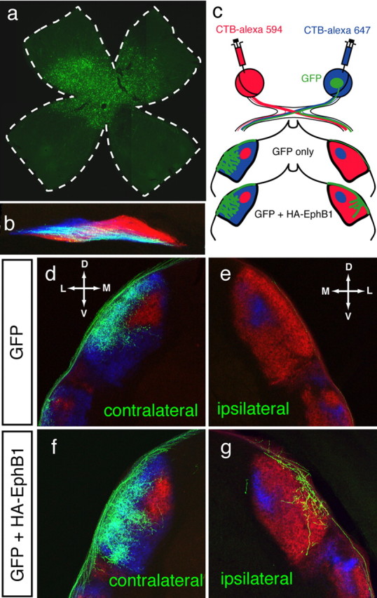
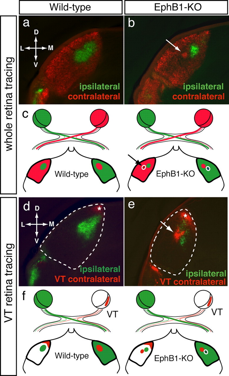
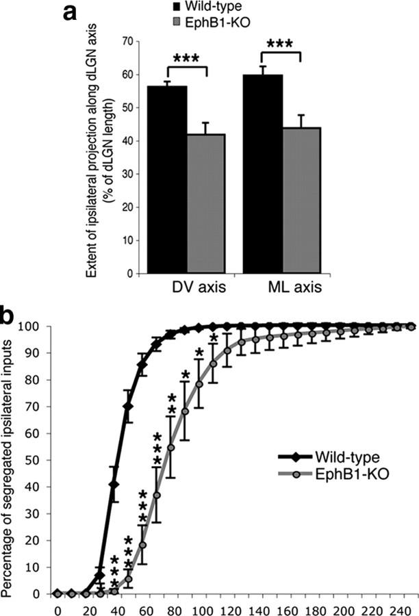
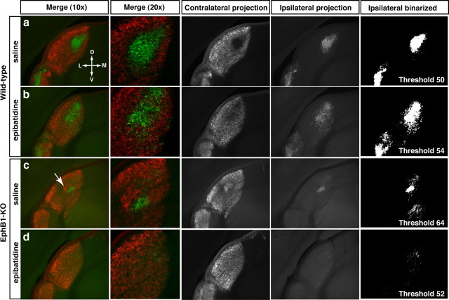
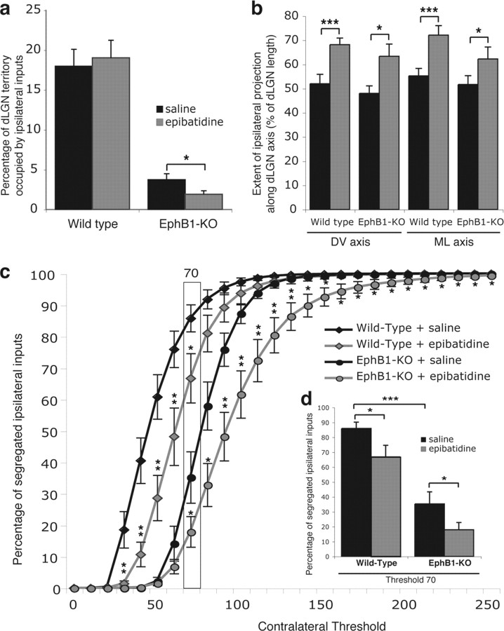
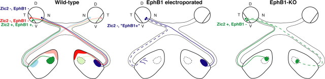
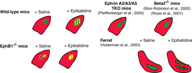
Partial decussation of sensory pathways allows neural inputs from both sides of the body to project to the same target region where these signals will be integrated. Here, to better understand mechanisms of eye-specific targeting, we studied how retinal ganglion cell (RGC) axons terminate in their thalamic target, the dorsal lateral geniculate nucleus (dLGN), when crossing at the optic chiasm midline is altered. In models with gain- and loss-of-function of EphB1, the receptor that directs the ipsilateral projection at the optic chiasm, misrouted RGCs target the appropriate retinotopic zone in the opposite dLGN. However, in EphB1(-/-) mice, the misrouted axons do not intermingle with normally projecting RGC axons and segregate instead into a distinct patch. We also revisited the role of retinal activity on eye-specific targeting by blocking correlated waves of activity with epibatidine into both eyes. We show that, in wild-type mice, retinal waves are necessary during the first postnatal week for both proper distribution and eye-specific segregation of ipsilateral axons in the mature dLGN. Moreover, in EphB1(-/-) mice, refinement of ipsilateral axons is perturbed in control conditions and is further impaired after epibatidine treatment. Finally, retinal waves are required for the formation of the segregated patch of misrouted axons in EphB1(-/-) mice. These findings implicate molecular determinants for targeting of eye-specific zones that are independent of midline guidance cues and that function in concert with correlated retinal activity to sculpt retinogeniculate projections.


Figures

Figure 1.
Figure 1.
Retinogeniculate targeting at P8 after in utero retinal electroporation of EphB1 constructs at E14.5. a, Retinal flat mount indicating GFP expressing RGCs in central retina. b, GFP+ RGC fibers at the optic chiasm. Retinal axons from each eye are in blue and red (see c) and GFP+ axons appear white. c, Scheme depicting the whole-eye labeling with CTB-Alexa Fluor dyes and GFP labeling along the retinogeniculate pathway. In GFP control electroporations, most GFP+ fibers project to the contralateral dLGN (d), with few (if any) GFP+ fibers projecting ipsilaterally (e). After GFP+ HA-EphB1 electroporations (f, g), there is an increase in GFP+ fibers in the ipsilateral dLGN. Both contralateral (f) and ipsilateral (g) GFP+ fibers project to the appropriate retinotopic region in the contralateral-recipient region (g). D, Dorsal; V, ventral; M, medial; L, lateral.
Figure 2.
Figure 2.
Retinogeniculate projections at P31 in wild-type and EphB1−/− mice. Coronal sections through the dLGN after whole-eye tracing in one eye and either whole eye (a, b) or VT retina (d, e) anterograde tracing from the other eye. a, d, In wild-type mice, the ipsilateral projection (green) forms a patch in the dorsocentral dLGN surrounded by contralateral terminations (red). Late-born VT RGCs project contralaterally to the dorsal tip of the dLGN (d, e, asterisk). b, e, In EphB1−/− mice, an ectopic patch (arrow) arising from the contralateral eye is adjacent to, but segregated from, the reduced ipsilateral patch. VT RGCs project contralaterally to two distinct sites (e): the dorsal tip of the dLGN, corresponding to the “normal” VT contralateral projection (asterisk), and the ectopic patch (arrow). c, f, Schemes depict dLGN termination regions after the anterograde tracing paradigms in wild-type and EphB1−/− mice. D, Dorsal; V, ventral; M, medial; L, lateral.
Figure 3.
Figure 3.
Distribution and segregation of ipsilateral retinal projections in wild-type and EphB1−/− mice. a, Extension of the ipsilateral projection expressed as a percentage of length along the DV and ML axes of the dLGN. Ipsilateral fibers cover less territory along the DV and ML dLGN axes in EphB1−/− mice compared with wild-type mice. b, Segregation plot. Percentage of pixels containing only ipsilateral signal (no contralateral signal) as a function of contralateral threshold (ipsilateral threshold is fixed). In EphB1−/− mice, ipsilateral fibers are less segregated from contralateral fibers (more overlap) than in wild-type mice. Error bars indicate SEM values. *p < 0.05, **p < 0.01, ***p < 0.001.
Figure 4.
Figure 4.
Retinogeniculate projections at P31 after binocular epibatidine treatment from P3 to P6. In wild-type (a, b) and EphB1−/− (c, d) mice, ipsilateral fibers are more scattered within the dLGN, cover an extended region of the dLGN, and overlap more with contralateral fibers after epibatidine treatment. In addition, aggregation of the abnormal contralateral patch adjacent to the ipsilateral patch and the optic tract is abolished after epibatidine treatment in EphB1−/− mice (c, arrow; compare with d). D, Dorsal; V, ventral; M, medial; L, lateral.
Figure 5.
Figure 5.
Comparison of ipsilateral projections in wild-type and EphB1−/− mice after epibatidine treatment. a, Binocular epibatidine treatment reduced the percentage of dLGN occupied by ipsilateral fibers in EphB1−/− mice but had no effect on wild-type mice. b, The ipsilateral projection extends more along the DV and ML axes of the dLGN after epibatidine treatment in both wild-type and EphB1−/− mice. c, Segregation plot. Percentage of pixels containing only ipsilateral signal (no contralateral signal), as a function of the contralateral threshold (ipsilateral threshold is fixed). The stars correspond to statistical significance between epibatidine and saline-treated animals. d, Columnal representation of the values in c, for the contralateral threshold set at 70. Segregation of ipsilateral axons in saline treated EphB1−/− mice is significantly impaired compared with wild-type mice, similar to the differences seen in nontreated animals. Segregation of ipsilateral fibers is further perturbed in the dLGN of both wild-type and EphB1−/− mice after epibatidine treatment. Error bars indicate SEM values. *p < 0.05, **p < 0.01, ***p < 0.001.
Figure 6.
Figure 6.
The retinogeniculate pathway in mouse models with impaired decussation at the optic chiasm. In wild-type mice, nasal RGCs (Zic2−/EphB1−; blue) project contralaterally to the lateral part of the dLGN. VT RGCs (Zic2+/EphB1+; green) project ipsilaterally to the dorsomedial part of the dLGN and later-born VT RGCs (Zic2−/EphB1−; red) project contralaterally to the dorsal tip of the dLGN. Ectopic EphB1 expression [Zic2−/EphB1+] in non-VT RGCs leads to the misrouting of some RGC fibers ipsilaterally (blue dashed line), which still target the contralateral-recipient region of the dLGN. In EphB1−/− mice, some VT RGCs (Zic2+/EphB1−) project contralaterally to the dorsomedial part of the dLGN (green dashed line), in a mirror image of the normal ipsilateral projection. D, Dorsal; V, ventral; T, temporal; N, nasal.
Figure 7.
Figure 7.
Schematic representation of eye-specific projection patterns to the dLGN. Retinogeniculate projections from the contralateral eye (red) and ipsilateral eye (green) in different mouse and ferret models. The extent of impairment in eye-specific segregation is represented as a grating of green and yellow, in proportion to the observed overlap.

References

    1. Badea TC, Cahill H, Ecker J, Hattar S, Nathans J. Distinct roles of transcription factors brn3a and brn3b in controlling the development, morphology, and function of retinal ganglion cells. Neuron. 2009;61:852–864. - PMC - PubMed
    1. Bansal A, Singer JH, Hwang BJ, Xu W, Beaudet A, Feller MB. Mice lacking specific nicotinic acetylcholine receptor subunits exhibit dramatically altered spontaneous activity patterns and reveal a limited role for retinal waves in forming ON and OFF circuits in the inner retina. J Neurosci. 2000;20:7672–7681. - PMC - PubMed
    1. Beg AA, Sommer JE, Martin JH, Scheiffele P. alpha2-Chimaerin is an essential EphA4 effector in the assembly of neuronal locomotor circuits. Neuron. 2007;55:768–778. - PubMed
    1. Bjartmar L, Huberman AD, Ullian EM, Rentería RC, Liu X, Xu W, Prezioso J, Susman MW, Stellwagen D, Stokes CC, Cho R, Worley P, Malenka RC, Ball S, Peachey NS, Copenhagen D, Chapman B, Nakamoto M, Barres BA, Perin MS. Neuronal pentraxins mediate synaptic refinement in the developing visual system. J Neurosci. 2006;26:6269–6281. - PMC - PubMed
    1. Butts DA, Kanold PO, Shatz CJ. A burst-based “Hebbian” learning rule at retinogeniculate synapses links retinal waves to activity-dependent refinement. PLoS Biol. 2007;5:e61. - PMC - PubMed

Publication types

MeSH terms