. 2009 Nov 25;29(47):14869-80.
doi: 10.1523/JNEUROSCI.4469-09.2009.

Peptidyl-prolyl isomerase 1 regulates protein phosphatase 2A-mediated topographic phosphorylation of neurofilament proteins

Affiliations

Peptidyl-prolyl isomerase 1 regulates protein phosphatase 2A-mediated topographic phosphorylation of neurofilament proteins

Parvathi Rudrabhatla et al. J Neurosci. .

Abstract

In normal neurons, neurofilament (NF) proteins are phosphorylated in the axonal compartment. However, in neurodegenerative disorders such as Alzheimer's disease (AD), Parkinson's disease (PD), and amyotrophic lateral sclerosis (ALS), NF proteins are aberrantly hyperphosphorylated within the cell bodies. The aberrant hyperphosphorylation of NF accumulations found in neurodegeneration could be attributable to either deregulation of proline-directed Ser/Thr kinase(s) activity or downregulation of protein phosphatase(s) activity. In this study, we found that protein phosphatase 2A (PP2A) expression is high in neuronal cell bodies and that inhibition of PP2A activity by okadaic acid (OA), microcystin LR (mLR), or fostriecin (Fos) leads to perikaryal hyperphosphorylation of NF. Peptidyl-prolyl isomerase Pin1 inhibits the dephosphorylation of NF by PP2A in vitro. In cortical neurons, Pin1 modulates the topographic phosphorylation of the proline-directed Ser/Thr residues within the tail domain of NF proteins by inhibiting the dephosphorylation by PP2A. Inhibition of Pin1 inhibits OA-induced aberrant perikaryal phosphorylation of NF. Treatment of cortical neurons with OA or Fos prevents the general anterograde transport of transfected green fluorescent protein-high-molecular-mass (NF-H) into axons caused by hyperphosphorylation of NF-H, and inhibition of Pin1 rescues this effect. Furthermore, inhibition of Pin1 inhibits the OA- or Fos-induced neuronal apoptosis. We show that OA-induced hyperphosphorylation of NF is a consequence of dephosphorylation of NF and is independent of c-Jun N-terminal protein kinase, extracellular signal-regulated kinase, and cyclin-dependent kinase-5 pathways. This study highlights a novel signaling role of PP2A by Pin1 and implicates Pin1 as a therapeutic target to reduce aberrant phosphorylation of NF proteins in neurodegenerative disorders such as AD, PD, and ALS.


Figures

Figure 1.
Figure 1.
Effect of protein phosphatase inhibitors on perikaryal phosphorylation of NF. A, Structure of rat NF-M/H representing KSP repeats in the tail domain. The KSP repeats are recognized by phospho-NF-M/H antibodies (SMI31, SMI34) and phospho-NF-H antibodies (RT97). The SMI32 antibodies recognize the non-phospho-KSP repeat region of NF-M/H. B, The dissociated E18 rat primary cortical neurons were cultured for 7 d in situ and then treated with or without different protein phosphatase PP2A inhibitors for 2.5 h [OA (b; 0.25 μm), microcystin LR (c; 15 μm), or Fos (d; 1 μm)] and then subjected to immunofluorescence with phospho-NF (SMI31) antibodies. Control (a) shows phosphorylated NF in processes with no phosphorylation in cell bodies. There was a significant increase of somatic phosphorylation in OA-treated (b), mLR-treated (c), and fostriecin-treated (d) neurons. C, The 7 DIC cortical neurons were subjected to the dose-dependent treatment of the PP2B-specific inhibitor cyclosporine A [0.5 μm (b), 1 μm (c), and 2.5 μm (d)]. Scale bar, 10 μm. D, Densitometry analyses of phospho-NF was performed with images captured using NIH Image software. To determine the relative intensity of SMI31 immunoreactivity within perikarya, representative areas of perikarya excluding obvious vesicles and the nucleus were quantified using the freehand selection tool. Similar results were obtained using RT97 and SMI34 antibodies. Intensity of SMI31 (blue), RT97 (red), and SMI34 (yellow) immunoreactivity (p-NF-H) in neuronal cell bodies was analyzed with the NIH ImageJ histogram from 50 to 100 individual cells from at least three experiments. The mean signal intensity (total pixel density per number pixels) is shown. Values in graphs are means ± fluorescence intensity (in arbitrary densitometric units) in perikarya from three different experiments. Mean fluorescence of phosphorylated NF-M/H immunoreactivity. *p < 0.01 and **p < 0.001 of phospho-NF-M/H in perikarya relative to fluorescence intensity values of nontreated neurons (NT).
Figure 2.
Figure 2.
Colocalization of PP2A and phospho-NF in neuronal perikarya in OA-treated cortical neurons. Colocalization of PP2A and phospho-NF in the nontreated (top; NT) and OA-treated (bottom) neurons. The E18 dissociated rat cortical neurons were cultured in vitro for 6 d, treated with OA, and subjected to immunofluorescence. The phospho-NF was visualized using the mouse phospho-NF-M/H monoclonal antibody SMI31 (red), and PP2A was visualized using goat polyclonal antibody (green). Scale bar, 10 μm.
Figure 3.
Figure 3.
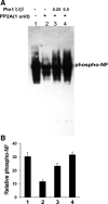
Dephosphorylation of NF by PP2A is inhibited by addition of Pin1 in vitro. A, NF triplet (NF-H/M/L) proteins were prepared from rat spinal cord and sciatic nerve as described in Material and Methods and was subjected to dephosphorylation with 1 U of PP2A (lane 2). The effect of Pin1 on NF-H dephosphorylation was tested by preincubating 2.5 μg of NF-triplet protein with GST–Pin1 (0.25 and 0.5 μg; lanes 3–4) at 30°C for 10 min, followed by addition of 1 U of PP2A. Reaction mixtures were incubated at 30°C for 30 min. Reactions were terminated by addition of SDS loading buffer, and proteins were analyzed by 4–20% SDS-PAGE. Blots were probed with monoclonal antibodies for phosphorylated NF-H (RT97). Data show the effect of Pin1 on NF-H dephosphorylation. PP2A dephosphorylates NF (lane 2). Addition of Pin1 prevents NF dephosphorylation in a dose-dependent manner [lane 3 (0.25 μg of Pin1); lane 4 (0.5 μg of Pin1)]. B, Densitometry analyses of phospho-NF obtained from A.
Figure 4.
Figure 4.
OA increases the NF phosphorylation in a time-dependent manner. A, The 7 DIC cortical neurons were treated with OA (0.25 μm) for 1 h (lane 2), 2 h (lane 3), and 4 h (lane 4) and subjected to Western blot analysis with phospho-NF (SMI31) antibodies. The OA induced hyperphosphorylation of NF in a time-dependent manner. B, Densitometry analyses of phospho-NF obtained from A. Mean density, Phospho-NF/actin is plotted on the y-axis. C, Five DIC neurons were treated with OA for 1, 2, and 4 h, and immunofluorescence was performed with SMI31 antibodies. In normal neurons, SMI31 is stained in axonal compartment (a). The phospho-NF accumulates in cell bodies in a time-dependent manner. Scale bar, 10 μm.
Figure 5.
Figure 5.
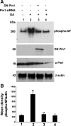
Pin1 expression and activity is critical toward OA-induced aberrant hyperphosphorylation of NF. A, Cortical neurons were subjected to treatment with OA (0.2 μm for 2.5 h), and Western blot analysis was performed with phospho-NF-M/H (SMI31) staining. The OA-induced hyperphosphorylation of NF is inhibited by knockdown of Pin1 by Pin1 siRNA and DN Pin1-transfected neurons. Lanes 1 and 2, Control scrambled siRNA-transfected neurons. Lanes 2–4, OA-treated neurons. Lane 3, Pin1 siRNA-transfected neurons. Lane 4, DN Pin1-transfected neurons. The transfected Pin1 is a GFP fusion protein and migrates at 50 kDa, at which the endogenous Pin1 (e Pin1) is 18 kDa. B, Densitometry analysis of phospho-NF immunoreactivity obtained from A. Mean density, Phospho-NF/actin is plotted on the y-axis. *p < 0.01 and **p < 0.001 of phospho-NF-M/H relative to nontreated neurons.
Figure 6.
Figure 6.
Knockdown of Pin1 by Pin1 siRNA inhibits OA-induced perikaryal phosphorylation of NF-M/H. A, Five DIC cortical neurons were transfected with either control (Ctrl) and scrambled siRNA (a–f) or Pin1 siRNA (g–i) and then treated with 0.25 μm OA (d–i) on day 7 for 2.5 h. Neurons were immunostained, p-NF-H was detected using SMI31 (red), and Pin1 was detected by using Pin1 antibody (green). Neurons transfected with Pin1 siRNA exhibited reduced p-NF-H in the cell body. Nontreated neurons exhibited SMI31 staining in the processes with little or no staining in the cell body (a–c), which is increased during OA treatment (d–f). Cell body SMI31 staining was reduced in Pin1 siRNA-treated neurons (g–i). Scale bar, 20 μm. B, Representative neurons are treated with OA as indicated. Intensity of SMI31 immunoreactivity (phospho-NF) in neuronal cell bodies was analyzed with the NIH ImageJ histogram. The mean signal intensity (total pixel density per number pixels) is shown. Values in graphs are means ± fluorescence intensity (in arbitrary densitometric units) in perikarya from four different experiments. Note the accumulation of phospho-NF in perikarya in OA-treated neurons and restoration of normal levels of p-NF-H in Pin1 siRNA-treated cortical neurons. C, Intensity of SMI31 immunoreactivity (phospho-NF) in neuronal cell bodies was analyzed with the NIH ImageJ histogram after treatment of cortical neurons with Fos. *p < 0.01, phospho-NF-M/H in perikarya relative to fluorescence intensity values of control. NT, Nontreated.
Figure 7.
Figure 7.
OA-mediated increase in phosphorylated NF is reduced by overexpression of DN Pin1. Five-day-old cortical neurons were transfected with GFP–DN Pin1 and after 24 h were treated with 0.25 μm OA for 2.5 h. Neurons were immunostained, phospho-NF was detected using SMI31 (red), and DN Pin1 was detected through expression of GFP. Note that only neurons that show DN Pin1 expression exhibited significant reduction of phosphorylated NF in the cell body. Scale bar, 20 μm.
Figure 8.
Figure 8.
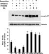
Pin1 modulates PP2A-mediated dephosphorylation of NF independent of ERK, JNK, and Cdk5 pathways. Seven DIC neurons were treated with PD98059 (ERK inhibitor; PD; lanes 2, 6), SP600125 (JNK inhibitor; SP; lanes 3, 7), and roscovitine (Cdk5 inhibitor; Ros; lanes 4, 8) for 1 h. The neurons were then subjected to treatment with OA (lanes 5–8) for 2 h. The phospho-NF was detected by SMI31 antibodies. The equal loading of the protein was confirmed by Western blot analysis with monoclonal β-tubulin antibodies. The bottom shows the densitometry analysis of mean phospho-NF/tubulin obtained from the top. *p < 0.01 and **p < 0.001 of phospho-NF-M/H intensity relative to nontreated neurons (NT).
Figure 9.
Figure 9.
Pin1 modulates NF transport of OA-treated neurons. The GFP–NF-H was transfected on 5 DIC cortical neurons. After 24 h, the NF translocation of transfected NF-H was monitored. The colocalization of GFP–NF-H-transfected neurons was observed by staining the cortical neurons with the neuronal marker TuJ1 (red) polyclonal antibodies. In normal neurons, GFP–NF-H is well translocated into axons (a–c). However, in cortical neurons treated with OA for 2 h, NF proteins are hyperphosphorylated in cell bodies, and therefore NF translocation is impaired. NF proteins are not translocated until the distal end of the axons. The GFP–NF-H remains in the cell bodies and proximal axons but not the distal part of the axons (d–f). Similarly, in fostriecin-treated cortical neurons, GFP–NF-H is not translocated into axons (g–i). However, knockdown of Pin1 rescues the normal NF translocation into the axons in OA-treated neurons (j–l) and Fos-treated neurons (m–o). Scale bar, 10 μm. NT, Nontreated.
Figure 10.
Figure 10.
Inhibition of PP2A by OA increases apoptosis in a time-dependent manner. The presence of apoptotic neurons examined by TUNEL–tetramethylrhodamine red staining in 7 DIC cortical neurons. Nuclei were counterstained using DAPI. TUNEL-positive cells were increased during treatment of cortical neurons with OA in a time-dependent manner. A, The neuronal death is increased by nearly 28% (2.5 h), 43% (5 h), and 78% (10%) after treatment with 0.25 μm OA. B, The quantization in the bar graph represents TUNEL-positive counts from four separate experiments, in which 14 independent fields were counted. Ctrl, Control.
Figure 11.
Figure 11.
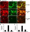
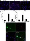
Inhibition of Pin1 reduces OA/Fos-induced neuronal death. A, Presence of apoptotic neurons examined by TUNEL–tetramethylrhodamine red staining in 7 DIC cortical neurons. Nuclei were counterstained using DAPI (blue). TUNEL-positive neurons were increased during OA treatment for 5 h and declined in OA-treated neurons transfected with Pin1 siRNA. Scale bar, 20 μm. The quantization in the bar graph (B) represents TUNEL-positive counts from four separate experiments, in which 12 independent fields were counted. NT (ctrl), Nontreated controls. B, The neuronal death is increased by 48% during exposure to OA (basal level, 5%), and this was reduced to 10% in neurons transfected with Pin1 siRNA and subjected to OA. C, The neuronal death is increased by 35% during exposure to Fos, and this was reduced to 12% in neurons transfected with Pin1 siRNA and subjected to Fos. D, OA-mediated neuronal apoptosis is reduced by overexpression of DN Pin1. Five-day-old cortical neurons were transfected with DN Pin1 and, after 24 h, were treated with 0.1 μm OA for 5 h. The GFP–DN Pin1-transfected neurons survive OA-induced cell death. Scale bar, 20 μm.
Figure 12.
Figure 12.
Role of Pin1 in the progression of compartment-specific NF-M/H phosphorylation. Top, In normal neurons, the NF phosphorylation is selective in the axonal compartment in normal neurons. After NF protein synthesis, higher activity of PP2A in cell bodies inhibits the NF tail domain phosphorylation. Although Pin1 is localized in cell bodies, it cannot act on the non-S/T-P phosphorylated proteins. During assembly of NF in axonal hillock region, NFs are transported by slow axonal transport. In the axonal compartment, the axonal–glial interaction activates the Ser/Thr kinases and phosphorylates some of pSer/Thr-Pro residues. The phosphorylation of these residues renders the phospho-Ser/Thr-Pro to remain in cis configuration and blocks additional phosphorylation. However, Pin1 induces cis-trans isomerization, increases the accessibility of the Ser/Thr kinases, and stabilizes the NF phosphorylation. Because of the high PP2A activity, there is no NF phosphorylation in the cell bodies. Bottom, Inhibition of PP2A activity leads to aberrant perikaryal hyperphosphorylation of NF proteins in cell bodies. Pin1 stabilizes the NF phosphorylation in the cell bodies, phospho-NF proteins are accumulated in the cell bodies and proximal axons, and NF transport is impaired. This is consistent with the observation that PP2A activity is reduced in AD brain and ALS spinal cord with higher accumulations of phospho-NF in the perikarya.

Similar articles

Cited by

References

    1. Ahlijanian MK, Barrezueta NX, Williams RD, Jakowski A, Kowsz KP, McCarthy S, Coskran T, Carlo A, Seymour PA, Burkhardt JE, Nelson RB, McNeish JD. Hyperphosphorylated tau and neurofilament and cytoskeletal disruptions in mice overexpressing human p25, an activator of cdk5. Proc Natl Acad Sci U S A. 2000;97:2910–2915. - PMC - PubMed
    1. Chao SH, Greenleaf AL, Price DH. Juglone, an inhibitor of the peptidyl-prolyl isomerase Pin1, also directly blocks transcription. Nucleic Acids Res. 2001;29:767–773. - PMC - PubMed
    1. Chin SS, Liem RK. Transfected rat high-molecular-weight neurofilament (NF-H) coassembles with vimentin in a predominantly nonphosphorylated form. J Neurosci. 1990;10:3714–3726. - PMC - PubMed
    1. Dashiell SM, Tanner SL, Pant HC, Quarles RH. Myelin-associated glycoprotein modulates expression and phosphorylation of neuronal cytoskeletal elements and their associated kinases. J Neurochem. 2002;81:1263–1272. - PubMed
    1. de Waegh SM, Lee VM, Brady ST. Local modulation of neurofilament phosphorylation, axonal caliber and slow axonal transport by mylenating Schwann cells. Cell. 1992;68:451–463. - PubMed

Publication types

MeSH terms

LinkOut - more resources