. 2009 Nov 25;29(47):14942-55.
doi: 10.1523/JNEUROSCI.2276-09.2009.

Distinct muscarinic acetylcholine receptor subtypes contribute to stability and growth, but not compensatory plasticity, of neuromuscular synapses

Affiliations

Distinct muscarinic acetylcholine receptor subtypes contribute to stability and growth, but not compensatory plasticity, of neuromuscular synapses

Megan C Wright et al. J Neurosci. .

Abstract

Muscarinic acetylcholine receptors (mAChRs) modulate synaptic function, but whether they influence synaptic structure remains unknown. At neuromuscular junctions (NMJs), mAChRs have been implicated in compensatory sprouting of axon terminals in paralyzed or denervated muscles. Here we used pharmacological and genetic inhibition and localization studies of mAChR subtypes at mouse NMJs to demonstrate their roles in synaptic stability and growth but not in compensatory sprouting. M(2) mAChRs were present solely in motor neurons, whereas M(1), M(3), and M(5) mAChRs were associated with Schwann cells and/or muscle fibers. Blockade of all five mAChR subtypes with atropine evoked pronounced effects, including terminal sprouting, terminal withdrawal, and muscle fiber atrophy. In contrast, methoctramine, an M(2/4)-preferring antagonist, induced terminal sprouting and terminal withdrawal, but no muscle fiber atrophy. Consistent with this observation, M(2)(-/-) but no other mAChR mutant mice exhibited spontaneous sprouting accompanied by extensive loss of parental terminal arbors. Terminal sprouting, however, seemed not to be the causative defect because partial loss of terminal branches was common even in the M(2)(-/-) NMJs without sprouting. Moreover, compensatory sprouting after paralysis or partial denervation was normal in mice deficient in M(2) or other mAChR subtypes. We also found that many NMJs of M(5)(-/-) mice were exceptionally small and reduced in proportion to the size of parental muscle fibers. These findings show that axon terminals are unstable without M(2) and that muscle fiber growth is defective without M(5). Subtype-specific muscarinic signaling provides a novel means for coordinating activity-dependent development and maintenance of the tripartite synapse.


Figures

Figure 1.
Figure 1.
Atropine causes diverse and severe effects on NMJ organization. Confocal images of LAL muscles treated twice daily for 7 d with saline or atropine. Preterminal and terminal axons were immunolabeled for neurofilaments (NF) and a synaptic vesicle protein, SV2 (green), nAChRs were labeled for α-bungarotoxin (red), and Schwann cells were labeled for S100 (blue). A, Low-magnification images of saline- or atropine-treated LAL muscles. In saline-treated muscles, axon terminals (green) precisely overlap brightly labeled nAChRs (red), making NMJs appear yellow. In atropine-treated muscles, axon terminals sprout (arrows) or retract (arrowheads), and postsynaptic nAChRs are faintly labeled at some NMJs. B, High-magnification images of NMJs showing diverse patterns of synaptic disorganization evoked by atropine. Saline-treated NMJs (e.g., junction 1) show precise alignment of axon terminals (green), tSCs (blue), and postsynaptic clusters of nAChRs (red). In atropine-treated muscles, at some NMJs (e.g., junction 2), nerve terminals extended sprouts (white arrows) that elongated along activated tSC processes (blue arrows), whereas nAChRs are unaffected. Another set of NMJs (e.g., junction 3) demonstrated selective, profound loss of postsynaptic nAChRs. Other NMJs (e.g., junction 4) displayed selective, complete loss of axon terminals and abnormally quiescent tSCs that formed no processes even in the absence of axon terminals. C–F, Quantification of diverse effects evoked by atropine at various doses. C, The percentage of NMJs exhibiting terminal sprouts. D, The percentage of NMJs exhibiting uniform, profound loss of postsynaptic nAChRs. E, The percentage of NMJs exhibiting selective loss of terminal arbors. F, The average diameter of muscle fibers. Atropine-treated muscle fibers are substantially atrophied. *p < 0.01. Scale bars: A, 30 μm; B, 10 μm.
Figure 2.
Figure 2.
Subtype-specific inhibition of mAChRs elicits subsets of atropine-evoked effects. Confocal views of representative synapses in the LAL muscles treated twice daily for 7 d with methoctramine or 4-DAMP. A, Methoctramine, an M2/4 receptor-preferring antagonist, induces terminal sprouting, tSC activation, and partial terminal loss. Note terminal sprouts (arrowheads; green) associated with leading tSC processes (arrowheads; blue). At some NMJs with sprouting (bottom), axon terminals are partially retracted as evidenced by clusters of nAChRs (arrow; red) unoccupied by terminal branches (arrow; green). B, 4-DAMP, which blocks the odd-numbered mAChRs and M4 receptors with high affinity, induces dramatic dying back of nerve terminal accompanied by abnormally quiescent tSCs. Retracting axons are present at the vacant endplates (arrowheads; green) or along the preterminal Schwann cells (arrows; green). Note that tSCs are abnormally quiescent as evidenced by bright S100 labeling without process extension (blue arrows). Postsynaptically, muscle fibers are normal and there is no loss of nAChRs or fiber atrophy. Schwann cells are presented in black and white (single images) as well as in blue (merged images) to illustrate better fine processes formed by tSCs, a hallmark of activated tSCs. Scale bar, 10 μm. NF, Neurofilaments.
Figure 3.
Figure 3.
Spontaneous sprouting and terminal loss at M2−/− NMJs. A, Low-magnification confocal views of M2+/+ and M2−/− NMJs. Terminal arbors of many M2−/− NMJs (arrows; green) do not have the compact oval shape of wild-type NMJs but display unusually long and dispersed branches in contact with fragmented patches of nAChRs. B, High-magnification view of M2−/− NMJs. Terminal sprouts form varicosities directly apposed to islands of nAChR patches (arrowheads), whereas parental terminal branches that covered original synaptic contacts are retracted, leaving abandoned, faintly labeled branches of parental clusters of nAChRs (e.g., area marked by an asterisk). Some M2−/− NMJs with no sprouting exhibit vacant branches of nAChRs and tSCs that are unoccupied by terminal branches (arrows in bottom), indicating that terminal loss precedes terminal sprouting in M2−/− muscles. Scale bars: A, 30 μm; B, 10 μm. NF, Neurofilaments.
Figure 4.
Figure 4.
Unlike paralysis-induced sprouting, sprouting in M2−/− NMJs is accompanied by parental terminal loss. Confocal views of a representative junction from an M2−/− LAL muscle (top) and a junction from an LAL muscle paralyzed for 7 d by botulinum toxin. Original synaptic contacts are identified by the presence of preterminal axons (arrowheads), terminal sprouts (arrows), and compact oval-shaped, postsynaptic clusters of nAChRs (asterisks in nAChR panels). Note that M2−/− NMJs display a cluster of nAChRs completely unoccupied by axons at original synaptic contacts (asterisks, top). In contrast, nerve terminals in paralyzed muscles sprout but maintained parental terminal branches (asterisks, bottom). Scale bars, 10 μm.
Figure 5.
Figure 5.
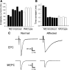
A subset of M2−/− NMJs shows markedly abnormal synaptic function. A, Mean endplate current amplitude for LAL muscles from seven M2−/− and four M2+/+ mice. Approximately 17 muscle fibers were recorded in each mouse. In two of the M2−/− mice, endplate current amplitude was reduced by one-third relative to M2+/+ mice. B, The time constant of endplate current decay was greatly prolonged in the two affected M2−/− mice. C, Representative EPCs and MEPCs for a wild-type endplate and an endplate from an affected M2−/− mouse. EPCs and MEPCs from unaffected M2−/− NMJs were indistinguishable from M2+/+ NMJs.
Figure 6.
Figure 6.
Nerve terminal shifting and fragmentation in M2/4−/− dKO NMJs. Confocal views of LAL muscles of 3-week-old (A, B) and 3-month-old (C, D) M2/4−/− dKO mice. Muscles were labeled as described in Figure 1. A, Portions of endplates are commonly unoccupied by terminal arbors (e.g., NMJs marked by arrows, asterisk area in A). B, Some terminal arbors appear to have shifted, leaving previous endplates completely abandoned (e.g., junctions 1 and 2 in B). The synaptic morphology of abandoned endplates closely resembles that of shifted terminal arbors. C, An example of 3-month-old M2/4−/− NMJs exhibiting extensively fragmented clusters of postsynaptic nAChRs. D, Relative distribution of NMJs displaying particular numbers of nAChR patches in M2/4+/+ and M2/4−/− dKO mice. Scale bars: A, B, 20 μm; C, 10 μm.
Figure 7.
Figure 7.
Parallel reduction of muscle fiber and synaptic size in M5−/− muscles. LAL muscles of adult M5+/+ and M5−/− mice were labeled as described in Figure 1. A, Low-magnification confocal views show numerous unusually small NMJs (arrows) in M5−/− muscles. Inset denotes an exceptionally small M5−/− NMJ comprising a terminal bouton and a tiny patch of nAChRs (arrows in inset). B, At some small M5−/− NMJs, postsynaptic clusters of nAChRs are fragmented and only faintly labeled (arrowheads), indicating postsynaptic disassembly. C, Comparative analysis of synaptic and muscle fiber size in M5+/+ and M5−/− muscles. Many muscle fibers and NMJs in M5−/− muscles, except slow-twitch soleus muscles, are substantially smaller than those of M5+/+ muscles. In addition, synaptic size is highly correlated with the size of parental muscle fibers, indicating that muscle fiber growth is the primary limitation in M5−/− muscles. Scale bars: A, 100 μm; B, 20 μm. STM, Sternomastoid muscle; SOL, soleus muscle; NF, neurofilaments.
Figure 8.
Figure 8.
Paralysis initiates normal sprouting in mAChR KO mice. LAL muscles of wild-type (+/+), M1/3−/−, M2/4−/−, and M5−/− mice were paralyzed for 7 d and labeled as described in Figure 1. A, Representative junctions of +/+, M2/4−/−, and M5−/− NMJs exhibiting sprouting. Paralysis induced normal sprouting in M1/3−/− (data not shown) and M2/4−/− NMJs. In M5−/− NMJs, terminal sprouts form numerous secondary and tertiary branches, elongating far more extensively than those observed at +/+ or M1/3−/− or M2/4−/− NMJs. B–E, Quantitative and comparative analysis of the sprouting in terms of the percentage of the junctions displaying sprouts (B), the number of primary branches at NMJs with sprouts (C), relative distribution of NMJs with different lengths of sprouts (D), and the average length of sprouts (E). Terminal sprouts become exceptionally lengthy in M5−/− NMJs attributable to elongation of secondary and tertiary branches. The initiation of paralysis-induced sprouting, however, was normal in M1/3−/−, M2/4−/−, and M5−/− NMJs. n = 3 or more LAL muscles, >100 NMJs per muscle were analyzed. **p < 0.0001. Scale bar, 30 μm.
Figure 9.
Figure 9.
Localization of mAChR subtypes at NMJs. A, Although an anti-M2 antibody labels axon terminals, tSCs, and postsynaptic gutters in wild-type NMJs, nerve terminal-associated labeling (arrow, inset) disappears in M2−/− NMJs, whereas labeling associated with tSC cell bodies and postsynaptic gutters remains. Thus, M2 mAChRs appear to be expressed selectively in motor neurons because only this binding is specific. B, Laser-assisted microdissection of spinal motor neurons (MN) and NMJs. C, RT-PCR analyses show that motor neurons express only M2 mAChRs, whereas Schwann cells and/or muscle fibers at NMJs are likely to express multiple odd-numbered receptors (M1, M3, M5). Scale bars: A, 10 μm; B, 50 μm. IR, Immunoreactivity; NF, neurofilaments.

Similar articles

Cited by

References

    1. Abrams P, Andersson KE, Buccafusco JJ, Chapple C, de Groat WC, Fryer AD, Kay G, Laties A, Nathanson NM, Pasricha PJ, Wein AJ. Muscarinic receptors: their distribution and function in body systems, and the implications for treating overactive bladder. Br J Pharmacol. 2006;148:565–578. - PMC - PubMed
    1. Albani M, Lowrie MB, Vrbová G. Reorganization of motor units in reinnervated muscles of the rat. J Neurol Sci. 1988;88:195–206. - PubMed
    1. Alexander SP, Mathie A, Peters JA. Guide to receptors and channels (GRAC), Ed 3. Br J Pharmacol. 2008;153(Suppl 2):S1–S209. - PMC - PubMed
    1. Angaut-Petit D, Molgó J, Faille L, Juzans P, Takahashi M. Incorporation of synaptotagmin II to the axolemma of botulinum type-A poisoned mouse motor endings during enhanced quantal acetylcholine release. Brain Res. 1998;797:357–360. - PubMed
    1. Argentieri TM, Aiken SP, Laxminarayan S, McArdle JJ. Characteristics of synaptic transmission in reinnervating rat skeletal muscle. Pflugers Arch. 1992;421:256–261. - PubMed

Publication types

MeSH terms

LinkOut - more resources