. 2009 Nov 25;29(47):15017-27.
doi: 10.1523/JNEUROSCI.3451-09.2009.

A rapamycin-sensitive signaling pathway is essential for the full expression of persistent pain states

Affiliations

A rapamycin-sensitive signaling pathway is essential for the full expression of persistent pain states

Sandrine M Géranton et al. J Neurosci. .

Abstract

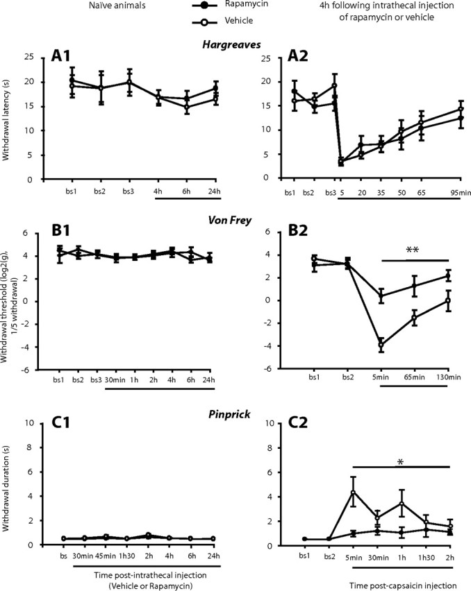
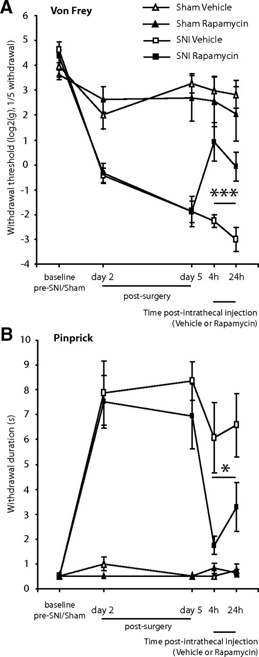
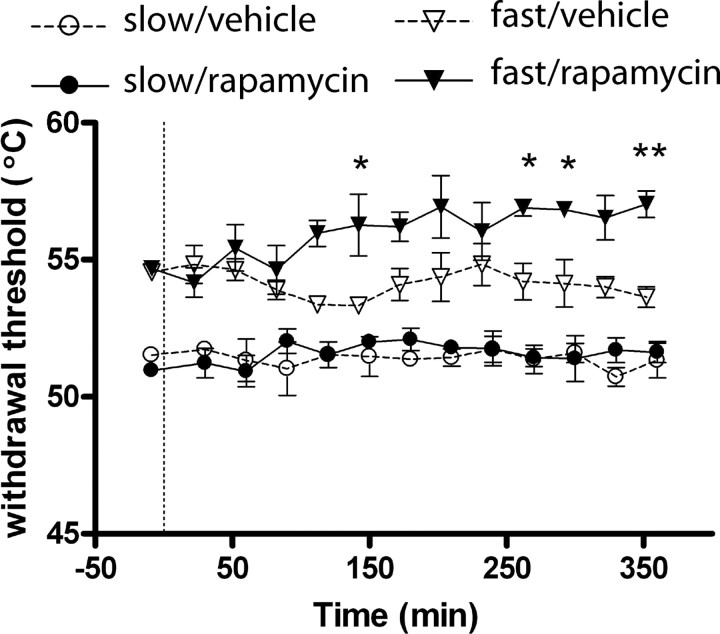
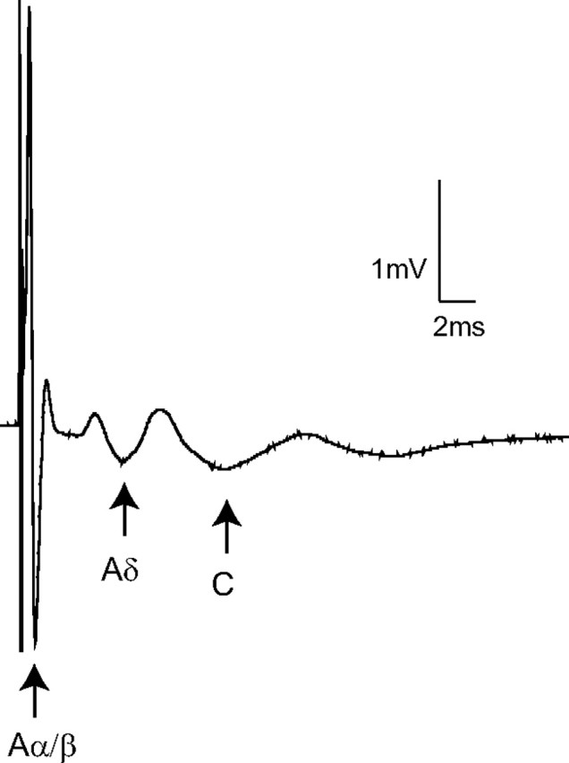
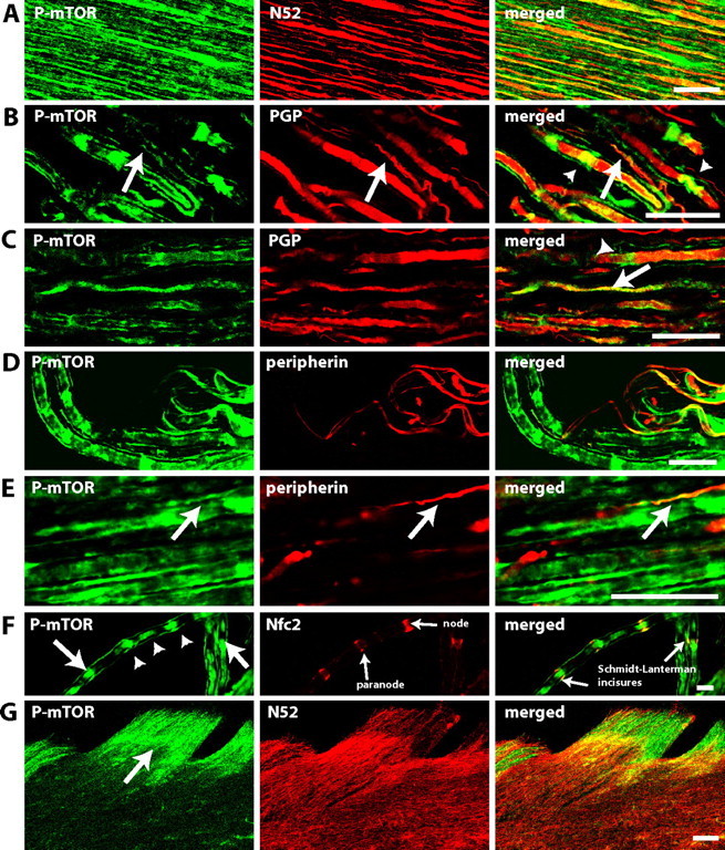
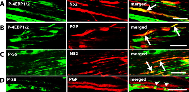
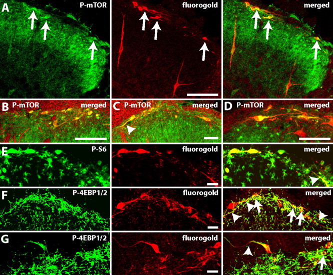
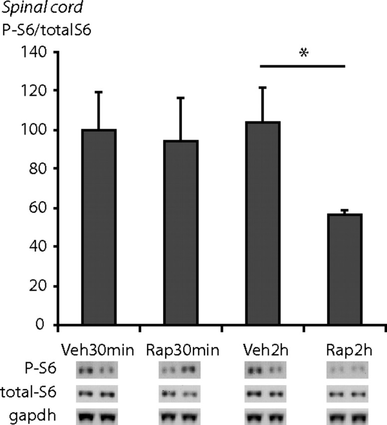
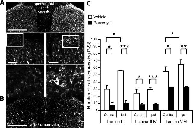
Translational control through the mammalian target of rapamycin (mTOR) is critical for synaptic plasticity, cell growth, and axon guidance. Recently, it was also shown that mTOR signaling was essential for the maintenance of the sensitivity of subsets of adult sensory neurons. Here, we show that persistent pain states, but not acute pain behavior, are substantially alleviated by centrally administered rapamycin, an inhibitor of the mTOR pathway. We demonstrate that rapamycin modulates nociception by acting on subsets of primary afferents and superficial dorsal horn neurons to reduce both primary afferent sensitivity and central plasticity. We found that the active form of mTOR is present in a subpopulation of myelinated dorsal root axons, but rarely in unmyelinated C-fibers, and heavily expressed in the dorsal horn by lamina I/III projection neurons that are known to mediate the induction and maintenance of pain states. Intrathecal injections of rapamycin inhibited the activation of downstream targets of mTOR in dorsal horn and dorsal roots and reduced the thermal sensitivity of A-fibers. Moreover, in vitro studies showed that rapamycin increased the electrical activation threshold of Adelta-fibers in dorsal roots. Together, our results imply that central rapamycin reduces neuropathic pain by acting both on an mTOR-positive subset of A-nociceptors and lamina I projection neurons and suggest a new pharmacological route for therapeutic intervention in persistent pain states.


Figures

Figure 1.
Figure 1.
Intrathecal administration of rapamycin does not affect acute pain but reduces capsaicin-induced secondary but not primary hyperalgesia. A1, B1, C1, Effects of intrathecal injection of rapamycin (or vehicle) in naive rats on the following: withdrawal latency to heat using the Hargreaves test (A1; N = 6 in each group), mechanical withdrawal threshold measured with Von Frey hairs (B1; N = 8 in each group), and withdrawal response duration after nociceptive mechanical stimulation (pinprick stimulus) of the plantar surface of the paw (C1; N = 4 in each group). A2, B2, C2, Rapamycin (or vehicle) was delivered 4 h before the administration of capsaicin in the center of the plantar surface of the paw. Effects of intrathecal injection of rapamycin (or vehicle) on the following: withdrawal latency to heat after capsaicin (A2, N = 6 in each group), lateral mechanical withdrawal threshold after capsaicin injection measured with Von Frey hairs (B2, N = 7 in each group), withdrawal response duration to lateral pinprick stimulation after capsaicin (C2, N = 9–10). Mean ± SEM for the injected (left) hindpaw is illustrated in each panel. *p < 0.05, **p < 0.01.
Figure 2.
Figure 2.
Intrathecal administration of rapamycin attenuates mechanical hyperalgesia in the SNI model. Effect of intrathecal injection of rapamycin, or its vehicle on, A, mechanical withdrawal threshold measured with Von Frey hairs, and B, withdrawal response duration (s) after nociceptive mechanical stimulation (pinprick stimulus) of the lateral plantar surface of the paw of SNI animals, or sham animals. Mean ± SEM is illustrated. N = 3–4 in sham groups, N = 7–8 in SNI groups. *p < 0.05, ***p < 0.001.
Figure 3.
Figure 3.
Intrathecal administration of rapamycin increases A- but not C-nociceptor-evoked paw withdrawal thresholds. Time course effects of intrathecal injection of rapamycin or vehicle on paw withdrawal thresholds to fast and slow heat ramps that preferentially activate A- and C-nociceptors respectively. N = 3 in each group. Mean ± SEM heat withdrawal threshold (°C) is illustrated in each panel. Vertical dashed line indicates the drug injection time. *p < 0.05; **p < 0.01.
Figure 4.
Figure 4.
Extracellular compound action potential recordings. Representative compound action potentials recorded from an isolated rapamycin-treated dorsal root illustrating the fast (Aα/β-), medium (Aδ-) and slow (C-) conducting components evoked by 500 μA stimulation (average of 10 traces shown). Arrows indicate the negative peak of each triphasic (positive-negative-positive) profile. In this example the last positive peak of the Aδ-component overlaps the first positive peak of the C-component. The threshold stimulation intensities for the Aα/β-, Aδ-, and C-fiber components were 5, 60, and 200 μA, respectively.
Figure 5.
Figure 5.
P-mTOR immunoreactivity in dorsal roots is largely found within myelinated fibers. A–F, Confocal images of longitudinal sections of dorsal roots. A, Colocalization of P-mTOR (green) and myelinated fiber marker N52 (red). B, C, Colocalization of P-mTOR (green) and nerve fibers marker PGP (red), a general marker of nerve fibers. P-mTOR staining can be seen along the nerve fibers (arrows) but also in accumulations along the fibers (arrow heads). D, E, Colocalization of P-mTOR (green) and peripherin (red), a marker for small fibers. F, Colocalization of P-mTOR (green) and neurofascin (red), a marker for nodes and paranodes. Axonal accumulations of P-mTOR (arrow head) can be seen in association with nodes of Ranvier but in some cases also localized adjacent to the Schmidt-Lanterman incisures (arrow) of the surrounding Schwann cells. G, Confocal images of horizontal sections of dorsal horn. Pictures show P-mTOR (green) and myelinated fiber marker N52 (red). P-mTOR staining in sensory axons does not extend beyond the dorsal root entry zone (arrow). In A–G the single staining for each antibody and the merged image are shown from left to right and double staining appears in yellow. A–F, Single plane picture. Scale bars: A–E, 25 μm; F, 50 μm; G, 100 μm.
Figure 6.
Figure 6.
P-4EBP1/2 and P-S6 immunoreactivity in dorsal roots is largely found within myelinated fibers. A–D, Confocal images of longitudinal sections of dorsal roots. A, Colocalization of P-4EBP1/2 (green) and myelinated fiber marker N52 (red). B, Colocalization of P-4EBP1/2 (green) and nerve fibers marker PGP (red). C, Colocalization of P-S6 (green) and myelinated fiber marker N52 (red). D, Colocalization of P-S6 (green) and nerve fibers marker PGP (red). P-4EBP1/2 and P-S6 staining can be seen along the nerve fibers but also in accumulations along the fibers. In A–D, the single staining for each antibody and the merged image are shown from left to right and double staining appears in yellow. A–D, Single plane picture. Scale bars: A–D, 25 μm.
Figure 7.
Figure 7.
P-mTOR, P-S6 protein, and P-4EBP1/2 are strongly expressed in lamina I/III projection neurons. Colocalization of projection neurons labeled with Fluorogold and an antibody against Fluorogold (red) and (green): A–D, P-mTOR; E, P-S6; and F, G, P-4EBP1/2. In A, E–G, the single staining for each antibody and the merged image are shown from left to right and double staining appears in yellow. Notice the extensive staining of dendrites (arrow heads) marked on merged pictures. The percentage of projections neurons labeled with P-mTOR and P-S6 was, respectively: 77 ± 3% and 80 ± 1%; B–D show the merged pictures. Arrows show double labeled neurons. Scale bars: A–C, 100 μm; D–G, 25 μm.
Figure 8.
Figure 8.
Intrathecal administration of rapamycin decreases phosphorylation of the downstream target of mTORC1 S6 protein (S6) in the spinal cord. Immunoblots probed with anti-P-S6 antibody and anti-S6 antibody after gel electrophoresis of lysates from spinal cord tissue. Animals received an intrathecal injection of rapamycin or vehicle 30 min or 2 h before killing. There was a significant reduction in S6 phosphorylation 2 h after rapamycin injection in spinal cord tissue. N = 3–4 in each condition. Mean ± SEM is illustrated. *p < 0.05.
Figure 9.
Figure 9.
Intrathecal administration of rapamycin reduces the capsaicin-induced increase in phosphorylation of S6 protein in the spinal cord. A, Confocal images of P-S6 immunostaining in the superficial dorsal horn 2 h after intraplantar injection of capsaicin in the center of the plantar surface of the hindpaw. Scale bar, 100 μm. B, Confocal images of P-S6 immunostaining in the superficial dorsal horn 4 h after intrathecal administration of rapamycin. Scale bar, 100 μm. C, Effects of intrathecal administration of rapamycin (or vehicle) before capsaicin injection in the hindpaw. Rapamycin (or vehicle) was administered 4 h before capsaicin and animals perfused 2 h following capsaicin injection. Rapamycin significantly reduced P-S6 protein levels in the three spinal cord domains. Furthermore, following rapamycin treatment, capsaicin did not increase P-S6 protein expression. *p < 0.05, **p < 0.01, ***p < 0.001. N = 3 in each group.

Similar articles

Cited by

References

    1. Alvarez J. The autonomous axon: a model based on local synthesis of proteins. Biol Res. 2001;34:103–109. - PubMed
    1. Antion MD, Merhav M, Hoeffer CA, Reis G, Kozma SC, Thomas G, Schuman EM, Rosenblum K, Klann E. Removal of S6K1 and S6K2 leads to divergent alterations in learning, memory, and synaptic plasticity. Learn Mem. 2008;15:29–38. - PMC - PubMed
    1. Asante CO, Wallace VC, Dickenson AH. Formalin-induced behavioural hypersensitivity and neuronal hyperexcitability are mediated by rapid protein synthesis at the spinal level. Mol Pain. 2009;5:27. - PMC - PubMed
    1. Banko JL, Poulin F, Hou L, DeMaria CT, Sonenberg N, Klann E. The translation repressor 4E-BP2 is critical for eIF4F complex formation, synaptic plasticity, and memory in the hippocampus. J Neurosci. 2005;25:9581–9590. - PMC - PubMed
    1. Breivik H, Collett B, Ventafridda V, Cohen R, Gallacher D. Survey of chronic pain in Europe: prevalence, impact on daily life, and treatment. Eur J Pain. 2006;10:287–333. - PubMed

Publication types

MeSH terms