. 2009 Dec 15;106(50):21167-72.
doi: 10.1073/pnas.0906436106. Epub 2009 Nov 25.

Molecular priming of Lyn by GPVI enables an immune receptor to adopt a hemostatic role

Affiliations

Molecular priming of Lyn by GPVI enables an immune receptor to adopt a hemostatic role

Alec A Schmaier et al. Proc Natl Acad Sci U S A. .

Abstract

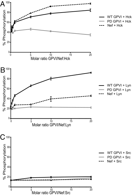
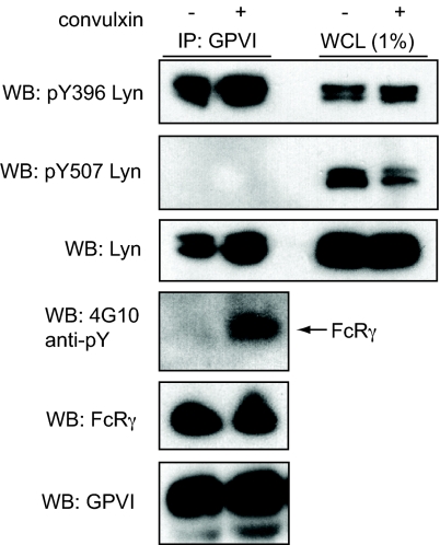
The immune receptor signaling pathway is used by nonimmune cells, but the molecular adaptations that underlie its functional diversification are not known. Circulating platelets use the immune receptor homologue glycoprotein VI (GPVI) to respond to collagen exposed at sites of vessel injury. In contrast to immune cell responses, platelet activation must take place within seconds to successfully form thrombi in flowing blood. Here, we show that the GPVI receptor utilizes a unique intracellular proline-rich domain (PRD) to accelerate platelet activation, a requirement for efficient platelet adhesion to collagen under flow. The GPVI PRD specifically binds the Src-family kinase Lyn and directly activates it, presumably through SH3 displacement. In resting platelets, Lyn is constitutively bound to GPVI in an activated state and platelets lacking Lyn exhibit defective collagen adhesion like that of platelets with GPVI receptors lacking the PRD. These findings define a molecular priming mechanism that enables an immune-type receptor to adopt a hemostatic function. These studies also demonstrate that active kinases can constitutively associate with immune-type receptors without initiating signal transduction before receptor ligation, consistent with a recent molecular model of immune receptor signaling in which receptor ligation is required to bring active kinases to their receptor substrates.


Conflict of interest statement

The authors declare no conflict of interest.

Figures

Fig. 1.
Fig. 1.
The intracellular PRD is required for rapid platelet activation by GPVI receptors but not sustained GPVI signaling. (A) Dose-response curve of platelet activation mediated by WT and PD GPVI signaling. Platelets from GPVI-deficient mice expressing WT GPVI or PD GPVI receptors were stimulated with the indicated concentrations of convulxin for 10 min, and activation was measured using flow cytometry to detect the expression of surface P-selectin. (B) Time-course of platelet fibrinogen binding by WT and PD GPVI-expressing platelets following convulxin stimulation. Platelets were stimulated with 10-nM convulxin in the presence of Alexa-Fluor 647-conjugated fibrinogen for times indicated, fixed, and analyzed by flow cytometry. (C) Time-course of JON-A binding following 10-nM convulxin stimulation. (D) Time-course of fibrinogen binding following stimulation with 10 μg/ml CRP. In all experiments, only GFP+, GPVI-expressing platelets were analyzed. *, P < 0.05; **, P < 0.01; error bars indicate mean ± SD; n = 3 to 6 experiments for each time-point indicated, using four animals for WT or PD GPVI.
Fig. 2.
Fig. 2.
PD GPVI receptors exhibit delayed Syk phosphorylation and calcium signaling. (A) Lentiviral vectors were used to express WT GPVI or PD GPVI or vector alone (vector) in RBL-2H3 cells and surface expression of GPVI was detected by flow cytometry. (B) Time-course of GPVI-mediated activation of Syk following convulxin stimulation in WT GPVI and PD GPVI-expressing RBL-2H3 cells. GPVI-expressing RBL-2H3 cells were stimulated with convulxin for the time-points indicated, and whole-cell lysate was analyzed by immunoblotting (WB) with antibodies against activated Syk (pSyk) and total Syk. (C) Ca2+ mobilization in RBL-2H3 cells expressing WT and PD GPVI and vector control. Cells were stimulated with convulxin and intracellular Ca2+ mobilization was measured by the ratio of Fura-2 emissions in a fluorimeter. Curves from representative experiments are shown; n = 3 measurements.
Fig. 3.
Fig. 3.
The GPVI PRD is required for efficient platelet adhesion to collagen under flow. (A) Adhesive efficiency to collagen under flow of WT or PD GPVI-expressing platelets or vector control platelets. (B) Rolling times on collagen for WT or PD GPVI platelets. The time each adherent platelet spent in the rolling phase was calculated based on the number of frames during which each platelet changed position. (i) Rolling times are shown divided into terciles (1st, shortest third of rolling times in each group; 2nd, middle third of rolling times in each group; 3rd, longest third of rolling times in each group). (ii) Percentage rolling times: <350 ms, 350 to 700 ms, and >700 ms. The same adherent platelets were analyzed in (i) and (ii). **, P < 0.01; error bars indicate mean ± SD; three to five movies using three to four animals for each GPVI variant or vector control were analyzed in a blinded fashion; n = total number of adherent platelets analyzed.
Fig. 4.
Fig. 4.
GPVI directly activates Lyn and Hck in a PRD-dependent manner. Recombinant Hck (A), Lyn (B), and c-Src (C) were purified from Sf9 insect cells in an inactive state and assayed for kinase activity with a peptide substrate in vitro in the presence of increasing concentrations of purified GST-WT GPVI, GST-PD GPVI, or recombinant HIV-1 Nef. Each condition was repeated in quadruplicate, and the extent of phosphorylation is expressed as mean percent-phosphorylation relative to a control phosphopeptide ± SD. The results shown are representative of two independent experiments.
Fig. 5.
Fig. 5.
GPVI-bound Lyn is held in the active state in unstimulated platelets. Washed human platelets were left unstimulated or stimulated with 3-nM convulxin for 15 s, lysed, and proteins immunoprecipitated (IP) with GPVI were immunoblotted (WB) with phosphospecific antibodies for Lyn. pY396 indicates Lyn phosphorylated at the activation loop, and pY507 indicates Lyn phosphorylated at the negative regulatory site. Phospho-Lyn Y396 antibody reacts with other SFKs phosphorylated at their activation loop, but the characteristic doublet at 53 kDa and 56 kDa identifies Lyn as the predominant species. Probing with 4G10 was used to detect phosphorylated FcRγ chain. An immunoblot of whole cell lysate (WCL), loaded at 1% of GPVI immunoprecipitation input, is also shown. Blots depicted are representative of three independent experiments.
Fig. 6.
Fig. 6.
Lyn-deficient platelets exhibit delayed GPVI-induced platelet activation and defective adhesion to collagen under flow. (A) Platelets from Lyn−/− mice or wild-type littermate controls (WT) were stimulated with the indicated concentrations of convulxin for 10 min, and P-selectin expression was determined by flow ctyometry. (B) Time-course of platelet fibrinogen binding following convulxin stimulation. Platelets were stimulated with 10-nM convulxin in the presence of Alexa-Fluor 647-conjugated fibrinogen for times indicated, fixed, and analyzed by flow cytometry. *, P < 0.05; **, P < 0.01; ***, P < 0.005; error bars indicate mean ± SD; n = 4 to 6 experiments using three mice of each genotype for each concentration of convulxin or time point. (C) Lyn-deficient platelets exhibit defective adhesion to collagen under flow. Blood from Lyn−/−, Fyn−/−, or wild-type control animals was flowed over a patterned type-I collagen strip at a shear rate of 1,000 s−1. Images depict adhesion of fluorescently labeled platelets after 5 min of flow. The direction of flow is from left to right. (D) Quantitation of percent of surface area coverage following 5 min of flow. ***, P < 0.005 for surface area coverage between WT and Lyn−/− platelets and between Fyn−/− and Lyn−/− platelets; error bars indicate mean ± SD; n = 6 to 8 experiments using four mice of each genotype.

References

    1. Lusis AJ. Atherosclerosis. Nature. 2000;407:233–241. - PMC - PubMed
    1. Savage B, Almus-Jacobs F, Ruggeri ZM. Specific synergy of multiple substrate-receptor interactions in platelet thrombus formation under flow. Cell. 1998;94:657–666. - PubMed
    1. Stoll G, Kleinschnitz C, Nieswandt B. Molecular mechanisms of thrombus formation in ischemic stroke: novel insights and targets for treatment. Blood. 2008;112:3555–3562. - PubMed
    1. Clemetson JM, Polgar J, Magnenat E, Wells TN, Clemetson KJ. The platelet collagen receptor glycoprotein VI is a member of the immunoglobulin superfamily closely related to FcalphaR and the natural killer receptors. J Biol Chem. 1999;274:29019–29024. - PubMed
    1. Watson SP, Auger JM, McCarty OJ, Pearce AC. GPVI and integrin alphaIIb beta3 signaling in platelets. J Thromb Haemost. 2005;3:1752–1762. - PubMed

Publication types