Review
. 2010 Mar;24(3):665-83.
doi: 10.1096/fj.09-145482. Epub 2009 Nov 25.

Calreticulin: non-endoplasmic reticulum functions in physiology and disease

Affiliations
Review

Calreticulin: non-endoplasmic reticulum functions in physiology and disease

Leslie I Gold et al. FASEB J. 2010 Mar.

Abstract

Calreticulin (CRT), when localized to the endoplasmic reticulum (ER), has important functions in directing proper conformation of proteins and glycoproteins, as well as in homeostatic control of cytosolic and ER calcium levels. There is also steadily accumulating evidence for diverse roles for CRT localized outside the ER, including data suggesting important roles for CRT localized to the outer cell surface of a variety of cell types, in the cytosol, and in the extracellular matrix (ECM). Furthermore, the addition of exogenous CRT rescues numerous CRT-driven functions, such as adhesion, migration, phagocytosis, and immunoregulatory functions of CRT-null cells. Recent studies show that topically applied CRT has diverse and profound biological effects that enhance cutaneous wound healing in animal models. This evidence for extracellular bioactivities of CRT has provided new insights into this classically ER-resident protein, despite a lack of knowledge of how CRT exits from the ER to the cell surface or how it is released into the extracellular milieu. Nonetheless, it has become clear that CRT is a multicompartmental protein that regulates a wide array of cellular responses important in physiological and pathological processes, such as wound healing, the immune response, fibrosis, and cancer.-Gold, L. I., Eggleton, P., Sweetwyne, M. T., Van Duyn, L. B., Greives, M. R., Naylor, S.-M., Michalak, M., Murphy-Ullrich, J. E. Calreticulin: non-endoplamic reticulum functions in physiology and disease.


Figures

Figure 1.
Figure 1.
Cell surface staining of CRT in human foreskin fibroblasts. Cells were transiently transfected with enhanced green fluorescent protein (EGFP) to label all cellular compartments (green). Nonpermeabilized cells were fixed with ice-cold 3% buffered formalin and then probed with a rabbit antibody to CRT (antibody 62, provided by P.E.) and a secondary Texas Red-labeled goat anti-rabbit antibody (red). Confocal images were collected in both the x-y and the y-z planes. Top panel: cell imaged at a slice from the most apical x-y section superimposed on a single midsection slice to show both the cell surface staining and the cytoplasm/nuclei. CRT is distributed as punctae at the cell surface. Dashed line indicates the y-z region imaged in the bottom panels. Panels 1, 3, 5, 7, 9: images of 5 of 12 slices imaged through the y-z plane from the apical to basal aspect of the cell. Small arrowheads indicate CRT punctae, which are apical to the cell membrane of the EGFP tagged cell. Panel 0–11: compilation of the 12 y-z sections.
Figure 2.
Figure 2.
CRT expression is increased in the endothelium and media of atherosclerotic arteries. New Zealand White rabbits were fed a 0.25% high-fat diet. Rabbits were treated with saline or a lipid-lowering synthetic peptide, hE-18A (as described in ref. 155) for 2 mo. CRT expression was assessed by immunohistochemical analysis using an antibody from Thermo Scientific Pierce Antibodies (Rockford, IL, USA). Arteries of the saline-treated atherosclerotic rabbit (c, d, g, h, j) had increased staining for CRT in the endothelium and media of the coronary arteries as compared to the hE-18A-treated rabbits (a, b, e, f, i). ECL, endothelium; IEL, internal elastic lamina; M, media; EEL, external elastic lamina; A, adventitia. Arteries were provided by Dr. G. M. Anantharamaiah and Dr. C. Roger White (Department of Medicine, University of Alabama at Birmingham, Birmingham, Alabama, USA).
Figure 3.
Figure 3.
CRT is localized to the detergent-insoluble ECM of fibroblasts. Human foreskin fibroblasts were treated for 72 h with 2 or 20 μM ascorbic acid. Cells were removed by trypsin-EDTA treatment. Remaining cell layer was scraped into 4% deoxycholate (DOC) to separate soluble proteins from the insoluble ECM. Supernatant was collected as the soluble fraction. DOC-insoluble pellets were washed again with 4% DOC, and the pellets were collected as the ECM fraction. DOC-soluble and -insoluble proteins were separated by SDS-PAGE and electrophoretically transferred to nitrocellulose membranes for immunoblotting for CRT using a rabbit anti-CRT antibody (SPA-600; Stressgen Biotechnologies, Ann Arbor, MI, USA). ECM from cells treated with 20 μM ascorbate have increased amounts of CRT in the ECM as compared ECM fractions obtained from cells treated with 2 μM ascorbate. Blots were stripped and reprobed with antibodies to β-tubulin and β-actin (not shown) to assess contamination of the ECM fraction with cellular components.
Figure 4.
Figure 4.
CRT enhances the rate and quality of wound closure in a murine diabetic wound model at 10 and 28 d postwounding. A) Gross wounds. Leptin receptor-deficient mice (db/db; BKS.Cg-m/leprdb) 8 to 12 wk old were used as a model of human type 2 diabetes mellitus; these mice have impaired rate and quality of wound healing. Unlike humans and pigs, rodents are loose-skinned haired animals with a panniculus carnosus muscle layer directly below the dermis that contracts following wounding, thereby causing a short window for the measurement of epithelial migration over the wound. To facilitate the measure of rate of epithelial migration (by digital imaging) over the granulation tissue (neodermis), 5.0-mm full-thickness excisional wounds (2/animal) were created on the dorsum of each mouse, and a silicon (orange) splint was centered on the wound to prevent wound contraction (wound resurfacing) . CRT (200 μg/d for 4 d) was applied to the wounds, and the wound tissue was excised at 3, 7, 5, 10, 14, 21, and 28 d postwounding. CRT-treated wounds show a marked increase in reepithelialization starting at d 3 compared to the buffer-treated controls (10 mM Tris and 3 mM Ca, pH 7.0; n=6 mice/time point). Accelerated wound reepithelialization of the CRT-treated wounds compared to controls is illustrated at 10 and 28 d postwounding. It is notable that at 28 d postwounding, hair has grown back in the CRT-treated but not the buffer-treated wounds. B) Histology of wounds. Histology of each hematoxylin-and-eosin-stained corresponding wound shown in A illustrates that whereas buffer-treated wounds have little reepithelialization and a paucity of granulation tissue, CRT-treated wounds have reepithelialized and contain a layer of granulation tissue over the fat layer in the 10-d wounds. At 28 d postwounding, the CRT-treated wounds are remarkably more mature, and hair follicles are observed directly in the wounded area, which is marked by the disrupted panniculus carnosus (arrowheads). The presence of hair follicles is unusual. However, it has been shown that Wnt-dependent de novo hair follicle regeneration can occur in adult mouse skin from migrating stem cells generated by epidermal cells outside the wound . Original view ×10.
Figure 5.
Figure 5.
CRT induces granulation tissue formation in a murine diabetic mouse wound model and up-regulates TGF-β3 protein in human dermal fibroblasts. A) Mice (db/db) were treated with CRT or buffer, and wound tissue was excised as described in Fig. 4. Formalin-fixed, paraffin-embedded wound tissue (5.0 μm) was stained with hematoxylin and eosin and evaluated by light microscopy. Topical application of CRT increased granulation tissue volume in a dose-dependent manner (dotted line indicates depth of granulation tissue). B) Immunoblot for expression of TGF-β3 by human primary dermal fibroblasts in vitro. Fibroblasts were treated with increasing concentrations of CRT in serum-free medium. After 24 h, cell lysates were analyzed for TGF-β isoform expression by immunoblotting with TGF-β isoform-specific IgG prepared to peptides of TGF-β isoforms (prepared by L.I.G.). Exogenous CRT induces TGF-β3 protein but not TGF-β1 or TGF-β2 in a dose-dependent manner with a peak response at 10 ng/ml. This result is consistent with the increased expression of only the TGFβ3 isoform shown in models of porcine and murine wound healing in vivo following topical application of 5.0 mg/ml CRT, observed at 5 d postwounding. Original view ×100.
Figure 6.
Figure 6.
CRT exerts diverse biological effects on human keratinocytes, fibroblasts, monocytes, and macrophages in vitro, consistent with its role in wound healing in vivo.
Figure 7.
Figure 7.
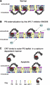
Schematic model of a potential means for CRT release during apoptotic stress. A) During oxidative stress, CRT expression increases. During such stress, CRT can retrotranslocate from the ER into the cytosol and associate with PS on the inner plasma membrane (PM) leaflet. B) Increased CRT production leads to activation of iNOS and NO production. Potentially, the increased NO production can nitrosylate the free cysteines on the flipase enzyme–aminophospholipid transferase (APLT). This blocks APLT, which retains PS on the inner leaflet of the PM. The majority of PS flips to the outer surface, and CRT is released at the same time. C) CRT has a lipophilic and hydrophobic region (red) that may allow CRT to associate with PS. Its interaction with PS is calcium dependent. Lowering the calcium concentration allows release of CRT from PS. SNCEE, S-nitroso-l-cysteine-ethyl ester.
Figure 8.
Figure 8.
Potential exit routes for CRT from the ER to the cytoplasm, cell surface, and extracellular space.

References

    1. Bedard K, Szabo E, Michalak M, Opas M. Cellular functions of endoplasmic reticulum chaperones calreticulin, calnexin, and ERp57. Int Rev Cytol. 2005;245:91–121. - PubMed
    1. Michalak M, Groenendyk J, Szabo E, Gold L I, Opas M. Calreticulin, a multi-process calcium-buffering chaperone of the endoplasmic reticulum. Biochem J. 2009;417:651–666. - PubMed
    1. Mesaeli N, Nakamura K, Zvaritch E, Dickie P, Dziak E, Krause K H, Opas M, MacLennan D H, Michalak M. Calreticulin is essential for cardiac development. J Cell Biol. 1999;144:857–868. - PMC - PubMed
    1. Tesniere A, Apetoh L, Ghiringhelli F, Joza N, Panaretakis T, Kepp O, Schlemmer F, Zitvogel L, Kroemer G. Immunogenic cancer cell death: a key-lock paradigm. Curr Opin Immunol. 2008;20:504–511. - PubMed
    1. Obeid M, Tesniere A, Ghiringhelli F, Fimia G M, Apetoh L, Perfettini J L, Castedo M, Mignot G, Panaretakis T, Casares N, Metivier D, Larochette N, van Endert P, Ciccosanti F, Piacentini M, Zitvogel L, Kroemer G. Calreticulin exposure dictates the immunogenicity of cancer cell death. Nat Med. 2007;13:54–61. - PubMed

Publication types