

Absence of biglycan accelerates the degenerative process in mouse intervertebral disc
- PMID: 19940720
- PMCID: PMC3125573
- DOI: 10.1097/BRS.0b013e3181b7c7ec
Absence of biglycan accelerates the degenerative process in mouse intervertebral disc
Abstract
Study design: A study of the histologic changes of the intervertebral discs (IVDs) in biglycan (Bgn)-deficient mice.
Objective: In this study, we investigate whether the absence of Bgn accelerates the degenerative process in mouse intervertebral disc (IVD).
Summary of background data: Proteoglycans and collagen fibrils are major components in the extracellular matrix (ECM) composition of IVD. The ECM of IVD contains several members of the small leucine repeat proteoglycans (SLRPs) family. Bgn is one member of SLRPs family, and showed a unique expression with age and degeneration in the human IVD. To date, there have been no in vivo studies to see whether SLRPs have a role in maintaining the structural integrity of IVD. To explore the functions of Bgn in the IVD, we examined discs in Bgn-deficient mice.
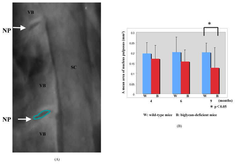
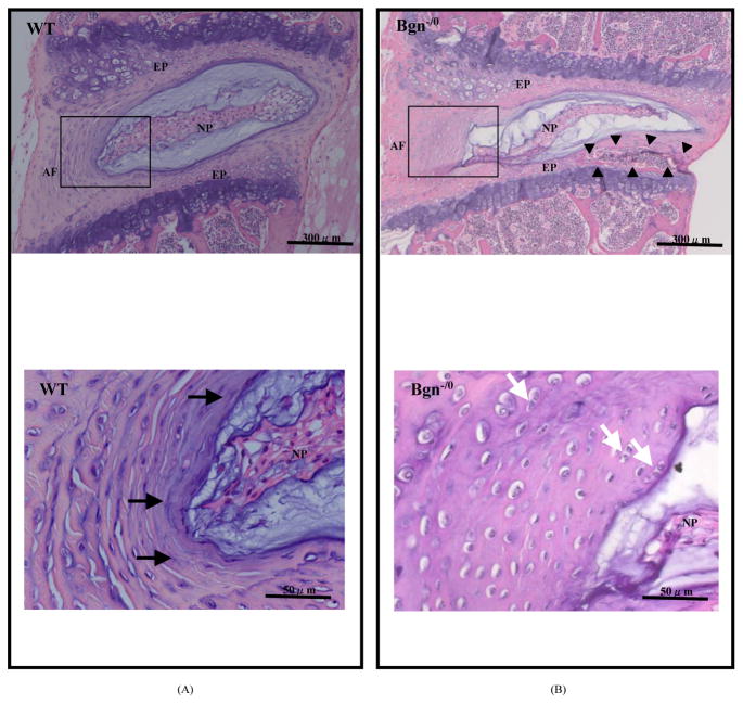
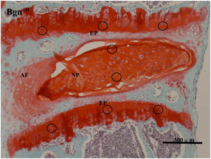
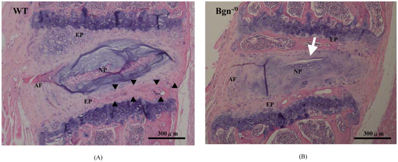
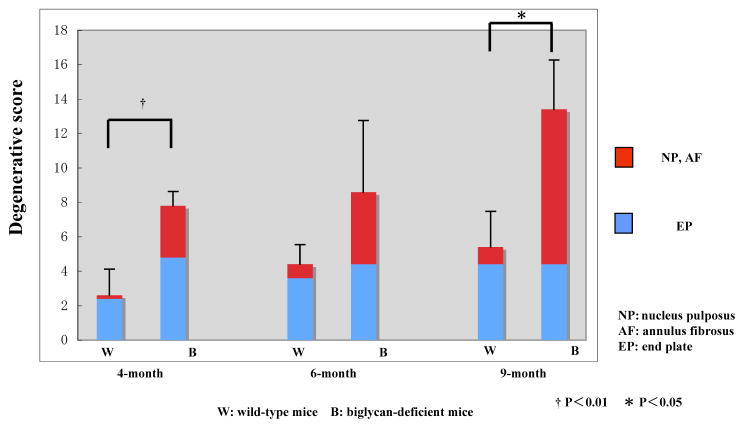
Methods: A total of 30 spine specimens were harvested from wild-type (WT) and Bgn-deficient mice. Five specimens for each genotype at 4-, 6-, and 9-month old were examined in the experiments. Histologic analysis of the IVD was performed. Histologic gradings were performed separately on nucleus pulposus, anulus fibrosus, and endplate according to the classification system proposed by Boos et al.
Results: We found that Bgn-deficient mice developed an early onset of disc degeneration compared with WT mice. The degenerative scores of Bgn-deficient mice were significantly higher than those of WT mice at 4- and 9-month-old. High scores for nucleus pulposus and anulus fibrosus in Bgn-deficient mice significantly affected the difference in total degenerative scores at 9 months of age.
Conclusion: Bgn deficiency significantly accelerated disc degeneration.
Figures
References
-
- Boden SD, Davis DO, Dina TS, et al. Abnormal magnetic-resonance scans of the lumbar spine in asymptomatic subjects. A prospective investigation. J Bone Joint Surg. 1990;72A:403–408. - PubMed
-
- Jensen MC, Brant-Zawadzki MN, Obuchowski N, et al. Magnetic resonance imaging of the lumbar spine in people without back pain. N Engl J Med. 1994;331:69–73. - PubMed
-
- Vernon-Rpberts Barrie. Age-related and degenerative pathology of intervertebral discs and apophyseal joints. In: Jayson MIV, editor. The Lumbar Spine and Back Pain. Edinburgh, Scotland: Churchill Livingstone; 1992. pp. 17–41.
-
- Heinegård D, Larsson T, Sommarin Y, et al. Two novel matrix proteins isolated from articular cartilage show wide distributions among connective tissues. J Biol Chem. 1986;261:13866–13872. - PubMed
-
- Hocking AM, Shinomura T, McQuillan DJ. Leucine-rich repeat glycoproteins of the extracellular matrix. Matrix Biol. 1998;17:1–19. - PubMed
Publication types
MeSH terms
Substances
Grants and funding
LinkOut - more resources
Full Text Sources
Research Materials
Miscellaneous
