

Review
doi: 10.1038/nature08602.
Designing materials to direct stem-cell fate
Affiliations
- PMID: 19940913
- PMCID: PMC2908011
- DOI: 10.1038/nature08602
Item in Clipboard
Review
Designing materials to direct stem-cell fate
Nature.
.
Abstract
Proper tissue function and regeneration rely on robust spatial and temporal control of biophysical and biochemical microenvironmental cues through mechanisms that remain poorly understood. Biomaterials are rapidly being developed to display and deliver stem-cell-regulatory signals in a precise and near-physiological fashion, and serve as powerful artificial microenvironments in which to study and instruct stem-cell fate both in culture and in vivo. Further synergism of cell biological and biomaterials technologies promises to have a profound impact on stem-cell biology and provide insights that will advance stem-cell-based clinical approaches to tissue regeneration.
Conflict of interest statement
The authors declare no competing financial interests.
Figures
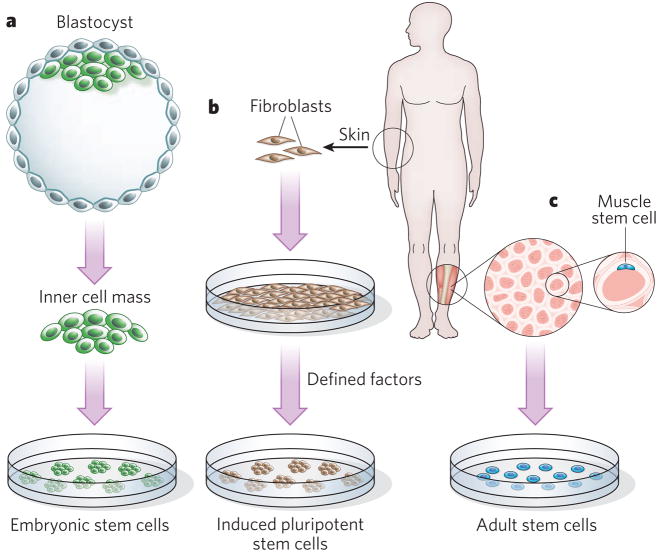
a, Embryonic stem cells, which are derived from blastocysts (formed at an early stage of embryogenesis), provided the first human source of pluripotent cells that could be differentiated to generate any cell type. b, Induced pluripotent stem cells, which have all of the properties of embryonic stem cells, were first generated by introducing genes encoding four proteins into somatic cells, such as skin fibroblasts. Embryonic stem cells and induced pluripotent stem cells have a seemingly unlimited self-renewal potential in culture, but the absence of methods to direct these cells into a single tissue-specific lineage robustly and reproducibly and to avoid the risk of tumour formation reliably have restricted their use in humans. Induced pluripotent stem cells overcome the problem of immune tolerance and the ethical issues faced by the use of embryonic stem cells and adult stem cells in patients, but current methods to reprogram somatic cells and to generate induced pluripotent stem cells are extremely slow and inefficient. c, Resident tissue-specific adult stem cells (for example muscle stem cells) lack the plasticity of embryonic stem cells and induced pluripotent stem cells but are not tumorigenic. They are primed for, and extremely efficient at, generating progeny that differentiate into specialized cell types. It is difficult to induce the self-renewal of adult stem cells in culture and to propagate the cells to yield clinically useful numbers in vitro, underscoring the importance of elucidating the role of the endogenous microenvironment in the regulation of stem-cell fate. A cross-sectional view of muscle fibres (red) surrounded by basement membrane (white) is shown, together with a muscle stem cell (blue); these stem cells reside on top of muscle fibres, beneath the basement membrane.
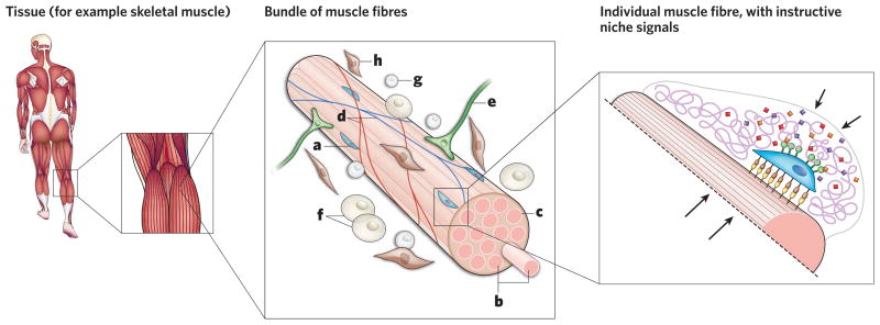
Adult stem cells reside in tissue-specific microenvironments, called niches. Niches protect stem cells and regulate their functions. First described in Drosophila melanogaster and Caenorhabditis elegans ovary and testis, niches have now been characterized in many other tissues, including skeletal muscle (left panel). Muscle stem cells (a) reside on post-mitotic, multinucleated muscle fibres (b) and are ensheathed by a basement membrane (c) (central panel). The complexity of this stem-cell niche is increased by the presence of many other, non-muscle, cell types, including endothelial and blood cells in the vasculature (d), motor neurons (e), adipocytes (f), and circulating immune cells (g) and fibroblasts (h). Within the niche (right panel), spatially and temporally controlled biochemical mixtures of soluble and tethered chemokines, cytokines and growth factors (diamonds), as well as ECM molecules (purple) and ligands presented by muscle fibres (yellow), interact with transmembrane receptors displayed by muscle stem cells (brown and green) to regulate stem-cell fate. It is also becoming clear that the biophysical properties of the stem-cell microenvironment are crucial components of the niche; arrows indicate forces imposed on stem cells by the resistance of the ECM and surrounding tissue.
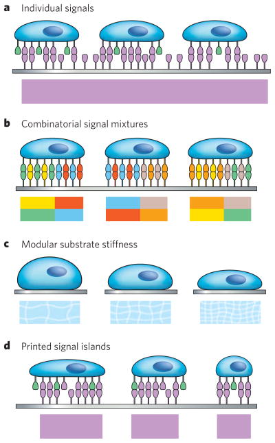
The top part of each panel shows stems cells exposed to a specific, engineered 2D microenvironment (viewed from the side), and the bottom part shows a schematic of the microenvironmental features (viewed from above), represented as blocks of colour matching the signals that are present. The substrates (grey) encompass various materials, such as plastics, glass or hydrogels, except for in panel c (in which soft materials such as hydrogels are depicted). a, Individual signal molecules are displayed on the substrate. b, Combinatorial mixtures of signals that are generated, for example, by robotic protein spotting can be presented to stem cells. c, The desired substrate stiffness can be controlled by, for example, differential crosslinking of hydrogel networks. d, Microcontact printing of cell-adhesion or cell-regulatory proteins on inert surfaces allows control of protein spot size and, therefore, cell shape.
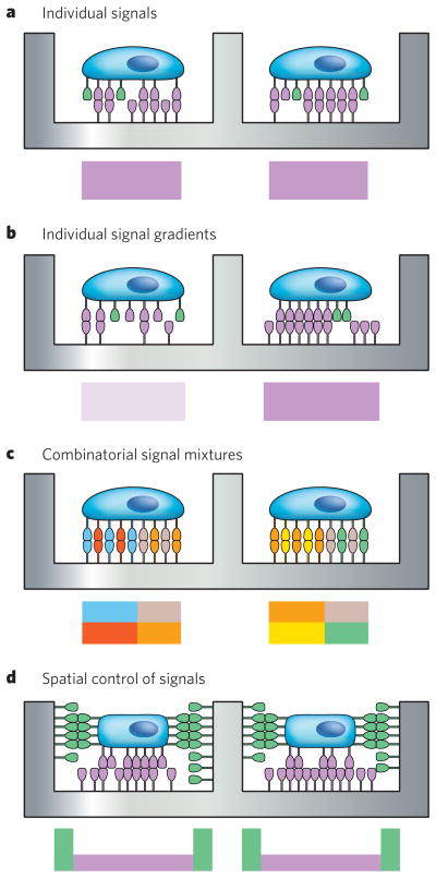
Microwell arrays allow the confinement of single stem cells and analysis of entire stem-cell populations at the individual cell level, overcoming the problem of heterogeneity of stem-cell populations. a, Microwell arrays can be readily engineered so that individual niche signals are presented at a certain concentration on the bottom of the well, by using manual microcontact printing. b, c, Robotic protein spotting on the microwell bottom should allow control of protein doses in each microwell, including the generation of protein gradients (b) or the production of combinatorial protein mixtures (c). d, Patterning approaches can be designed to allow the spatial arrangement of niche cues at the level of an individual, encapsulated stem cell. The top part of each panel shows stem cells exposed to a specific, engineered pseudo-3D microenvironment (viewed from the side), and the bottom part shows a schematic of the particular microenvironmental features (viewed from above (a–c) or from the side (d)).

Mild and selective hydrogel-crosslinking chemistries are necessary for a true 3D embedding of stem cells in an artificial microenvironment that more closely mimics natural stem-cell niches. Polymer-hydrogel networks can be engineered with tailor-made biochemical and biophysical characteristics. a, Individual niche signals can be tethered to gel networks to probe their function in stem-cell behaviour. b, Three-dimensional micropatterning technologies such as electropatterning allow the arrangement of cells in 3D hydrogels in a spatially well-controlled manner. Using this technique, single stem cells could be patterned in three dimensions in contact with support cells (pink) that provide many regulatory niche cues. c, Niche cues could be displayed as large-scale gradients (which is currently only possible with non-tethered signals). d, Hydrogel networks can now be precisely micropatterned in three dimensions; for example, by light-controlled modification of biochemical gel characteristics (such as niche-signal availability) or biophysical gel characteristics (such as gel-crosslink density). The laser from a confocal microscope allows high spatial resolution, as well as dynamic control of 3D gel patterning. The top part of each panel shows cells exposed to a specific, engineered 3D microenvironment (viewed from the side), and the bottom part shows a schematic of the particular microenvironmental features (viewed from above (a–c) or from the side (d)).
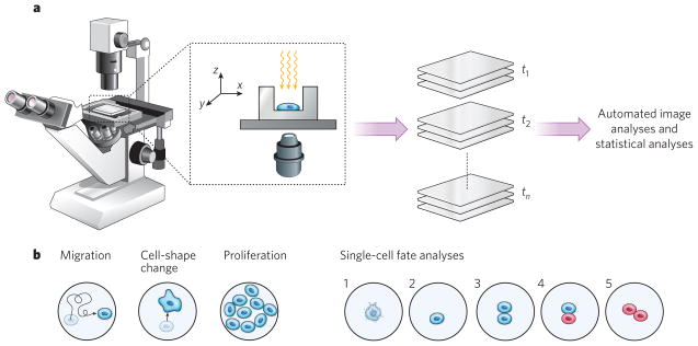
a, Time-lapse microscopy is a powerful way of probing the behaviour of live stem cells in artificial niches. Stem cells are imaged at various time points (t1 to tn) and locations to generate time-lapse movies, and automated image analysis and statistical analyses are used to quantify the dynamic cells’ behaviour. b, A number of different read-outs, corresponding to different stem-cell functions, are available. Together with cell migration, changes in cell shape and changes in proliferation kinetics, the recording and automated analyses of changes in the fate of individual stem cells are crucial. Illustrated are cell death (1); quiescence (that is, non-cycling; 2); symmetrical self-renewal divisions (proliferation behaviour imposed in response to stress or trauma; 3); asymmetrical self-renewal divisions generating one daughter cell that retains stem-cell identity and one already partly differentiated (a behaviour thought to be dominant during homeostatic conditions; 4); and symmetrical depletion divisions, in which both daughter cells lose stem-cell function (the default behaviour of adult stem cells grown in vitro; 5).
References
-
- Blau HM, Sacco A, Gilbert PM. In: Essentials of Stem Cell Biology. 2. Lanza R, et al., editors. Academic; pp. 249–257. in the press.
-
- Blau HM, Sacco A, Gilbert PM. In: Encyclopedia of Stem Cell Research. Svendsen C, Ebert A, editors. SAGE; in the press.
-
- Daley GQ, Scadden DT. Prospects for stem cell-based therapy. Cell. 2008;132:544–548. - PubMed
-
- Lutolf MP, Hubbell JA. Synthetic biomaterials as instructive extracellular microenvironments for morphogenesis in tissue engineering. Nature Biotechnol. 2005;23:47–55. - PubMed
-
- Scadden DT. The stem-cell niche as an entity of action. Nature. 2006;441:1075–1079. - PubMed
Publication types
MeSH terms
Substances
Grants and funding
LinkOut - more resources
Full Text Sources
Other Literature Sources
Medical
