. 2010 Apr;201(4):701-17.
doi: 10.1007/s00221-009-2090-0. Epub 2009 Nov 26.

The macaque midbrain reticular formation sends side-specific feedback to the superior colliculus

Affiliations

The macaque midbrain reticular formation sends side-specific feedback to the superior colliculus

Niping Wang et al. Exp Brain Res. 2010 Apr.

Abstract

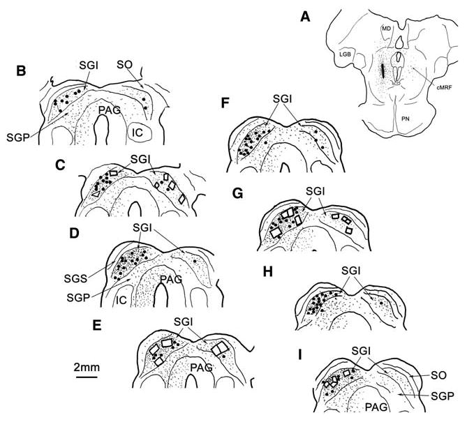
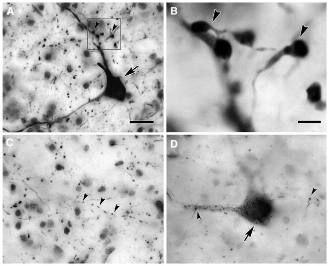
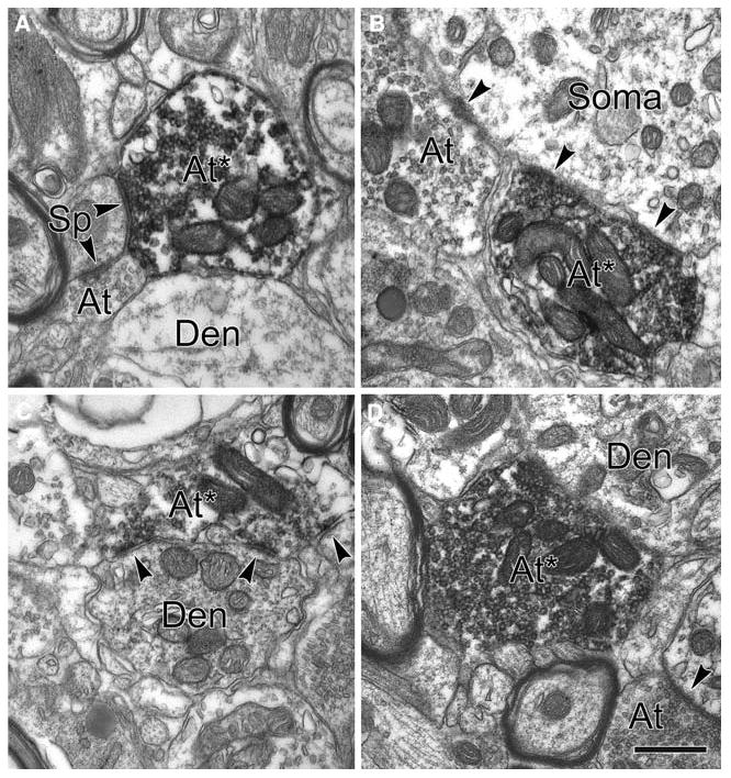
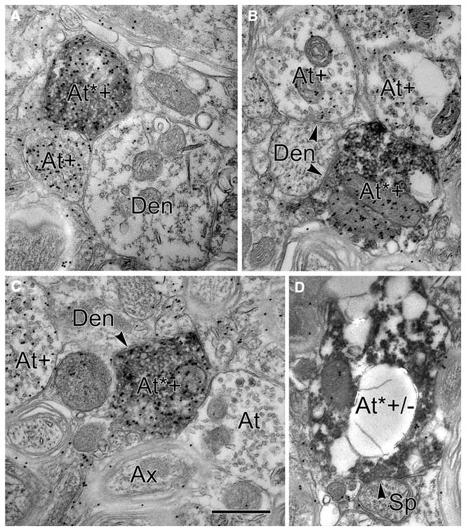
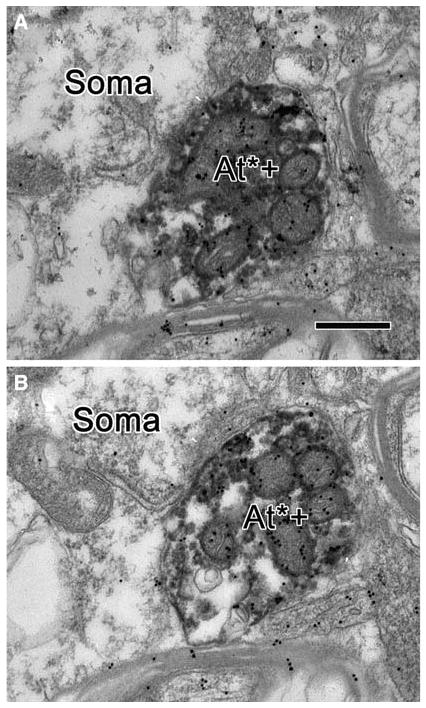
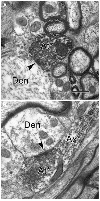
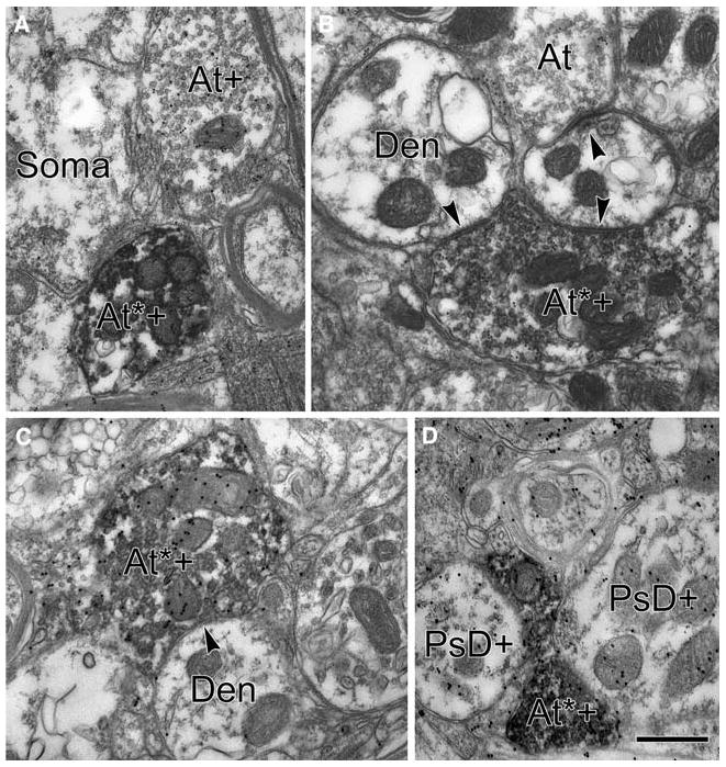
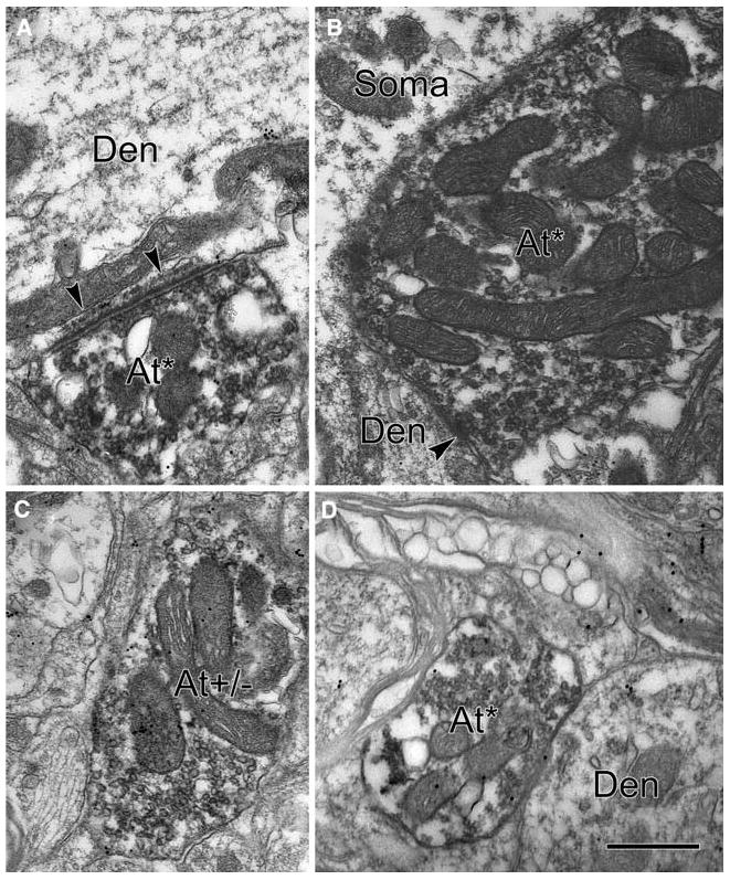
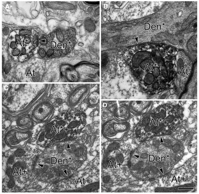
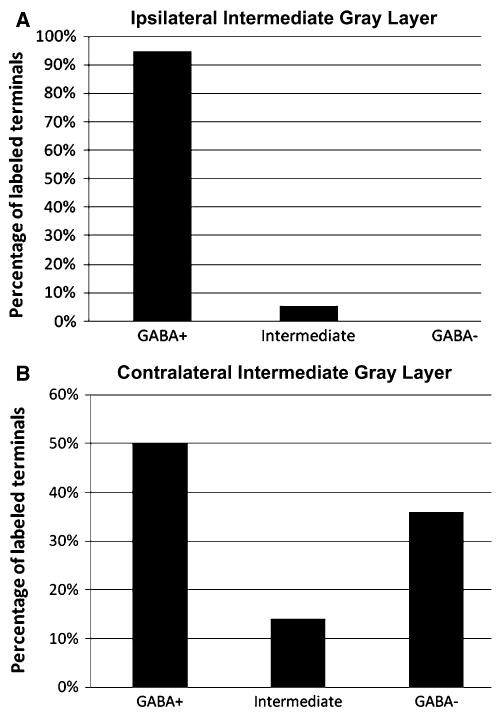
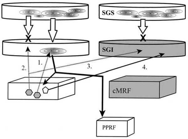
The central mesencephalic reticular formation (cMRF) likely plays a role in gaze control, as cMRF neurons receive tectal input and provide a bilateral projection back to the superior colliculus (SC). We examined the important question of whether this feedback is excitatory or inhibitory. Biotinylated dextran amine (BDA) was injected into the cMRF of M. fascicularis monkeys to anterogradely label reticulotectal terminals and retrogradely label tectoreticular neurons. BDA labeled profiles in the ipsi- and contralateral intermediate gray layer (SGI) were examined electron microscopically. Postembedding GABA immunochemistry was used to identify putative inhibitory profiles. Nearly all (94.7%) of the ipsilateral BDA labeled terminals were GABA positive, but profiles postsynaptic to these labeled terminals were exclusively GABA negative. In addition, BDA labeled terminals were observed to contact BDA labeled dendrites, indicating the presence of a monosynaptic feedback loop connecting the cMRF and ipsilateral SC. In contrast, within the contralateral SGI, half of the BDA labeled terminals were GABA positive, while more than a third were GABA negative. All the postsynaptic profiles were GABA negative. These results indicate the cMRF provides inhibitory feedback to the ipsilateral side of the SC, but it has more complex effects on the contralateral side. The ipsilateral projection may help tune the "winner-take-all" mechanism that produces a unified saccade signal, while the contralateral projections may contribute to the coordination of activity between the two colliculi.


Figures

Fig. 1
Fig. 1
Distribution of anterogradely labeled reticulotectal axonal terminals (stipple) and retrogradely labeled tectoreticular neurons (dots) following a BDA injection into monkey cMRF. a A small BDA injection site is shown within cMRF. b-i In the midbrain, BDA labeled terminals were distributed the superior colliculus (SC), especially the intermediate gray layer (SGI), and the periaqueductal gray (PAG). BDA labeled neurons are seen bilaterally in the SC (b–i). Small windows in figure c, e, g and i demonstrate the areas where samples were taken for EM. IC inferior colliculus, SGP stratum griseum profundum, SO stratum opticum, SGS stratum griseum superficiale, LGB lateral geniculate body, MD medial dorsal nucleus, PN pontine nuclei. Scale bar 2 mm
Fig. 2
Fig. 2
Photomicrographs of BDA labeled terminals and retrogradely labeled tectoreticular cells within the ipsilateral and contralateral superior colliculi in the macaque monkey. a Labeled tectoreticular neuron (arrow) in ipsilateral SGI. Note the homogeneous labeling of its cytoplasm. The BDA labeled reticulotectal axons exhibit numerous en passant boutons along their course. Close associations (arrowheads) were present between the labeled tectoreticular dendrites and the labeled reticulotectal axon terminals. b Higher magnification of close associations indicated by frame in a. c When compared with the ipsilateral SGI, the neuropil in the contralateral SC exhibits fewer labeled reticulotectal axons and boutons (arrowheads). d A BDA labeled tectoreticular neuron (arrow) in contralateral SGI shows less reaction product in its cytoplasm. BDA labeled boutons (arrowheads) of passing reticulotectal axons are arranged in close association with its dendrites. Scale bar in a for a, c, d = 20 μm, scale bar in b = 5 μm
Fig. 3
Fig. 3
Electron micrographs of BDA labeled axon terminals in ipsilateral SGI. a–d All from material processed for BDA only. BDA labeled terminals (At*) make symmetric synapses (arrowheads) with non-labeled dendrites (Den) (c, d) or putative spines (Sp) (a) or non-labeled somata (b). At unlabeled axon terminal in this and following figures. Scale bar 0.5 μm
Fig. 4
Fig. 4
GABA labeling in electron micrographs showing BDA labeled axon terminals in the ipsilateral SGI. BDA labeled terminals (At*) are predominately GABA+ (At*+), as shown in a–c. They contact (arrowhead) GABA dendrites (Den). d A BDA labeled terminal contacting a small profile which is a putative dendritic spines (Sp). Based on the gold particle density, this terminal is categorized in the intermediate, non-classified category (At*+/−). Ax axon. Scale bar 0.5 μm
Fig. 5
Fig. 5
Electron micrographs of semiserial sections through a BDA labeled, GABA+ cMRF terminal. a, b Two semiserial sections (spaced 100 nm apart) through a terminal in ipsilateral SGI. The terminal has a dark cytoplasm, due to BDA labeling, and numerous gold particles, indicating it is GABA+. It lies adjacent to an unlabeled soma. Scale bar 0.5 μm
Fig. 6
Fig. 6
BDA labeled axon terminals in contralateral SGI from material processed for BDA only. a A BDA labeled terminal (At*) containing pleomorphic vesicles making a symmetric synapse (arrowhead) onto a dendritic profile (Den). b A BDA labeled preterminal axonal profile (Ax*) continuing into a BDA labeled terminal (At*). This terminal contains spherical vesicles and forms an asymmetric synapse (arrowhead) onto a dendritic profile (Den). Scale bar 0.5 μm
Fig. 7
Fig. 7
BDA labeled, GABA+ axon terminals in contralateral SGI. Most BDA labeled, crossed reticulotectal terminals were GABA+ (At*+), like those shown in a–d. The BDA labeled reticulotectal terminal in b contacts (arrowhead) two distal dendrites, one of which is also contacted by a BDA/GABA terminal (At). The other examples of BDA labeled, GABAergic terminals appose a GABA soma (a) and dendrite (Den c). In d, the BDA labeled, GABA+ terminal lies adjacent to GABA+ profiles that appear to be presynaptic dendrites (PsD+), but no synaptic contacts are observable. Ax axon, At+ GABA+ terminal. Scale bar 0.5 μm
Fig. 8
Fig. 8
BDA labeled, GABA axon terminals in the contralateral SGI. A minority of the BDA labeled terminals were GABA (At*), like those shown in a–d. a An asymmetric synapse (arrowheads) between a BDA labeled, GABA terminal and a GABA dendrite. The BDA labeled, GABA terminal in b apposes a GABA soma and contacts its dendrite (Den). The BDA labeled, GABA terminal in d is only associated with a GABA dendrite (Den), no contact is visible. c An example of a BDA labeled terminal that fell into the intermediate category (At+/−). Ax axon. Scale bar 0.5 μm
Fig. 9
Fig. 9
BDA labeled terminals contact (arrowheads a–d) BDA labeled dendrites. a, b BDA single label material in ipsilateral SGI. The BDA labeled terminals (At*) contact the BDA labeled dendrites (Den*) with symmetric synapses (arrowheads), c, d are from double label material cut from contralateral SGI. These examples are semiserial sections through a BDA labeled, GABA+ cMRF terminal (At*+), which contacts a dendritic profile with a symmetric synapse. The dendrite appears to be lightly labeled with BDA reaction product (Den*), and it is contacted by other GABA+ terminals (At+), c, d are spaced 200 nm apart. Scale bar 0.5 μm
Fig. 10
Fig. 10
Histogram of percentage of reticulotectal axonal terminals that are GABA+ or GABA in SGI (n = 3); a greater than 90% of the BDA labeled reticulotectal axonal terminal profiles within the ipsilateral SGI are GABA+, and about 5% of the terminal profiles fall into the intermediate category, but none are GABA. b Half of the BDA labeled reticulotectal axonal terminal profiles within the contralateral SGI are GABA+, while 36% of them are GABA. The rest fall into the intermediate category
Fig. 11
Fig. 11
Schematic diagram of reciprocal connections of central mesencephalic reticular formation (cMRF) and intermediate gray layer (SGI) of superior colliculi. The predorsal bundle sends excitatory projections (thick solid arrow) to the horizontal gaze center (paramedian pontine reticular formation—PPRF) to activate horizontal saccades. Collaterals emitted before the predorsal bundle crosses the midline, provide excitatory input to the ipsilateral cMRF neurons. Inhibitory cMRF neurons (gray hexagons) issue recticulotectal axons (dash arrow) to the ipsilateral SC to either turn off the predorsal bundle cells that directly activated them (1), or suppress other predorsal bundle neurons (2), and keep them from being excited (X) by activity in the superficial gray layer (SGS). The cMRF neurons send both inhibitory (dashed arrow) and excitatory (continuous arrow) projections to the contralateral SGI. The inhibitory projection (3) presumably helps keep the contralateral SGS from exciting the contralateral SGI (X). The role of excitatory reticulotectal neurons (pentagon) that send a crossed projection to SGI (4) is unclear. Gray shading in SGI and cMRF indicates this side is suppressed during rightward saccades

Similar articles

Cited by

References

    1. Appell PP, Behan M. Sources of subcortical GABAergic projections to the superior colliculus in the cat. J Comp Neurol. 1990;302:143–158. - PubMed
    1. Arai K, Das S, Keller EL, Aiyoshi E. A distributed model of the saccade system: simulations of temporally perturbed saccades using position and velocity feedback. Neural Netw. 1999;12:1359–1375. - PubMed
    1. Araki M, McGeer PL, McGeer EG. Presumptive γ-aminobutyric acid pathways from the midbrain to the superior colliculus studied by a combined horseradish peroxidase- γ-aminobutyric acid transaminase pharmacohistochemical method. Neuroscience. 1984;13:433–439. - PubMed
    1. Basso MA, Wurtz RH. Modulation of neuronal activity in superior colliculus by changes in target probability. J Neurosci. 1998;18:7519–7534. - PMC - PubMed
    1. Beckstead RM. Long collateral branches of substantia nigra pars reticulata axons to thalamus, superior colliculus and reticular formation in monkey and cat. Multiple retrograde neuronal labeling with fluorescent dyes. Neuroscience. 1983;10:767–779. - PubMed

Publication types

MeSH terms

LinkOut - more resources