. 2010 Feb 1;518(3):255-76.
doi: 10.1002/cne.22225.

D(2) receptors receive paracrine neurotransmission and are consistently targeted to a subset of synaptic structures in an identified neuron of the crustacean stomatogastric nervous system

Affiliations

D(2) receptors receive paracrine neurotransmission and are consistently targeted to a subset of synaptic structures in an identified neuron of the crustacean stomatogastric nervous system

Max F Oginsky et al. J Comp Neurol. .

Abstract

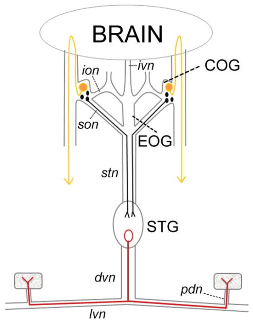
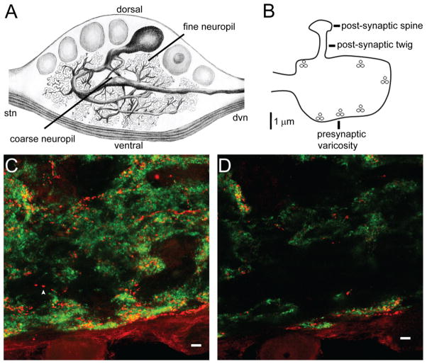
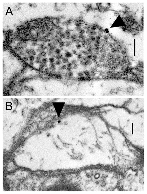
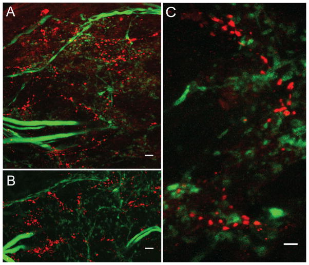
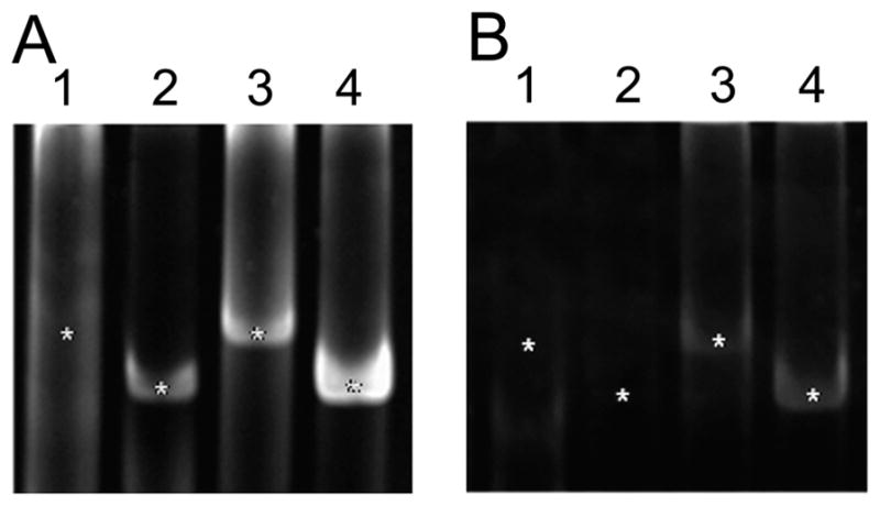
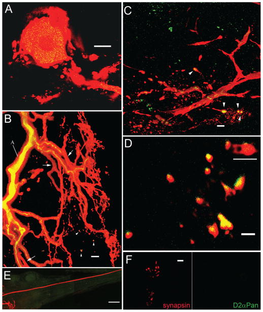
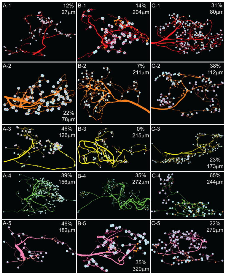
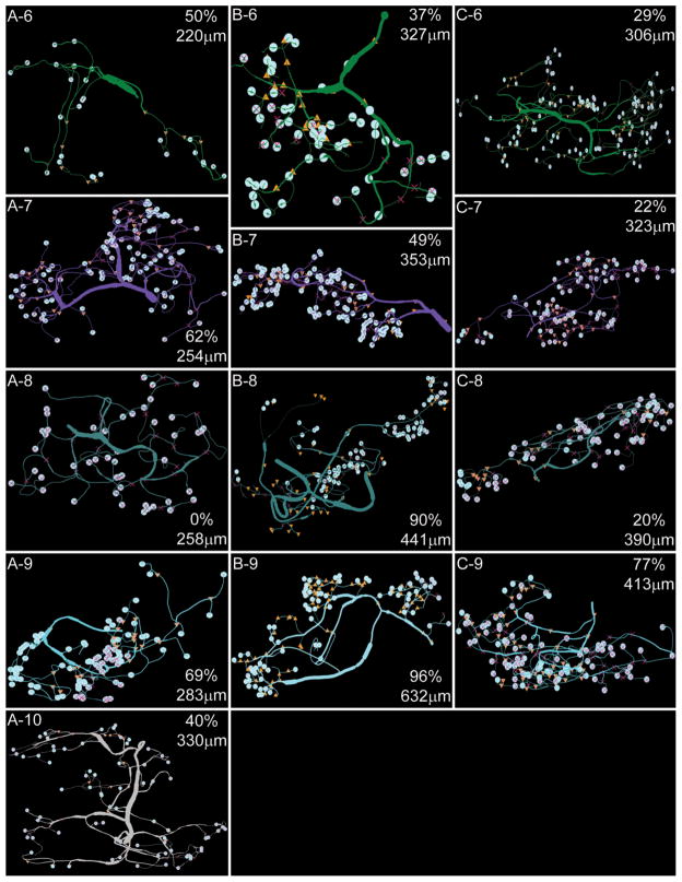
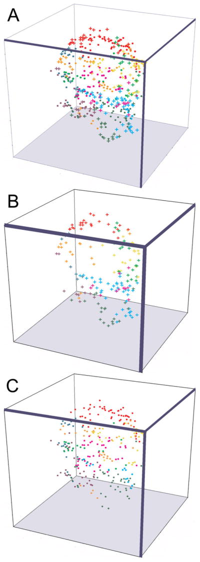
Dopamine (DA) modulates motor systems in phyla as diverse as nematodes and arthropods up through chordates. A comparison of dopaminergic systems across a broad phylogenetic range should reveal shared organizing principles. The pyloric network, located in the stomatogastric ganglion (STG), is an important model for neuromodulation of motor networks. The effects of DA on this network have been well characterized at the circuit and cellular levels in the spiny lobster, Panulirus interruptus. Here we provide the first data about the physical organization of the DA signaling system in the STG and the function of D(2) receptors in pyloric neurons. Previous studies showed that DA altered intrinsic firing properties and synaptic output in the pyloric dilator (PD) neuron, in part by reducing calcium currents and increasing outward potassium currents. We performed single cell reverse transcriptase-polymerase chain reaction (RT-PCR) experiments to show that PD neurons exclusively expressed a type 2 (D(2alphaPan)) DA receptor. This was confirmed by using confocal microscopy in conjunction with immunohistochemistry (IHC) on STG whole-mount preparations containing dye-filled PD neurons. Immunogold electron microscopy showed that surface receptors were concentrated in fine neurites/terminal swellings and vesicle-laden varicosities in the synaptic neuropil. Double-label IHC experiments with tyrosine hydroxylase antiserum suggested that the D(2alphaPan) receptors received volume neurotransmissions. Receptors were further mapped onto three-dimensional models of PD neurons built from Neurolucida tracings of confocal stacks from the IHC experiments. The data showed that D(2alphaPan) receptors were selectively targeted to approximately 40% of synaptic structures in any given PD neuron, and were nonuniformly distributed among neurites.


Figures

Figure 1
Figure 1
The stomatogastric nervous system (STNS). Diagram of the STNS and connection to the brain. Nerves are drawn as lines and rectangles, muscles as stippled squares, dopamine-containing neurons as filled circles, and the pyloric dilator (PD) neuron as an open circle. The STNS comprises four ganglia: the two commissural ganglia (COGs), the esophageal ganglion (EOG), and the stomatogastric ganglion (STG). The PD neuron, located in the STG, projects an axon down the dorsal ventricular nerve (dvn). The axon bifurcates prior to entering the lateral ventricular nerves (lvn) and continues to run through the pyloric dilator nerves (pdn) to innervate the two PD muscles. Small DA-containing neurons in the COGs can project axons through the superior esophageal nerve (son) and down the stomatogastric nerve (stn) to terminate in the STG. The large L-cells in the COGs project to the ventral surface of the brain, whereupon axons reverse direction and ultimately terminate in ipsilateral pericardial organs, which release neuromodulators into the hemolymph (indicated by arrowheads). Not shown are the many dopaminergic cells in the brain (Tierney et al., 2003). In some species, neurons in the STG may also contain DA (Pulver et al., 2003). Dopamine has not been observed in the EOG, the inferior esophageal nerve (ion), or the inferior ventricular nerve (ivn) that connects the brain to the STNS.
Figure 2
Figure 2
D2αPan receptors co-localize with synapsin in the fine neuropil. A: Illustration of a filled PD neuron in a sagittal section of the STG. A single primary process extends from the soma and exits the STG via the dvn. Before leaving the STG, the primary neurite branches to produce 9–10 large-diameter secondary neurites, that can continue to branch beyond the 16th branch order. Large-diameter neurites are found in the central, coarse neuropil. Small-diameter neurites and synapses lie in the fine neuropil. B: STG synaptic terminal drawn to scale. The presynaptic varicosity contains six release sites, indicated by vesicle clusters. A fingerlike postsynaptic twig extends from the presynaptic compartment and ends in a bulbous postsynaptic spine. C,D: A merged, 18-μm confocal projection (C) and an individual merged 1-μm optical slice (D) from a double-label IHC experiment indicate that D2αPan (red) and synapsin (green) can be either juxtaposed (red and green puncta) or co-localized (yellow puncta). Isolated D2αPan can also be observed (arrowhead). Magenta-green panels for C and D may be seen in Supplementary Figure 4. (Illustration by Sonia Hilliard, DVM.) Scale bar = 10 μm in C,D.
Figure 3
Figure 3
D2αPan receptors can be located on presynaptic varicosities and terminal swellings. Electron micrographs showing anti-D immunogold staining (arrows) on a vesicle-containing varicosity (A) and a terminal swelling lacking vesicles (B). T-bars and postsynaptic densities indicative of synapses are not observed in these micrographs, suggesting that D2αPan receptors can be extrasynaptic. Scale bar = 0.25 μm in A,B.
Figure 4
Figure 4
D2αPan receptors appear to receive volume transmissions. Three double-label IHC experiments showing that D2αPan receptors (red) and tyrosine hydroxylase-containing terminals (green) are not arranged in classical point-to-point synapses. Merged 23-μm (A) and 40-μm (B) confocal projections from the synaptic neuropil showing that clusters of red puncta are associated with green puncta. C: High-magnification 16-μm merged confocal projection showing a lack of co-localization (yellow) and that red and green puncta are not in a 1:1 relationship. Magenta-green panels may be seen in Supplementary Figure 5. Scale bar = 10 μm in A,B; 5 μm in C.
Figure 5
Figure 5
PD neurons express D2 but not D1 receptors. Individual PD neurons, physically isolated with (A) versus without (B) their glial caps, served as templates in RT-PCRs. PCR products were visualized on ethidium bromide-stained polyacrylamide gels. The RT-PCRs tested each PD neuron for the expression of four transcripts: D1αPan, lane 1; D1βPan, lane 2; D2αPan, lane 3; and α-tubulin, lane 4. The expected sizes of the PCR products are indicated with asterisks.
Figure 6
Figure 6
The D2αPan receptor is restricted to the PD somatodendritic compartment. A: Merged 1-μm optical section from a wholemount anti-D (green) IHC preparation containing a Texas red-filled PD neuron. Yellow staining indicates D2αPan receptor expression in the PD soma and primary neurite. B: A 34-μm merged confocal projection showing the same PD neuron as in A branching throughout the STG neuropil. D2αPan receptors are in cytoplasmic transport vesicles in the primary neurite (labeled A), and some higher order neurites. Arrows indicate higher order neurites lacking the receptor. Arrowheads show D2αPan receptors in PD terminals. The green channel was set to maximum intensity for this panel in order to detect receptors in the finest neurites. At lower intensity, vesicles in the primary neurite showed a punctuate distribution, as in A. C: A 29-μm merged confocal projection from deep within the synaptic neuropil showing a single tertiary and higher order neurites. Arrowheads show D2αPan receptors in PD putative synaptic terminals. Green staining represents D2αPan receptors in unidentified neurons. D: High-magnification 4-μm projection from the synaptic neuropil showing a cluster of PD terminals, some of which contain D2αPan receptors. A putative synaptic structure containing both pre- and postsynaptic elements, as illustrated in Figure 2B, is underlined in white. Note that the two postsynaptic twigs end in bulbous structures, consistent with previously described postsynaptic spines (King, 1976a). Also note that the ridge of green observed on some putative synaptic processes does not represent a technical artifact (i.e., red-green channel offset), and is consistent with the fact that the antibody recognizes an extracellular N-terminal epitope. E: Montage of confocal projections showing only background levels of anti-D immunoreactivity in/along the PD axon as it leaves the STG and projects down the dvn. F: An unmerged 1-μm confocal slice showing PD NMJs. The presence of synapsin (red) and the absence of D2αPan receptor (green) staining indicates that there are no detectable D2αPan receptors in PD axon terminals. A magenta-green version may be seen in Supplementary Figure 6. Scale bar = 20 μm in A; 10 μm in B,C; 2 μm in D; 50 μm in E; 5 μm in F.
Figure 7
Figure 7
PD neurons from two different animals. 3D renderings of PD1 (A) and PD2 (B) neurons are displayed in 2D. Secondary branch clusters are color coded according to order of emergence from the primary neurite: red-orange-yellow-light green-pink-dark green-light purple-teal-cyan-gray. In all panels: left = anterior, right = posterior. Varicosities are marked with pale blue circles, and D2αPan receptors in varicosities are marked by orange triangles.
Figure 8
Figure 8
A1–C5: D2αPan receptor distribution in PD neurites and varicosities. Secondary branch clusters for the three PD neurons are displayed according to order of emergence, where branch 1 is closest to the soma. The point of emergence from the primary neurite is shown in microns in the right corner of each panel, where 0 μm is the point where the primary neurite emerges from the soma. Varicosities are marked by pale blue circles. The percent of varicosities containing D2αPan receptors is also listed in the right corner of each panel. ▲ = marker for D2αPan immuno-staining in neurite/varicosity; X = marker for absence of D2αPan immunostaining in branch or varicosity. Note that some markers may not be visible, as they are behind the pale blue circle.
Figure 8
Figure 8
A1–C5: D2αPan receptor distribution in PD neurites and varicosities. Secondary branch clusters for the three PD neurons are displayed according to order of emergence, where branch 1 is closest to the soma. The point of emergence from the primary neurite is shown in microns in the right corner of each panel, where 0 μm is the point where the primary neurite emerges from the soma. Varicosities are marked by pale blue circles. The percent of varicosities containing D2αPan receptors is also listed in the right corner of each panel. ▲ = marker for D2αPan immuno-staining in neurite/varicosity; X = marker for absence of D2αPan immunostaining in branch or varicosity. Note that some markers may not be visible, as they are behind the pale blue circle.
Figure 9
Figure 9
PD terminals are largely non-overlapping and restricted within the STG. Dendritic terminals are represented as colored objects. The 3D position of every terminal on a PD neuron (PD3) was plotted in 3D space relative to the origin of the dvn. All terminals from a given secondary branch cluster share the same color, which corresponds to the color of the secondary branch clusters shown in Figures 7 and 8. Filled circles and crosses represent terminals bearing and lacking D2αPan receptors, respectively. The terminal field maps contain either (A) all terminals, (B) only terminals bearing D2αPan receptors or (C) only terminals lacking D2αPan receptors. As shown in the Supplemental Movie, when the plots were continuously rotated about all axes by using3D Grapher software, it became clear that the terminals containing D2αPan receptors (B) formed a patchy, hollow shell that encapsulated the terminals lacking D2 receptors (C).

Similar articles

Cited by

References

    1. Agnati LF, Leo G, Zanardi A, Genedani S, Rivera A, Fuxe K, Guidolin D. Volume transmission and wiring transmission from cellular to molecular networks: history and perspectives. Acta Physiol (Oxf) 2006;187:329–344. - PubMed
    1. Barker DL, Kushner PD, Hooper NK. Synthesis of dopamine and octopamine in the crustacean stomatogastric nervous system. Brain Res. 1979;161:99–113. - PubMed
    1. Baro DJ, Cole CL, Harris-Warrick RM. RT-PCR analysis of shaker, shab, shaw, and shal gene expression in single neurons and glial cells. Receptors Channels. 1996;4:149–159. - PubMed
    1. Baro DJ, Levini RM, Kim MT, Willms AR, Lanning CC, Rodriguez HE, Harris-Warrick RM. Quantitative single-cell-reverse transcription-PCR demonstrates that A-current magnitude varies as a linear function of shal gene expression in identified stomatogastric neurons. J Neurosci. 1997;17:6597–6610. - PMC - PubMed
    1. Baro DJ, Ayali A, French L, Scholz NL, Labenia J, Lanning CC, Graubard K, Harris-Warrick RM. Molecular underpinnings of motor pattern generation: differential targeting of shal and shaker in the pyloric motor system. J Neurosci. 2000;20:6619–6630. - PMC - PubMed

Publication types

MeSH terms