. 2010 May-Sep;104(3-4):203-14.
doi: 10.1016/j.jphysparis.2009.11.017. Epub 2009 Nov 23.

Functional specializations of primary auditory afferents on the Mauthner cells: interactions between membrane and synaptic properties

Affiliations

Functional specializations of primary auditory afferents on the Mauthner cells: interactions between membrane and synaptic properties

Sebastian Curti et al. J Physiol Paris. 2010 May-Sep.

Abstract

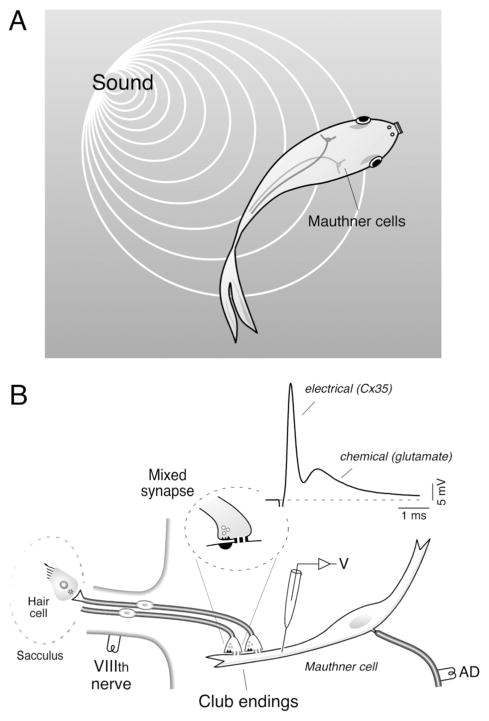
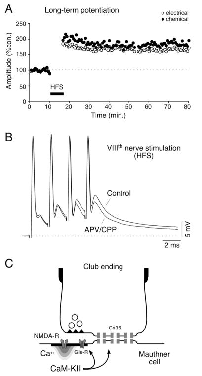
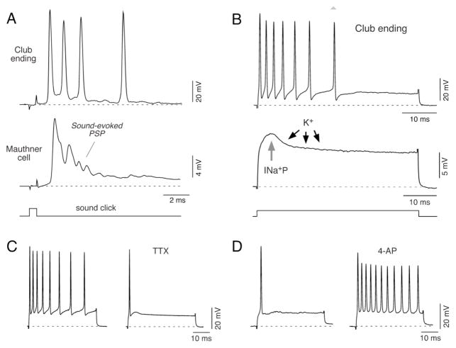
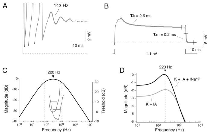
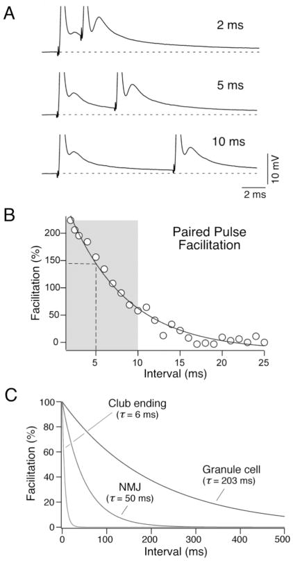
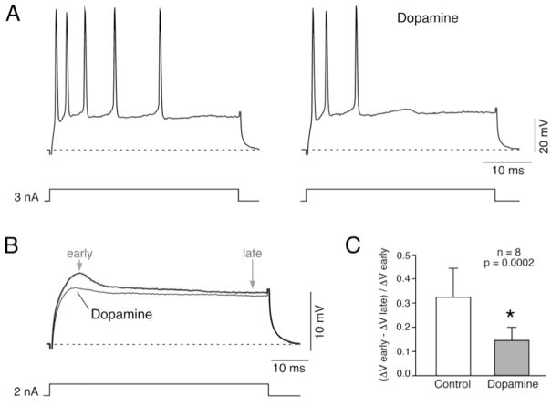
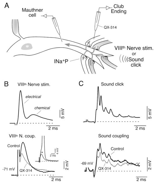
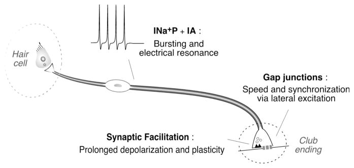
Primary auditory afferents are usually perceived as passive, timing-preserving, lines of communication. Contrasting this view, a special class of auditory afferents to teleost Mauthner cells, a command neuron that organizes tail-flip escape responses, undergoes potentiation of their mixed (electrical and chemical) synapses in response to high frequency cellular activity. This property is likely to represent a mechanism of input sensitization as these neurons provide the Mauthner cell with essential information for the initiation of an escape response. We review here the anatomical and physiological specializations of these identifiable auditory afferents. In particular, we discuss how their membrane and synaptic properties act in concert to more efficaciously activate the Mauthner cells. The striking functional specializations of these neurons suggest that primary auditory afferents might be capable of more sophisticated contributions to auditory processing than has been generally recognized.


Figures

Figure 1
Figure 1. The Mauthner cell system and the Club ending afferents
A, The M-cells mediate sound-evoked tail-flip escapes responses in teleost fish. B, Large identifiable auditory afferents innervate the rostral portion of the saccular macula (Sacculus), the main auditory component of the goldfish ear, and terminate as mixed (electrical and chemical) synapses known as Large Myelinated Club endings (“Club endings”) on the lateral dendrite of the M-cell. These terminals form mixed, electrical and chemical, synapses (Mixed synapse) with the distal portion of the M-cell lateral dendrite. Experimental arrangement used to obtain in vivo dendritic intracellular recordings of the M-cell. Stimulation of the VIIIth nerve, where Club endings run, elicits a mixed excitatory postsynaptic potential (EPSP) composed of an early, fast electrical component (electrical) which is followed by a delayed, longer lasting glutamatergic component (chemical).
Figure 2
Figure 2. Activity-dependent modulation of synaptic transmission in Club endings
A, Discontinuous high frequency stimulation (HFS; trains of 6 pulses at 500 Hz, every 2 seconds for 4 minutes) of the VIIIth nerve evokes persistent potentiation of both components of the EPSP. Plot illustrates the amplitudes of the electrical (open circles) and chemical (filled circles) components versus time (each point represents the average of 20 traces) for one experiment. B, Induction of the short-term potentiations of the synaptic responses depends on NMDA receptor activation. Superimposed synaptic responses evoked by a train of four VIIIth nerve stimuli, in control and after superfusing with saline containing the antagonists (APV/CPP). Note that while the first chemical synaptic response produced by the train does not produce a significant NMDA receptor mediated response the late facilitated components do. C, Schematic representation of the proposed potentiating mechanism for Club endings. Influx of Ca2+ through NMDA receptors activates CaM-KII that phosphorylates either glutamate receptors and connexins or regulatory molecules. Modified from Pereda et al. (2004), with permission.
Figure 3
Figure 3. Intrinsic membrane properties underlying repetitive firing in Club ending afferents
A, Electrophysiological responses obtained sequentially from the M-cell lateral dendrite (middle trace) and a Club ending (upper trace) in response to brief acoustic stimulation (500 μs duration sound click, lower trace). This stimulus evokes a high frequency burst of action potentials in the Club ending that temporally correlates with the sound-evoked synaptic potential (Sound-evoked PSP) recorded in the M-cell lateral dendrite, produced by the activity of this and an undetermined number of these afferents. B, Top: Direct intracellular activation of Club ending afferents with depolarizing current pulses (50 ms duration, lower trace) at 1.5 times its threshold (1.5 T) evokes a repetitive discharge consisting of a train of action potentials that exhibits marked frequency adaptation. Middle: Active mechanisms involved in near-threshold membrane responses. The figure shows a representative response of a Club ending to a depolarizing current pulse (represented by the lower trace; magnitude is different for upper and middle trace). At the beginning of the pulse the response is dominated by the activation of a persistent Na+ current (INa+P). The amplifying action of this Na+ current is counterbalanced by the delayed activation of an A-type K+ current (K+). C, Firing responses evoked by a depolarizing current pulse (3.3 nA, 50 ms duration) before (left) and after extracellular application of 1 μM TTX (right; TTX). The effects of TTX were observed within the time window in which the amplitude of the action potential of the afferents remained largely unaffected by the drug, suggesting that only persistent sodium channels were affected. D, Membrane responses to current pulses obtained before (left) and after extracellular application of 5mM 4-AP (right; 4-AP). The current pulse, which in control conditions was just sufficient to evoke a single action potential, was capable of inducing a vigorous repetitive discharge 5 minutes after 4-AP application. Modified from Curti et al. (2008), with permission.
Figure 4
Figure 4. Club ending afferents exhibit resonant membrane properties
A, Electrophysiological recordings revealed the presence of subthreshold membrane oscillations underlying repetitive responses. Trace illustrates the membrane response to a depolarizing current pulse, consisting of four action potentials followed by a damped oscillation of the membrane potential (trace represents the average of 5 single responses centered in the oscillation; action potentials are truncated). The frequency of this subthreshold oscillation was estimated fitting a function consisting in the sum of a sinusoidal and an exponential function (143 Hz, thicker trace). B, Indirect estimates of electrical resonance were obtained by calculating cutoff frequencies of the low-pass and high-pass filtering properties of the membrane from kinetics of subthreshold membrane responses. The time constant of a high-pass filter representing the activation of the A-type current (τk) was estimated by fitting a double-exponential function to the decaying portion of the near-threshold response about 5 ms after the pulse onset. Only the faster and most prominent time constant (30 fold the magnitude and 10 times faster than the second one), which likely represents the activation of the A-type current, is represented here and its value used for the estimates of electrical resonance. The time constant of a low-pass filter, representing passive membrane properties (τm), was accurately estimated by fitting a single exponential function to the decay that followed the cessation of current pulses of different polarities and amplitudes (only one of these pulses is illustrated in this example and indicated as τm). This estimate is influenced by both the passive membrane properties and the spread of the injected current along the axon, and represents the effective time constant of the afferent fiber. C, Bode plot (magnitude versus frequency) constructed using a linear model that combines both high- and low-pass filter properties of the membrane. The combination of membrane mechanisms with high-pass and low-pass filtering properties determines a band-pass filter with a bandwidth of 742 Hz and a maximum at 220 Hz. For comparison, the tuning curves of two representative saccular afferents are also illustrated in the same graph (gray traces; curves originally illustrated in Fay, 1995). Threshold in dB (right side ordinates) is plotted against sound frequency. Note that the characteristic frequencies and Q10dB responses (horizontal lines) of both tuning curves (corresponding to the responses of the two types of afferents found in goldfish) matched the estimated bandwidth of the electrical resonance. D, Computer simulations with NEURON revealed the relative contributions of A-type (IA) and persistent Na+ (INa+P) currents to membrane resonance. Based on available anatomical data, an ideal Club ending afferent fiber was modeled as a section consisting of a cylindrical process with passive properties (see Curti et al., 2008). Plot illustrates the computed input impedance (ordinates, normalized to the magnitude of the steady-state membrane response in the absence of any active mechanism) versus frequency (abscissa). When a delayed rectifier and an A-type current with the kinetics estimated experimentally for the ventral cochlear nucleus (Rothman and Manis, 2003) were added to the model, a clear resonant behavior appeared centered at 220 Hz (K + IA, gray trace). Note that the addition of a persistent Na+ current produces a significant amplification of the membrane resonance (K + IA + INa+P, black trace), without modifying the resonant frequency. Modified from Curti et al. (2008), with permission.
FIGURE 5
FIGURE 5. Matching of membrane and synaptic properties
A, Time course of the frequency-dependent facilitation of the chemical component with a two stimuli protocol (paired pulse facilitation). Stimuli were delivered at different intervals (2 ms, 5 ms and 10 ms are illustrated; the electrical components of the synaptic responses appear truncated). B, Facilitation of the chemical component, estimated as ((second EPSP amplitude − first EPSP amplitude)/first EPSP amplitude)*100, is plotted as a function of the paired pulse interval in a representative experiment. The data were fitted to a single exponential function (solid line) with a time constant in this case of 6.9 ms. Vertical dashed line and the gray rectangular area approximately indicate the peak value and bandwidth of the estimated electrical resonance, respectively. C, Time constants of paired pulse facilitation of chemical EPSP at Club endings, the neuromuscular junction (NMJ; Magleby 1987) and cerebellar granule to Purkinje cell synapse (granule cell; Atluri and Regehr 1996). Modified from Curti et al. (2008), with permission.
Figure 6
Figure 6. Effect of Dopamine on Club ending repetitive firing and near-threshold membrane responses
A, Firing responses evoked by a depolarizing current pulse (3 nA, 50 ms duration) before (left) and after (right) extracellular application of 100 μM Dopamine. B, Near-threshold voltage response (2 nA, 50 ms duration) obtained before and after Dopamine application. The characteristic initial non-linear membrane response in the form of an apparent increase in the slope resistance, attributed to the activation of INa+P (Curti and Pereda, 2004; Curti et al, 2008) was substantially reduced by application of Dopamine, suggesting the down-regulation of INa+P by this modulator. C, This effect was quantified by comparing the ratio between early and late responses during control conditions and after Dopamine application. This ratio, estimated as (ΔV early − ΔV late)/ΔV early, averaged 0.33 ± 0.12 in control, and was significantly affected by dopamine application averaging 0.15 ± 0.05 (mean ± S.D.; p= 0.0002; n=8).
Figure 7
Figure 7. A mechanism of lateral excitation contributes to afferent synchronization
A, Dendritic synaptic potentials (Mauthner cell) evoked by suprathreshold electrical or natural stimulation of VIIIth nerve afferents (darker afferents; VIIIth Nerve stim. and Sound click; upper traces in B and C) can be recorded as coupling potentials, in neighbouring subthreshold terminals (light terminals; lower traces in B and C, respectively). INa+P (persistent, subthreshold, Na+ current. B, Mixed synaptic response (electrical and chemical) produced by extracellular electrical stimulation of the posterior branch of the VIIIth nerve (Upper, VIIIth Nerve stim.) evokes a retrograde coupling potential in a subthreshold terminal (lower, VIIIth Nerve coup.). Inset: amplitude of the VIIIth nerve coupling (asterisk) was adjusted to be at the threshold of the presynaptic afferent (truncated spike: 100 mV). Superimposed traces in the lower panel represent the amplitude of these retrograde responses obtained right after (control) and 5 minutes after the penetration of the terminal with an electrode containing QX-314. C, Sound-evoked synaptic potential (Sound click; 500 μs pulse) can also be recorded as a coupling potential in neighbouring inactive terminals (bottom trace; Sound coupling). Superimposed traces represent the retrograde responses obtained right after (Control) and 5 min. after the penetration of the synaptic terminal with an electrode containing QX-314. Note the reduction in amplitude of both retrograde coupling potentials observed after injection of QX-314 (grey traces). Modified from Curti and Pereda (2004), with permission.
Figure 8
Figure 8. Membrane and synaptic specializations of Club ending afferents
Anatomically simple Club ending afferents (dendritic arborization is not represented in this cartoon) are endowed with electrophysiological properties that allow these neurons to translate behaviorally relevant acoustic signals into patterns of activity that match the requirements of their fast and highly modifiable synapses. The properties of both electrical and chemical synapses contribute to generate adequate synaptic activation of the M-cell.

Similar articles

Cited by

References

    1. Atluri PP, Regehr WG. Determinants of the time course of facilitation at the granule cell to Purkinje cell synapse. J Neurosci. 1996;16:5661–71. - PMC - PubMed
    1. Auerbach AA, Bennett MVL. A rectifying synapse in the central nervous system of a vertebrate. J Gen Physiol. 1969;53:183–210. - PMC - PubMed
    1. Bartelmez GW. Mauthner’s cell and the nucleus motorius tegmenti. J Comp Neurol. 1915;25:87–128.
    1. Beccari N. Richerche sulle cellule e fibre del Mauthner e sulle loro conessioni in pesci ed anfibii. Arch Ital Anay E Embr. 1907;T.6:660–705.
    1. Bodian D. The structure of the vertebrate synapse. A study of the axon endings on Mauthner’s cell and neighboring centers in the goldfish. J Comp Neurol. 1937;1:117–160.

Publication types

LinkOut - more resources