

Age-related defects in the cytoskeleton signaling pathways of CD4 T cells
- PMID: 19941976
- PMCID: PMC2888705
- DOI: 10.1016/j.arr.2009.11.003
Age-related defects in the cytoskeleton signaling pathways of CD4 T cells
Abstract
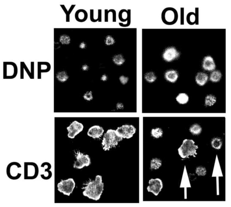
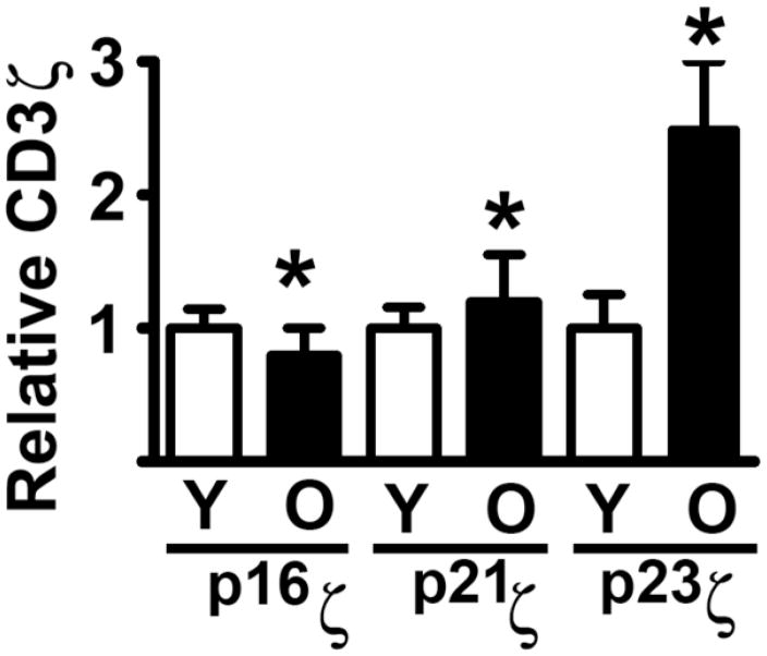
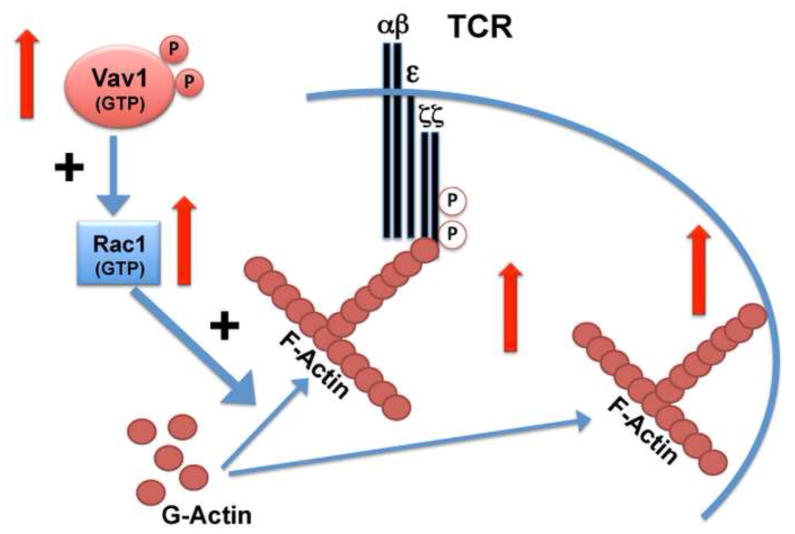
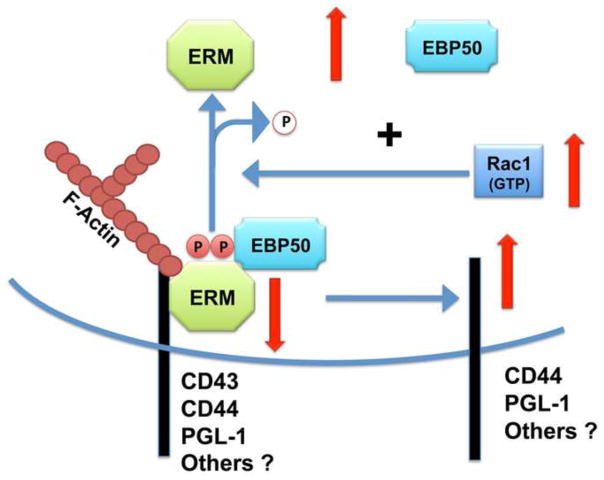
It has been postulated that the cytoskeleton controls many aspects of T cell function, including activation, proliferation and apoptosis. Recent advances in our understanding of F-actin polymerization and the Ezrin-Radixin-Moesin (ERM) family of cytoskeleton signal proteins have provided new insights into immunological synapse formation during T cell activation. During aging there is a significant decline of T cell function largely attributable to declines in activation of CD4 T cells and defects in the formation of the immunological synapse. Here we discuss recent progress in the understanding of how aging alters F-actin and ERM proteins in mouse CD4 T cells, and the implications of these changes for the T cell activation process.
Copyright © 2009 Elsevier Ireland Ltd. All rights reserved.
Figures
Similar articles
-
Age-related defects in moesin/ezrin cytoskeletal signals in mouse CD4 T cells.J Immunol. 2007 Nov 15;179(10):6403-9. doi: 10.4049/jimmunol.179.10.6403. J Immunol. 2007. PMID: 17982027 Free PMC article.
-
ERM (ezrin/radixin/moesin) family: from cytoskeleton to signal transduction.Curr Opin Cell Biol. 1997 Feb;9(1):70-5. doi: 10.1016/s0955-0674(97)80154-8. Curr Opin Cell Biol. 1997. PMID: 9013673 Review.
-
Role of ERM (ezrin-radixin-moesin) proteins in T lymphocyte polarization, immune synapse formation and in T cell receptor-mediated signaling.Front Biosci. 2006 May 1;11:1987-97. doi: 10.2741/1940. Front Biosci. 2006. PMID: 16368573 Review.
-
Ezrin and moesin function together to promote T cell activation.J Immunol. 2009 Jan 15;182(2):1021-32. doi: 10.4049/jimmunol.182.2.1021. J Immunol. 2009. PMID: 19124745 Free PMC article.
-
Ezrin/radixin/moesin proteins differentially regulate endothelial hyperpermeability after thrombin.Am J Physiol Lung Cell Mol Physiol. 2013 Aug 1;305(3):L240-55. doi: 10.1152/ajplung.00355.2012. Epub 2013 May 31. Am J Physiol Lung Cell Mol Physiol. 2013. PMID: 23729486 Free PMC article.
Cited by
-
Signaling pathways in aged T cells - a reflection of T cell differentiation, cell senescence and host environment.Semin Immunol. 2012 Oct;24(5):365-72. doi: 10.1016/j.smim.2012.04.003. Epub 2012 May 4. Semin Immunol. 2012. PMID: 22560928 Free PMC article. Review.
-
Impact of aging on calcium influx and potassium channel characteristics of T lymphocytes.Oncotarget. 2015 May 30;6(15):13750-6. doi: 10.18632/oncotarget.3808. Oncotarget. 2015. PMID: 25948778 Free PMC article.
-
Ex vivo enzymatic treatment of aged CD4 T cells restores cognate T cell helper function and enhances antibody production in mice.J Immunol. 2012 Dec 15;189(12):5582-9. doi: 10.4049/jimmunol.1200487. Epub 2012 Nov 7. J Immunol. 2012. PMID: 23136198 Free PMC article.
-
Nuclear envelope lamin-A couples actin dynamics with immunological synapse architecture and T cell activation.Sci Signal. 2014 Apr 22;7(322):ra37. doi: 10.1126/scisignal.2004872. Sci Signal. 2014. PMID: 24757177 Free PMC article.
-
L-Deprenyl reverses age-associated decline in splenic norepinephrine, interleukin-2 and interferon-γ production in old female F344 rats.Neuroimmunomodulation. 2013;20(2):72-8. doi: 10.1159/000345043. Epub 2012 Nov 29. Neuroimmunomodulation. 2013. PMID: 23207416 Free PMC article.
References
-
- Allenspach EJ, Cullinan P, Tong J, Tang Q, Tesciuba AG, Cannon JL, Takahashi SM, Morgan R, Burkhardt JK, Sperling AI. ERM-dependent movement of CD43 defines a novel protein complex distal to the immunological synapse. Immunity. 2001;15 (5):739–750. - PubMed
-
- Anvari B, Torres JH, McIntyre BW. Regulation of pseudopodia localization in lymphocytes through application of mechanical forces by optical tweezers. J Biomed Opt. 2004;9 (5):865–872. - PubMed
-
- Auvinen E, Kivi N, Vaheri A. Regulation of ezrin localization by Rac1 and PIPK in human epithelial cells. Exp Cell Res. 2007;313 (4):824–833. - PubMed
-
- Berger SB, Sadighi Akha AA, Miller RA. A glycoprotein endopeptidase enhances calcium influx and cytokine production by CD4+ T cells of old and young mice. Int Immunol. 2005;17 (8):983–991. - PubMed
Publication types
MeSH terms
Substances
Grants and funding
LinkOut - more resources
Full Text Sources
Medical
Research Materials
