

Muscle fatigue does not lead to increased instability of upper extremity repetitive movements
- PMID: 19942220
- PMCID: PMC2834814
- DOI: 10.1016/j.jbiomech.2009.11.001
Muscle fatigue does not lead to increased instability of upper extremity repetitive movements
Abstract
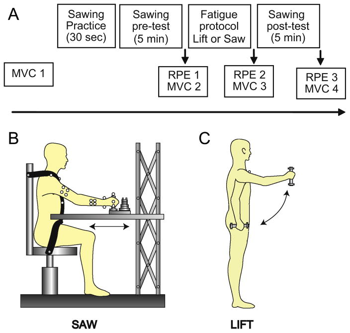
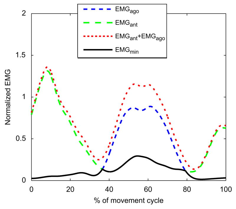
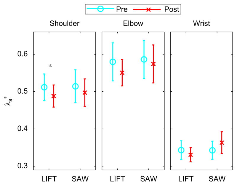
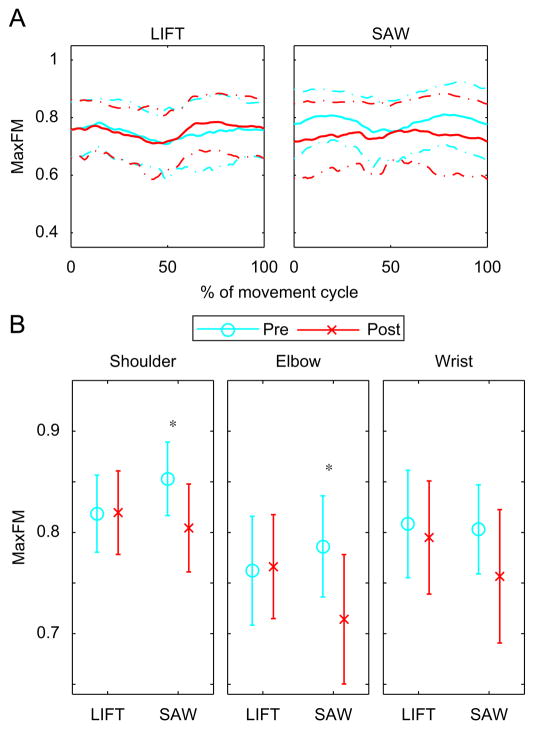
Muscle fatigue alters neuromuscular responses. This may lead to increased sensitivity to perturbations and possibly to subsequent injury risk. We studied the effects of muscle fatigue on movement stability during a repetitive upper extremity task. Twenty healthy young subjects performed a repetitive work task, similar to sawing, synchronized with a metronome before and after performing each of two fatiguing tasks. The first fatigue task (LIFT) primarily fatigued the shoulder flexor muscles, while the second fatigue task (SAW) fatigued all of the muscles of the arm. Subjects performed each task in random order on two different days at least seven days apart. Instantaneous mean EMG frequencies (IMNF) decreased over both fatiguing tasks indicating that subjects did experience significant muscle fatigue. The slopes of the IMNF over time and the decreases in maximum force measurements demonstrated that the LIFT fatigue task successfully fatigued the shoulder flexors to a greater extent than any other muscle. On average, subjects exhibited more locally stable shoulder movements after the LIFT fatigue task (p=0.035). They also exhibited more orbitally stable shoulder (p=0.021) and elbow (p=0.013) movements after the SAW fatigue task. Subjects also had decreased cocontraction at the wrist post-fatigue for both tasks (p=0.001) and at the shoulder (p<0.001) for the LIFT fatigue task. Therefore, increased dynamic stability of these repeated movements cannot be explained by increased muscle cocontraction. Possible alternative mechanisms are discussed.
Copyright (c) 2009 Elsevier Ltd. All rights reserved.
Figures


Similar articles
-
The effects of muscle fatigue and movement height on movement stability and variability.Exp Brain Res. 2011 Apr;209(4):525-36. doi: 10.1007/s00221-011-2580-8. Epub 2011 Feb 18. Exp Brain Res. 2011. PMID: 21331526 Free PMC article.
-
The effects of neuromuscular fatigue on task performance during repetitive goal-directed movements.Exp Brain Res. 2008 Jun;187(4):573-85. doi: 10.1007/s00221-008-1326-8. Epub 2008 Mar 8. Exp Brain Res. 2008. PMID: 18327575 Free PMC article.
-
Upper extremity kinematic and kinetic adaptations during a fatiguing repetitive task.J Electromyogr Kinesiol. 2014 Jun;24(3):404-11. doi: 10.1016/j.jelekin.2014.02.001. Epub 2014 Feb 13. J Electromyogr Kinesiol. 2014. PMID: 24642235 Clinical Trial.
-
Influence of muscle fatigue on motor task performance of the hand and wrist: A systematic review.Hum Mov Sci. 2022 Feb;81:102912. doi: 10.1016/j.humov.2021.102912. Epub 2021 Dec 17. Hum Mov Sci. 2022. PMID: 34929434
-
Sex differences and mechanisms of task-specific muscle fatigue.Exerc Sport Sci Rev. 2009 Jul;37(3):113-22. doi: 10.1097/JES.0b013e3181aa63e2. Exerc Sport Sci Rev. 2009. PMID: 19550202 Free PMC article. Review.
Cited by
-
Changes in Shoulder Rotator Strength After Arthroscopic Capsulolabral Reconstruction in Patients With Anterior Shoulder Instability.Orthop J Sports Med. 2021 Jan 20;9(1):2325967120972052. doi: 10.1177/2325967120972052. eCollection 2021 Jan. Orthop J Sports Med. 2021. PMID: 33786332 Free PMC article.
-
Effects of motor fatigue on walking stability and variability during concurrent cognitive challenges.PLoS One. 2018 Jul 26;13(7):e0201433. doi: 10.1371/journal.pone.0201433. eCollection 2018. PLoS One. 2018. PMID: 30048551 Free PMC article.
-
Influence of exercise order on electromyographic activity during upper body resistance training.J Hum Kinet. 2014 Dec 30;44:203-10. doi: 10.2478/hukin-2014-0127. eCollection 2014 Dec 9. J Hum Kinet. 2014. PMID: 25713681 Free PMC article.
-
Effects of fatigue on synergies in a hierarchical system.Hum Mov Sci. 2012 Dec;31(6):1379-98. doi: 10.1016/j.humov.2012.06.008. Epub 2012 Nov 20. Hum Mov Sci. 2012. PMID: 23182434 Free PMC article.
-
The Impact of Physiological and Psychological Fatigue on Work Efficiency: A Case Study of Parcel Sorting Work.Sensors (Basel). 2024 Sep 15;24(18):5989. doi: 10.3390/s24185989. Sensors (Basel). 2024. PMID: 39338736 Free PMC article.
References
-
- Alizadehkhaiyat O, Fisher AC, Kemp GJ, Frostick SP. Strength and fatigability of selected muscles in the upper limb: Assessing muscle imbalance relative to tennis elbow. Journal of Electromyography and Kinesiology. 2007;4:428–36. - PubMed
-
- Borg GA. Psychophysical bases of perceived exertion. Medicine and Science in Sports and Exercise. 1982;14 (5):377–381. - PubMed
-
- Bruijn SM, van Dieën JH, Meijer OG, Beek PJ. Statistical precision and sensitivity of measures of dynamic gait stability. Journal of Neuroscience Methods. 2009;178 (2):327–333. - PubMed
-
- DeLuca CJ. The use of surface electromyography in biomechanics. Journal of Applied Biomechanics. 1997;13:135–163.
Publication types
MeSH terms
Grants and funding
LinkOut - more resources
Full Text Sources
