

Intravital imaging of stromal cell dynamics in tumors
- PMID: 19942428
- PMCID: PMC2821950
- DOI: 10.1016/j.gde.2009.10.011
Intravital imaging of stromal cell dynamics in tumors
Abstract
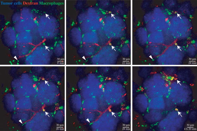
Tumor stroma, consisting of the extracellular matrix and multiple cell types such as immune cells, fibroblasts and vascular cells, contributes to the malignancy of solid tumors by a variety of mechanisms. Intravital imaging by different microscopy techniques, especially by confocal and multi-photon microscopy, has proven to be a powerful method for analyzing the cell-cell and cell-matrix interactions in the dynamic tumor microenvironments. Intravital imaging has fostered the acquisition of data on parameters such as motility of different cell types in distinct tumor regions or manipulated with defined challenges, kinetics of tumor cell killing by T cells or macrophage-assisted tumor cell extravasation, functionality of the vasculature, protease activity and metabolic state. Achieving the direct observation of intact tumors offered by intravital imaging provides unique insights into tumor biology that will continue to deepen our understanding of the processes leading to malignancy and of the ways they can be targeted.
Copyright 2009 Elsevier Ltd. All rights reserved.
Figures
References
-
- Tlsty TD, Coussens LM. Tumor stroma and regulation of cancer development. Annu Rev Pathol. 2006;1:119–150. - PubMed
-
- Schafer M, Werner S. Cancer as an overhealing wound: an old hypothesis revisited. Nat Rev Mol Cell Biol. 2008;9:628–638. - PubMed
-
- de Visser KE, Eichten A, Coussens LM. Paradoxical roles of the immune system during cancer development. Nat Rev Cancer. 2006;6:24–37. - PubMed
-
- Kalluri R, Zeisberg M. Fibroblasts in cancer. Nat Rev Cancer. 2006;6:392–401. - PubMed
Publication types
MeSH terms
Substances
Grants and funding
LinkOut - more resources
Full Text Sources
Other Literature Sources
