Case Reports
. 2010 Apr;31(4):696-705.
doi: 10.3174/ajnr.A1884. Epub 2009 Nov 26.

Negative CT angiography findings in patients with spontaneous subarachnoid hemorrhage: When is digital subtraction angiography still needed?

Affiliations
Case Reports

Negative CT angiography findings in patients with spontaneous subarachnoid hemorrhage: When is digital subtraction angiography still needed?

R Agid et al. AJNR Am J Neuroradiol. 2010 Apr.

Abstract

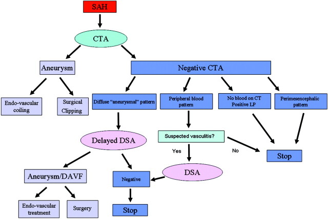
Background and purpose: CTA is becoming the frontline modality to reveal aneurysms in patients with SAH. However, in about 20% of SAH patients no aneurysm is found. In these cases, intra-arterial DSA is still performed. Our aim was to evaluate whether negative findings on CTA can reliably exclude aneurysms in patients with acute SAH.

Materials and methods: We conducted a retrospective analysis of all negative findings on CTAs performed from 2005 to 2009 in patients with spontaneous SAH. Findings were compared with DSA. CTAs were performed with a 64-section multidetector row CT scanner.

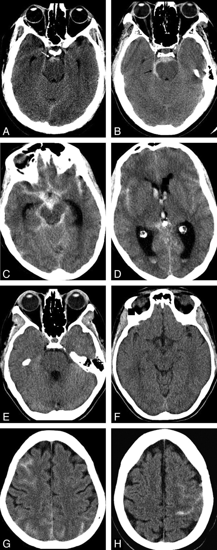
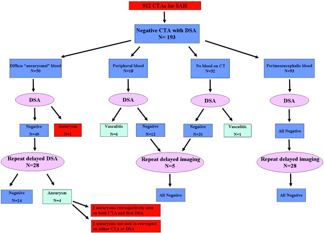
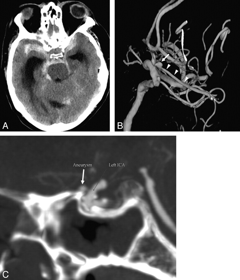
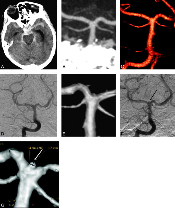
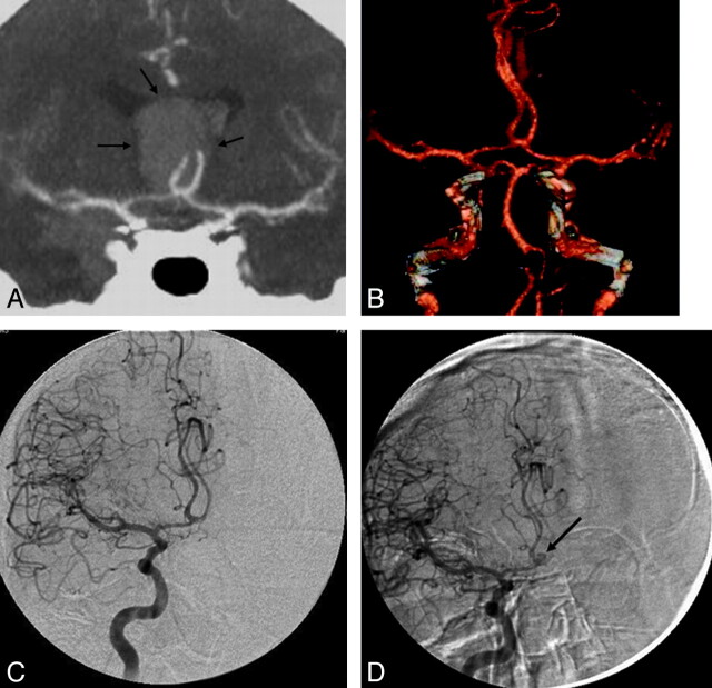
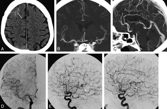
Results: One hundred ninety-three patients with SAH and negative findings on CTA who underwent subsequent DSA were identified. The distribution of blood on unenhanced CT was the following: PMH in 93 patients, diffuse aneurysmal pattern in 50, no blood on CT (xanthochromic LP) in 32, and peripheral sulcal distribution in 18. All patients with PMH had negative findings on DSA. One patient with no blood on CT had vasculitis on DSA. Six of 18 (33%) patients with peripheral blood had vasculitis on DSA. Three of these were also diagnosed by CTA. All except 1 patient with diffuse aneurysmal blood had negative findings on DSA. One patient was diagnosed with an aneurysm on DSA (1/50, 0.5%). Repeat delayed DSA performed in 28 of these patients revealed a small aneurysm in 4 (14%). Five patients had a complication of DSA (2.6%); 1 was a clinical stroke (0.5%).

Conclusions: In patients with SAH, negative CTA findings are reliable in ruling out aneurysms in the PMH pattern or no blood on CT. DSA is indicated in the diffuse aneurysmal pattern of SAH, and repeat delayed DSA is required if the initial DSA findings are negative. When the blood is peripheral, CTA should be scrutinized for vasculitis and DSA is recommended for confirmation.


Figures

Fig 1.
Fig 1.
Four groups of patients with a negative CTA findings according to the distribution of subarachnoid blood on the initial plain CT of the brain. A and B, Group 1: typical perimesencephalic distribution of blood as seen in 2 of our patients. Blood is confined to the prepontine and interpeduncular cisterns (white arrow, A). Extension into the right ambient cistern (black arrow, B) which is still accepted as PMH. C and D, Group 2: two CT images from 1 patient with a diffuse aneurysmal pattern of blood distribution. Blood is diffusely distributed throughout the basal cisterns and extends distally into the Sylvian fissures and ventricles with mild hydrocephalus. E and F, Two CT images of a patient from group 3 who presented with thunderclap headaches and a positive LP but no subarachnoid blood noted on CT. G and H, Two CT images from 2 patients in group 4, with blood located peripherally in the sulci of the brain and no blood in the basal cisterns.
Fig 2.
Fig 2.
Diagram represents the results of DSA in all patients included in this study, according to patterns of blood distribution on the initial CT.
Fig 3.
Fig 3.
False-negative CTA findings in a 58-year-old man who presented with SAH and a GCS score of 4. A, Axial nonenhanced CT at presentation demonstrates a diffuse aneurysmal pattern of SAH and hydrocephalus. CTA did not reveal an aneurysm; however, an ophthalmic aneurysm measuring 2.9 mm in diameter was demonstrated on DSA. B, 3D reconstructions of DSA of the left ICA shows the aneurysm (arrow) located proximally on the ophthalmic artery (arrowheads). C, Retrospective analysis of the CTA revealed the ophthalmic artery aneurysm as seen on the sagittal MIP reconstruction (arrow). A second small extradural cavernous carotid aneurysm is noted as well (B), which is of no clinical significance.
Fig 4.
Fig 4.
A 64-year-old woman who presented with SAH. A, Plain axial CT of the brain at presentation demonstrates a diffuse aneurysmal pattern of blood. B and C, CTA images of the posterior intracranial circulation, including a coronal MIP reconstruction (B) and a 3D reconstruction (C), show an intact basilar tip with no evidence of an aneurysm. The remainder of the CTA is also unremarkable. D and E, A conventional catheter angiogram (DSA) of the right vertebral artery from the same day in an anteroposterior projection (D) and a 3D reconstruction (E) confirm the results of CTA. F and G, A repeat delayed DSA 1 week later reveals a 1.4-mm aneurysm at the basilar tip on both the anteroposterior and 3D views (arrows). This aneurysm was likely partially thrombosed at the acute stage. Note a tiny “bump” at the basilar tip on the 3D reconstructions of both the CTA and first DSA (C and E). In retrospect, this is the location of the aneurysm; however, there was not enough evidence for it to be called an aneurysm on the original scans.
Fig 5.
Fig 5.
A 53-year-old man presented with a diffuse aneurysmal pattern of SAH. A and B, A thick clot at the anterior interhemispheric fissure raised suspicion of an ICA aneurysm responsible for the hemorrhage (arrows, A). However, CTA at presentation did not reveal an aneurysm, as seen on the coronal MIP (A) and 3D reconstructions (B). C, DSA is also negative for aneurysms. D, A repeat delayed DSA performed 1 week later reveals a 3-mm aneurysm at the ICA (arrow), which was likely thrombosed at presentation.
Fig 6.
Fig 6.
A 48-year-old man who presented with severe sudden headache. A, Axial nonenhanced CT at presentation demonstrates peripheral sulcal blood (arrow) with no hemorrhage at the basal cisterns and no history of trauma. B and C, Coronal (B) and sagittal (C) MIPs of the CTA reveal no aneurysm. D−F, Anteroposterior and lateral projections of a catheter angiogram of the right ICA (D and E) and a lateral projection of the left ICA angiogram (F) demonstrate multiple irregularities and narrowings in medium-sized vessels, including the M1 segment of the right MCA (arrows in D), A3 branches of the ACAs (arrows in E and F), P3 branches of the left PCA (black arrowheads in F), and M3 branches of the right and left MCA (white arrowheads in E and F). These findings on DSA suggest underlying vasculitis as the cause of hemorrhage. Retrospective analysis of coronal and sagittal MIP reconstructions of the CTA reveals reciprocal irregularities and narrowings at the M1 segment of the right MCA and at the A3 branches of the ACAs (arrows in B and C).
Fig 7.
Fig 7.
Suggested imaging approach to patients presenting with acute nontraumatic SAH based on the current study.

Similar articles

Cited by

References

    1. Goddard AJ, Tan G, Becker J. Computed tomography angiography for the detection and characterization of intra-cranial aneurysms: current status. Clin Radiol 2005; 60: 1221–36 - PubMed
    1. Agid R, Lee SK, Willinsky RA, et al. . Acute subarachnoid hemorrhage: using 64-slice multidetector CT angiography to “triage” patients’ treatment. Neuroradiology 2006; 48: 787–94 - PubMed
    1. Westerlaan HE, Gravendeel J, Fiore D, et al. . Multislice CT angiography in the selection of patients with ruptured intracranial aneurysms suitable for clipping or coiling. Neuroradiology 2007; 49: 997–1007 - PMC - PubMed
    1. Lubicz B, Levivier M, François O, et al. . Sixty-four-row multisection CT angiography for detection and evaluation of ruptured intracranial aneurysms: interobserver and intertechnique reproducibility. AJNR Am J Neuroradiol 2007; 28: 1949–55 - PMC - PubMed
    1. Willinsky RA, Taylor SM, TerBrugge K, et al. . Neurologic complications of cerebral angiography: prospective analysis of 2,899 procedures and review of the literature. Radiology 2003; 227: 522–28 - PubMed

Publication types