Review
. 2009 Dec 31;41(12):849-57.
doi: 10.3858/emm.2009.41.12.103.

Hypoxia and angiogenesis: regulation of hypoxia-inducible factors via novel binding factors

Affiliations
Review

Hypoxia and angiogenesis: regulation of hypoxia-inducible factors via novel binding factors

Li Chen et al. Exp Mol Med. .

Abstract

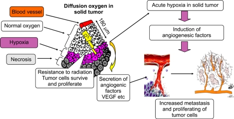
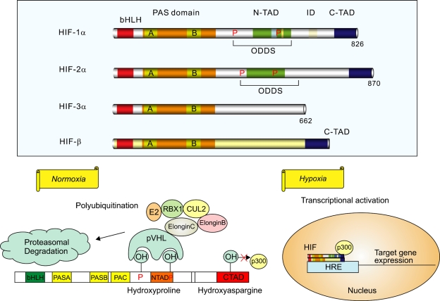
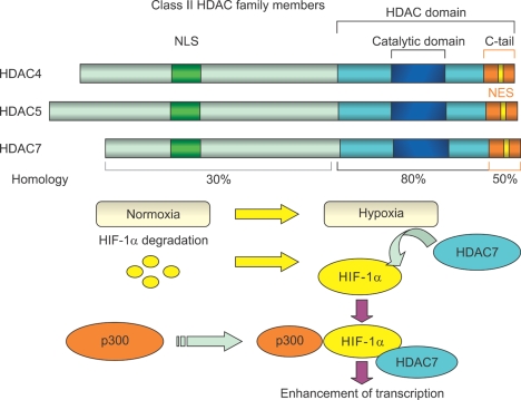
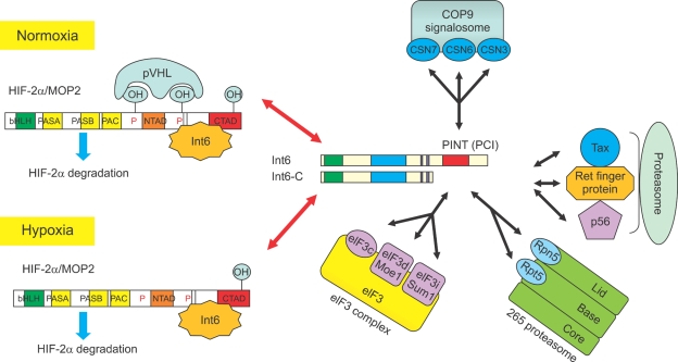
The mechanisms that regulate angiogenesis in hypoxia or hypoxic microenvironment are modulated by several pro- and antiangiogenic factors. Hypoxia-inducible factors (HIFs) have been established as the basic and major inducers of angiogenesis, but understanding the role of interacting proteins is becoming increasingly important to elucidate the angiogenic processes of a hypoxic response. In particular, with regard to wound healing and the novel therapies for vascular disorders such as ischemic brain and heart attack, it is essential to gain insights in the formation and regulation of HIF transcriptional machineries related to angiogenesis. Further, identification of alternative ways of inhibiting tumor growth by disrupting the growth-triggering mechanisms of increasing vascular supply via angiogenesis depends on the knowledge of how tumor cells develop their own vasculature. Here, we review our findings on the interactions of basic HIFs, HIF-1 alpha and HIF-2 alpha, with their regulatory binding proteins, histone deacetylase 7 (HDAC7) and translation initiation factor 6 (Int6), respectively. The present results and discussion revealed new regulatory interactions of HIF-related mechanisms.


Figures

Figure 1
Figure 1
Hypoxia and tumor angiogenesis: Hypoxia occurs in chronic and acute vascular diseases and tumor formation. It is toxic to normal cells, but cancer cells can survive and continue to proliferate in hypoxia. Human tumors grew like cords around blood vessels, and tumor cells located > 180 µm from the blood vessels were observed to become necrotic. Hypoxia induces angiogenesis during tumor growth; after tumor growth progresses, an oxygen gradient develops from the oxygen source to the periphery of the tumor. Cells lacking oxygen and nutrition because of their distance from the blood supply become necrotic, whereas those closer to the blood supply begin sensing hypoxia and secrete angiogenic factors. As a result, angiogenesis occurs and the tumor develops its own vasculature, independent of the original tissue.
Figure 2
Figure 2
HIF pathway: Upper panel: Known HIF-α isoforms and their cofactor HIF-β. HIF-1β and HIF-2α appear closely related, whereas HIF-3α seems to be involved in negative regulation of hypoxia response. Lower panel: Schematic diagram of HIF-1α regulation. In normoxia, the proline residues are hydroxylated and recognized by pVHL, which targets HIF-1α for degradation. pVHL is part of a large complex that comprises elongin B, elongin C, CUL2, RBX1, and a ubiquitin-conjugating enzyme (E2). The asparagine residue in the C-TAD of HIF-1α is an oxygen-dependent hydroxylation-regulated binding site for p300, which is inactive in its hydroxylated state. In hypoxia, prolyl hydroxylase cannot modify HIF-1α, and the protein remains stable. The stabilized HIF-1α is translocated to the nucleus, where it interacts with cofactors HIF-1β and p300 and transcribes hypoxia-related target genes. HIF, hypoxia-inducible factor; bHLH, basic helix-loop-helix; PAS, Per-ARNT-SIM; N-TAD, N-terminal transactivation domain; C-TAD, C-terminal transactivation domain; ID, inhibitory domain; ODDs, oxygen-dependent degradation domains; pVHL, von Hippel-Lindau protein; CUL2, cullin 2; RBX1, RING-box protein 1; E2, ubiquitin-conjugating enzyme; HRE, hypoxia-responsive element.
Figure 3
Figure 3
Class II HDAC family members and interaction of HDAC7 with HIF-1α in its activated state in hypoxia: HDAC4, HDAC5, and HDAC7 contain a highly homologous conserved catalytic domain (HDAC domain) in the C-terminal region. The N-terminal region and the C-terminal tail of HDAC7 are less homologous to the corresponding regions of HDAC4 and HDAC5. HDAC4, HDAC5, and HDAC7 also contain N-terminal NLS sequences and C-terminal signal-responsive NES sequences. In addition, the 3 proteins are known to shuttle between the cytoplasm and nucleus in a process regulated by calcium/calmodulin-dependent protein kinase. In hypoxia, HDAC7 forms a complex with HIF-1α and p300 in the nucleus, resulting in enhanced transcription of HIF-1α target genes. HDAC, histone deacetylase; NLS, nuclear localization signal; NES, nuclear export signal; HIF, hypoxia-inducible factor.
Figure 4
Figure 4
Multiple functions of Int6: Left panel: Schematic diagram of Int6-HIF-2α interaction leading to oxygen-independent HIF-2α degradation. The N-terminal region of Int6 specifically binds to the C-terminal inhibitory domain of HIF-2α. Binding of Int6 induced HIF-2α instability, whereas that of the dominant-negative mutant Int6-ΔC with deleted C-terminal PINT domain induced stable HIF-2α expression, even under normoxic conditions. Right panel: Known function of Int6, which may also be indirectly involved in proteolysis via at least 3 Int6-binding proteins, namely, Tax, Ret finger protein, and p56. Int6 has a C-terminal PCI domain, which has also been found in the subunits of the following 3 complexes: proteasome regulatory lid, CSN, and eIF3 complex. HIF, hypoxia-inducible factor; MOP2, member of PAS protein 2; pVHL, von Hippel-Lindau protein; bHLH, basic helix-loop-helix; PAS, Per-ARNT-SIM; N-TAD, N-terminal transactivation domain; C-TAD, C-terminal transactivation domain; Int6, translation initiation factor 6; CSN, constitutive photomorphogenesis (COP9) signalosome; PINT, proteasome/Int6/Nip1/thyroid receptor-interacting protein 15 (TRIP-15); PCI, proteasome/CSN/eIF3; eIF3, eukaryotic translation initiation factor 3; Moe1, microtubule over extended; Sum1, suppressor of uncontrolled mitosis 1; Tax, transactivator x; Ret, receptor tyrosine kinase; Rpn5, regulatory particle non-ATPase 5; Rpt5, regulatory particle, ATPase like.

Similar articles

Cited by

References

    1. Asano K, Merrick WC, Hershey JW. The translation initiation factor eIF3-p48 subunit is encoded by int-6, a site of frequent integration by the mouse mammary tumor virus genome. J Biol Chem. 1997;272:23477–23480. - PubMed
    1. Bertos NR, Wang AH, Yang XJ. Class II histone deacetylases: Structure, function, and regulation. Biochem Cell Biol. 2001;79:243–252. - PubMed
    1. Brown JM, Giaccia AJ. The unique physiology of solid tumors: Opportunities (and problems) for cancer therapy. Cancer Res. 1998;58:1408–1416. - PubMed
    1. Brunnberg S, Pettersson K, Rydin E, Matthews J, Hanberg A, Pongratz I. The basic helix-loop-helix-PAS protein ARNT functions as a potent coactivator of estrogen receptor-dependent transcription. Proc Natl Acad Sci USA. 2003;100:6517–6522. - PMC - PubMed
    1. Carroll VA, Ashcroft M. Role of hypoxia-inducible factor (HIF)-1α versus HIF-2α in the regulation of HIF target genes in response to hypoxia, insulin-like growth factor-I, or loss of von Hippel-Lindau function: Implications for targeting the HIF pathway. Cancer Res. 2006;66:6264–6270. - PubMed

MeSH terms

Substances

LinkOut - more resources