. 2010 Jan 20;29(2):469-81.
doi: 10.1038/emboj.2009.339. Epub 2009 Nov 26.

PKA phosphorylates and inactivates AMPKalpha to promote efficient lipolysis

Affiliations

PKA phosphorylates and inactivates AMPKalpha to promote efficient lipolysis

Nabil Djouder et al. EMBO J. .

Abstract

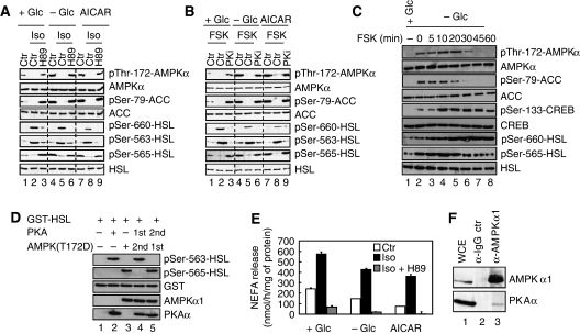
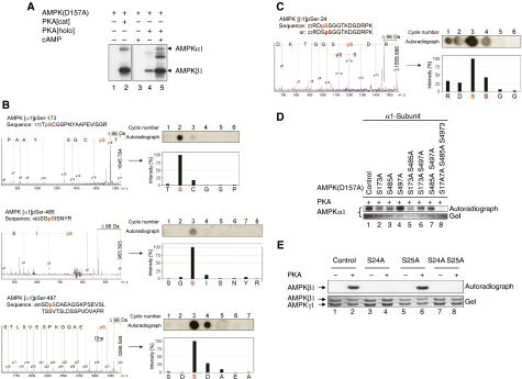
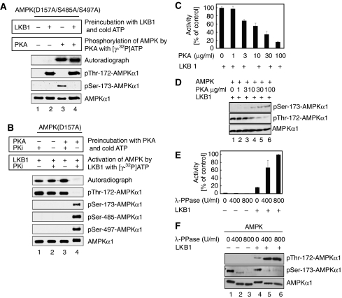
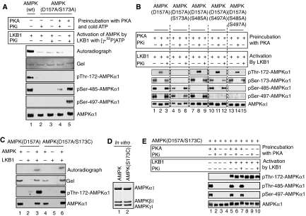
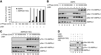
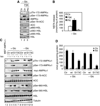
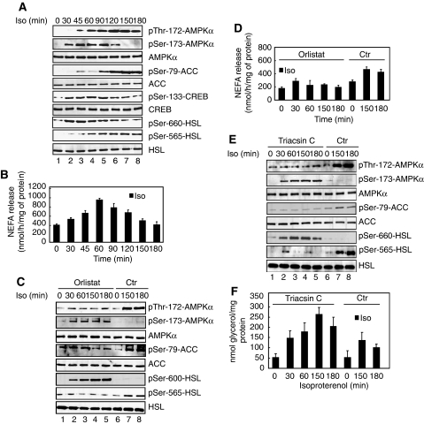
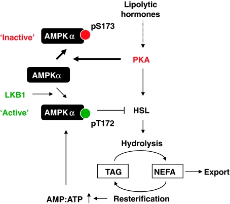
The mobilization of metabolic energy from adipocytes depends on a tightly regulated balance between hydrolysis and resynthesis of triacylglycerides (TAGs). Hydrolysis is stimulated by beta-adrenergic signalling to PKA that mediates phosphorylation of lipolytic enzymes, including hormone-sensitive lipase (HSL). TAG resynthesis is associated with high-energy consumption, which when inordinate, leads to increased AMPK activity that acts to restrain hydrolysis of TAGs by inhibiting PKA-mediated activation of HSL. Here, we report that in primary mouse adipocytes, PKA associates with and phosphorylates AMPKalpha1 at Ser-173 to impede threonine (Thr-172) phosphorylation and thus activation of AMPKalpha1 by LKB1 in response to lipolytic signals. Activation of AMPKalpha1 by LKB1 is also blocked by PKA-mediated phosphorylation of AMPKalpha1 in vitro. Functional analysis of an AMPKalpha1 species carrying a non-phosphorylatable mutation at Ser-173 revealed a critical function of this phosphorylation for efficient release of free fatty acids and glycerol in response to PKA-activating signals. These results suggest a new mechanism of negative regulation of AMPK activity by PKA that is important for converting a lipolytic signal into an effective lipolytic response.


Conflict of interest statement

The authors declare that they have no conflict of interest.

Figures

Figure 1
Figure 1
PKA signalling inhibits Thr-172 phosphorylation of AMPKα. (A) Primary adipocytes were cultivated for 30 min either in the presence of 25 mM glucose (+Glc) (lanes 1–3 and 7–9) or the absence of glucose (−Glc) (lanes 4–6) and treated with 1 mM AICAR (lanes 7–9), 200 nM isoproterenol (Iso) (lanes 2, 5 and 8) or 200 nM isoproterenol in combination with 1 μM H89 (lanes 3, 6 and 9). Protein lysates were prepared and probed with the indicated antibodies. (B) Similar experiment as in (A) except that 20 μM Forskolin (FSK) and 100 μM myristoylated PKi were added instead of isoproterenol and H89, respectively. (C) Primary adipocytes were glucose starved for 60 min (−Glc) and incubated with 20 μM Forskolin (FSK) for indicated times. FSK was either present ab initio (lane 8), added during the last 5, 10, 20, 30 or 45 min of starvation (lanes 3–8) or omitted (lane 2). As control, cells were grown in glucose-rich medium (+Glc) (lane 1). Equal amounts of protein lysates were subjected to immunoblotting with the indicated antibodies. (D) Phosphorylation of HSL by PKA and AMPK at Ser-563 and Ser-565, respectively, is mutually exclusive. In vitro kinase assay of GST-HSL in the presence of PKA (lanes 2, 4 and 5) and constitutively active AMPK(T172D) (lanes 3, 4 and 5). PKA was either added before (lane 4) or after incubation of HSL with AMPK(T172D) (lane 5). Proteins were subjected to immunoblotting with the indicated antibodies. (E) NEFA release in response to PKA signalling. Primary adipocytes were incubated for 30 min with 200 nM isoproterenol alone (Iso) or in combination with 1 μM H89. Furthermore, this treatment was done in the presence (+Glc) or absence (−Glc) of glucose or in the presence of glucose and 1 mM AICAR. NEFA were measured in the incubation medium. Bars represent the mean NEFA release from three independent experiments. (F) Whole-cell extracts of primary mouse adipocytes (WCE, lane 1) were prepared and aliquots were subjected to immunoprecipitation with control IgG (lane 2) or anti-AMPKα1 antibodies (lane 3) and immunoblotted for AMPKα1 and PKAα. Immunoblots are representative of at least three independent experiments.
Figure 2
Figure 2
PKA phosphorylates catalytic α- and regulatory β-subunits of AMPK at multiple sites. (A) PKA phosphorylates AMPK in vitro. Autoradiograph of α1β1γ1 catalytically inactive AMPK(D157A) complexes incubated in the presence of [γ-32P]ATP with either the cAMP-dependent PKA holoenzyme, PKA[holo], or the constitutively active, cAMP independent, catalytic domain of PKA, PKA[cat]. Arrows indicate phosphorylated AMPK(D157) at the α1- and β1-subunits. (B) Mass fingerprinting of isolated phosphopeptides derived from HPLC was performed by MALDI-ToF MS. Peptides differing by +80 Da (HPO3) from computed masses were then selected for MS/MS and phosphorylation was confirmed by a neutral loss of −98 Da (H3PO4) during fragmentation. Further verification of the phosphosites was attempted by solid-phase sequencing of the radiolabelled peptides. After each cycle of N-terminal Edman degradation, liberated single amino acids were collected and spotted onto DEAE cellulose. The respective phosphorylated residues were then detected by autoradiography. The annotated mass spectra indicates the phosphorylated residues of AMPKα1 pSer-173, pSer-485 and pSer-497. (C) As in (B) but related to AMPKβ1 pSer-24. Dha, dehydroala. (D) Analysis of AMPKα1 phosphorylation site mutants. Purified AMPK(D157A) (control), or the indicated serine to alanine mutants thereof, were incubated with PKA in the presence of [γ-32P]ATP and processed for SDS–PAGE and autoradiography. (E) Verification of in vitro AMPK β1-phosphorylation sites by mutational analysis. Catalytically inactive AMPK(D157A) complexes containing a regulatory β1-subunit mutated at Ser-24 and/or Ser-25 were incubated with [γ-32P]ATP in the presence or absence of PKA. Phosphorylation was visualized by SDS–PAGE and autoradiography. PKA was unable to phosphorylate AMPKβ1 mutated in Ser-24 (lane 4) but not Ser-25 (lane 6), showing that only Ser-24 is targeted by PKA. Control, AMPK(D157A) containing no additional mutations. Immunoblots are representative of at least three independent experiments.
Figure 3
Figure 3
PKA-mediated phosphorylation of AMPKα1 prevents its phosphorylation by LKB1 at Thr-172 in vitro and phosphorylation of AMPK at Thr172 and Ser-173 is not mutually exclusive. (A) LKB1 was allowed to phosphorylate AMPKα1(D157S485A/S497A) in the presence of ATP before addition of PKA and [γ-32P]ATP. Signals obtained by autoradiography indicate phosphorylation at Ser-173 (lanes 3 and 4). Phosphorylation of Thr-172 and Ser-173 is coexistent, if PKA is allowed to phosphorylate AMPK after preincubation with LKB1 (lane 4). In this double-phosphorylated form, phosphorylation of Thr-172 but not Ser-173 was recognized by the corresponding antibodies. PKi, PKA inhibitor. Samples were processed for immunoblotting with the specified antibodies. (B) PKA phosphorylation of AMPKα1 prevents LKB1-mediated Thr-172 phosphorylation in vitro. AMPKα1(D157A) was preincubated with/without PKA and non-radioactive ATP as indicated, followed by LKB1 assays in the presence of [γ-32P]ATP. LKB1 was unable to phosphorylate AMPK after PKA phosphorylation. Samples were processed for immunoblotting with the specified antibodies. (C) PKA-mediated AMPKα1 phosphorylation inhibits AMPK activity. AMPK was preincubated with different amounts of PKA before in vitro kinase reactions with LKB1. AMPK activity assessed as the amount of phosphate incorporated into the synthetic peptide substrate SAMS. (D) Immunoblot corresponding to (B) probed with the indicated antibodies. (E) PKA-phosphorylated AMPK was either left untreated or dephosphorylated with λ-PPase using the indicated concentrations, then processed for in vitro kinase assays with or without LKB1. AMPK activity quantified using the SAMS assay. (F) Immunoblot corresponding to (E) probed with the indicated antibodies. All activity measurements were done in triplicates. Error bars represent±s.e.m. Immunoblots are representative of at least three independent experiments.
Figure 4
Figure 4
Phosphorylation of AMPKα1 at Ser-173 mediates the inhibitory effect by PKA. (A) Mutagenesis of Ser-173 to alanine blocks phosphorylation of AMPK at Thr-172 by LKB1. AMPK(D157A/S173A) was phosphorylated by PKA with non-radioactive ATP and subsequently by LKB1 in the presence of [γ-32P]ATP in the absence or presence of PKi as indicated. Note that LKB1 is unable to phosphorylate this mutant AMPK protein, suggesting that Ser-173 is a key residue in the recognition sequence for LKB1. As control, AMPK(wt) was used in the assay (lane 1). (B) Mutation of Ser-485 and/or Ser-497 on AMPKα1 does not abolish the inhibitory effect of PKA. AMPK mutants were incubated with PKA and LKB1. Reactions were performed in the absence or presence of PKi as indicated. In vitro kinase assays were processed for immunoblotting with the indicated antibodies. Neither mutation of Ser-485 nor Ser-497 was able to nullify the inhibitory action of PKA on LKB1-mediated Thr-172 phosphorylation (lanes 7–15). Note that no phosphorylation of Thr-172 by LKB1 was observed when Ser-173 was exchanged to alanine (lanes 4–6). (C) AMPK(D157A) (lanes 1–3) and AMPK(D157A/S173C) (lanes 4–6) complexes were either incubated with LKB1 or not, in the presence of [γ-32P]ATP. AMPKα1(D157A) and AMPKα1(D157A/S173C) protein phosphorylation assessed by autoradiography (upper panel) and immunoblotting (lower panel). (D) AMPK(S173C) forms a 1:1:1 complex with the regulatory β- and γ-subunits in vitro. Histidine-tagged wild-type AMPKα1 or the (S173C) mutant thereof were coexpressed with the AMPK β- and γ-subunits, followed by affinity chromatography with Ni2+-NTA, SDS–PAGE and Coomassie blue staining. (E) AMPK(D157A/S173C) complexes were incubated with or without PKA and then treated with LKB1. Reactions were performed in the absence or presence of PKi as indicated. In vitro kinase assays were processed for immunoblotting with the indicated antibodies. Immunoblots are representative of at least three independent experiments.
Figure 5
Figure 5
PKA-mediated phosphorylation of AMPKα1 at Ser-173 efficiently interferes with LKB1-dependent activation of AMPK. (A) AMPK(S173C) complex is refractory to inhibition by PKA. PKA-phosphorylated AMPK and AMPK(S173C) complexes were dephosphorylated with λ-PPase using the indicated concentrations, then incubated with or without LKB1. Subsequently, AMPK activity was quantified using the SAMS assay. Assays were done in triplicate and errors bars represent±s.e.m. (B, C) Immunoblots corresponding to (A) probed with indicated antibodies. (D) LKB1-activated AMPK or AMPK(S173C) complexes were incubated with GST-HSL and processed for immunoblotting with the indicated antibodies. Immunoblots represent one experiment of three independent experiments.
Figure 6
Figure 6
PKA-mediated phosphorylation of AMPKα at Ser-173 is critical for efficient lipolysis. (A) Ser-173 of AMPKα is phosphorylated in vivo by PKA. Primary adipocytes were glucose starved (−Glc) for 30 min (lanes 1–3) and then treated with 20 μM forskolin (FSK) alone (lane 2) or in combination with 100 μM PKi (lane 3). Protein lysates were processed for immunoblotting with the indicated antibodies. (B) NEFA release in response to PKA signalling. Primary adipocytes were glucose starved (−Glc) for 30 min (lanes 1–3) and then treated with 20 μM forskolin (FSK) alone (lane 2) or in combination with 100 μM PKi (lane 3). NEFA were measured in the incubation medium. Bars represent the mean NEFA release from three independent experiments. (C) AMPKα1(S173C) is refractory to inhibition by PKA. Immortalized control AMPKα+/− and AMPKα−/− MEFs were differentiated into adipocytes as described in the Materials and methods section. AMPKα−/− adipocytes were infected with an adenovirus containing either an empty plasmid (lanes 2, 3, 8, 9), a plasmid encoding for myc-AMPKα1(wt) (lanes 4, 5, 10, 11) or myc-AMPKα1(S173C) (lanes 6, 7, 12, 13). Before immunoblotting experiments and lipolysis assays, adipocytes were grown for 30 min in the presence (+Glc) (lanes 2–7) or absence (−Glc) (lanes 8–13) of glucose and simultaneously treated with or without 200 nM isoproterenol (Iso). Aliquots of protein lysates were immunoblotted with the indicated antibodies. (D) Incubation medium of adipocytes described in (C) were processed for glycerol measurements. Bars represent the mean of glycerol release from three independent experiments and immunoblots are representative of at least three independent experiments.
Figure 7
Figure 7
Sustained PKA signalling activates and phosphorylates AMPKα at Thr-172. (A) Long-term isoproterenol treatment activates and phosphorylates AMPKα. Primary adipocytes were cultivated in the presence of 200 nM of isoproterenol (Iso) in a time-dependent manner. Protein lysates were prepared and probed with the indicated antibodies. (B) Incubation medium of primary adipocytes described in (A) were processed for NEFA measurements. Bars represent the mean NEFA release from three independent experiments. (C) Primary adipocytes were treated with 50 μM orlistat for 180 min and incubated with 200 nM isoproterenol for indicated times, in a serum-free DMEM and presence of glucose. Isoproterenol was either present ab initio (lane 5), added during the last 30, 60 or 150 min of treatment (lanes 1–5). As control, cells were cultivated without orlistat but in the presence of 200 nM isoproterenol for 150 and 180 min in a serum-free DMEM and presence of glucose (lanes 6–8). Equal amounts of protein lysates were subjected to immunoblotting with the indicated antibodies. (D) Incubation medium of primary adipocytes described in (C) were processed for NEFA measurements. Bars represent the mean NEFA release from three independent experiments. (E) Primary adipocytes were treated with 10 μM triacsin C for 180 min and incubated with 200 nM isoproterenol for indicated times, in a serum-free DMEM and in the presence of glucose. Like in (C), isoproterenol was either present ab initio (lane 5), added during the last 30, 60 or 150 min of treatment (lanes 1–5). As control, cells were cultivated in the presence of 200 nM isoproterenol for 150 and 180 min in a serum-free DMEM and presence of glucose (lanes 6–8). Equal amounts of protein lysates were subjected to immunoblotting with the indicated antibodies. (F) Incubation medium of primary adipocytes described in (E) were processed for glycerol measurements as described in the Materials and methods section. Bars represent the mean NEFA or glycerol release from three independent experiments. Immunoblots are representative of at least three independent experiments.
Figure 8
Figure 8
Model proposing a function of PKA in phosphorylating and inactivating AMPKα and its potential effects on lipolysis (for details see discussion).

Similar articles

Cited by

References

    1. Anthonsen MW, Ronnstrand L, Wernstedt C, Degerman E, Holm C (1998) Identification of novel phosphorylation sites in hormone-sensitive lipase that are phosphorylated in response to isoproterenol and govern activation properties in vitro. J Biol Chem 273: 215–221 - PubMed
    1. Bergeron R, Previs SF, Cline GW, Perret P, Russell RR III, Young LH, Shulman GI (2001) Effect of 5-aminoimidazole-4-carboxamide-1-beta-D-ribofuranoside infusion on in vivo glucose and lipid metabolism in lean and obese Zucker rats. Diabetes 50: 1076–1082 - PubMed
    1. Brooks B, Arch JR, Newsholme EA (1982) Effects of hormones on the rate of the triacylglycerol/fatty acid substrate cycle in adipocytes and epididymal fat pads. FEBS Lett 146: 327–330 - PubMed
    1. Carling D, Sanders MJ, Woods A (2008) The regulation of AMP-activated protein kinase by upstream kinases. Int J Obes (Lond) 32(Suppl 4): S55–S59 - PubMed
    1. Collins SP, Reoma JL, Gamm DM, Uhler MD (2000) LKB1, a novel serine/threonine protein kinase and potential tumour suppressor, is phosphorylated by cAMP-dependent protein kinase (PKA) and prenylated in vivo. Biochem J 345(Pt 3): 673–680 - PMC - PubMed

Publication types