

Somatosensory system deficits in schizophrenia revealed by MEG during a median-nerve oddball task
- PMID: 19943100
- PMCID: PMC2816821
- DOI: 10.1007/s10548-009-0122-5
Somatosensory system deficits in schizophrenia revealed by MEG during a median-nerve oddball task
Abstract
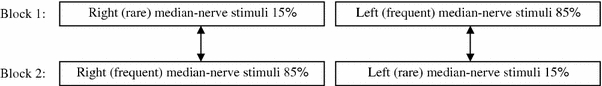
Although impairments related to somatosensory perception are common in schizophrenia, they have rarely been examined in functional imaging studies. In the present study, magnetoencephalography (MEG) was used to identify neural networks that support attention to somatosensory stimuli in healthy adults and abnormalities in these networks in patient with schizophrenia. A median-nerve oddball task was used to probe attention to somatosensory stimuli, and an advanced, high-resolution MEG source-imaging method was applied to assess activity throughout the brain. In nineteen healthy subjects, attention-related activation was seen in a sensorimotor network involving primary somatosensory (S1), secondary somatosensory (S2), primary motor (M1), pre-motor (PMA), and paracentral lobule (PCL) areas. A frontal-parietal-temporal "attention network", containing dorsal- and ventral-lateral prefrontal cortex (DLPFC and VLPFC), orbitofrontal cortex (OFC), anterior cingulate cortex (ACC), superior parietal lobule (SPL), inferior parietal lobule (IPL)/supramarginal gyrus (SMG), and temporal lobe areas, was also activated. Seventeen individuals with schizophrenia showed early attention-related hyperactivations in S1 and M1 but hypo-activation in S1, S2, M1, and PMA at later latency in the sensorimotor network. Within this attention network, hypoactivation was found in SPL, DLPFC, orbitofrontal cortex, and the dorsal aspect of ACC. Hyperactivation was seen in SMG/IPL, frontal pole, and the ventral aspect of ACC in patients. These findings link attention-related somatosensory deficits to dysfunction in both sensorimotor and frontal-parietal-temporal networks in schizophrenia.
Figures
Similar articles
-
A parietal-frontal network studied by somatosensory oddball MEG responses, and its cross-modal consistency.Neuroimage. 2005 Oct 15;28(1):99-114. doi: 10.1016/j.neuroimage.2005.05.036. Epub 2005 Jun 23. Neuroimage. 2005. PMID: 15979344
-
Neural circuit of verbal humor comprehension in schizophrenia - an fMRI study.Neuroimage Clin. 2017 Jun 3;15:525-540. doi: 10.1016/j.nicl.2017.06.005. eCollection 2017. Neuroimage Clin. 2017. PMID: 28652967 Free PMC article.
-
Spatiotemporal imaging of electrical activity related to attention to somatosensory stimulation.Neuroimage. 2002 Nov;17(3):1347-57. doi: 10.1006/nimg.2002.1222. Neuroimage. 2002. PMID: 12414274
-
Auditory mismatch impairments are characterized by core neural dysfunctions in schizophrenia.Brain. 2015 May;138(Pt 5):1410-23. doi: 10.1093/brain/awv049. Epub 2015 Mar 4. Brain. 2015. PMID: 25743635 Free PMC article.
-
Integrated technology for evaluation of brain function and neural plasticity.Phys Med Rehabil Clin N Am. 2004 Feb;15(1):263-306. doi: 10.1016/s1047-9651(03)00124-4. Phys Med Rehabil Clin N Am. 2004. PMID: 15029909 Review.
Cited by
-
Intersensory attention deficits in schizophrenia relate to ongoing sensorimotor beta oscillations.Schizophrenia (Heidelb). 2025 Feb 17;11(1):19. doi: 10.1038/s41537-025-00571-8. Schizophrenia (Heidelb). 2025. PMID: 39962042 Free PMC article.
-
Neuropsychological dysfunctions among chronic schizophrenia patients, alcohol dependence cases, and normal subjects: A comparative study.Ind Psychiatry J. 2020 Jan-Jun;29(1):105-122. doi: 10.4103/ipj.ipj_70_20. Epub 2020 Nov 7. Ind Psychiatry J. 2020. PMID: 33776284 Free PMC article.
-
The selective impairment of resting-state functional connectivity of the lateral subregion of the frontal pole in schizophrenia.PLoS One. 2015 Mar 6;10(3):e0119176. doi: 10.1371/journal.pone.0119176. eCollection 2015. PLoS One. 2015. PMID: 25748858 Free PMC article. Clinical Trial.
-
Altered Hemispheric Asymmetry of Functional Hierarchy in Schizophrenia.Brain Sci. 2025 Mar 16;15(3):313. doi: 10.3390/brainsci15030313. Brain Sci. 2025. PMID: 40149834 Free PMC article.
-
Association between auditory P300, psychopathology, and memory function in drug-naïve schizophrenia.Kaohsiung J Med Sci. 2014 Mar;30(3):133-8. doi: 10.1016/j.kjms.2013.10.003. Epub 2013 Dec 3. Kaohsiung J Med Sci. 2014. PMID: 24581213 Free PMC article.
References
-
- Achim A. Statistical detection of between-group differences in event-related potentials. Clin Neurophysiol. 2001;112:1023–1034. - PubMed
-
- Alain C, Hargrave R, Woods DL. Processing of auditory stimuli during visual attention in patients with schizophrenia. Biol Psychiatry. 1998;44:1151–1159. - PubMed
-
- Alho K. Cerebral generators of mismatch negativity (MMN) and its magnetic counterpart (MMNm) elicited by sound changes. Ear Hear. 1995;16:38–51. - PubMed
-
- Anderer P, Pascual-Marqui RD, Semlitsch HV, Saletu B. Differential effects of normal aging on sources of standard N1, target N1 and target P300 auditory event-related brain potentials revealed by low resolution electromagnetic tomography (LORETA) Electroencephalogr Clin Neurophysiol. 1998;108:160–174. - PubMed
-
- Andreasen NC. A unitary model of schizophrenia: Bleuler’s “fragmented phrene” as schizencephaly. Arch Gen Psychiatry. 1999;56:781–787. - PubMed
Publication types
MeSH terms
Grants and funding
LinkOut - more resources
Full Text Sources
Other Literature Sources
Medical
