. 2010 Feb;75(3):658-75.
doi: 10.1111/j.1365-2958.2009.06983.x. Epub 2009 Nov 25.

Septins enforce morphogenetic events during sexual reproduction and contribute to virulence of Cryptococcus neoformans

Affiliations

Septins enforce morphogenetic events during sexual reproduction and contribute to virulence of Cryptococcus neoformans

Lukasz Kozubowski et al. Mol Microbiol. 2010 Feb.

Abstract

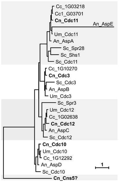
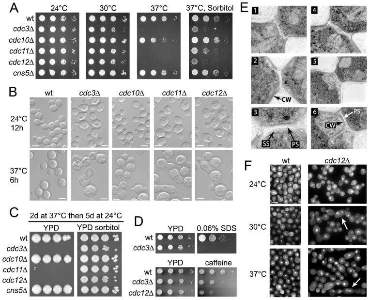
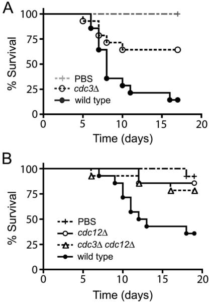
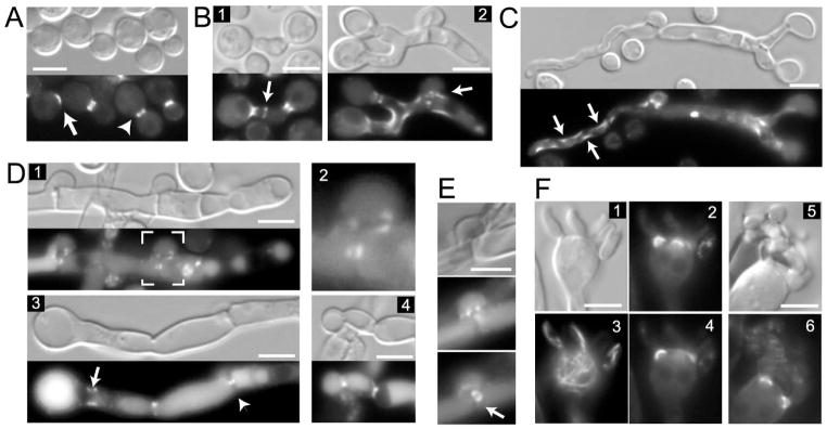
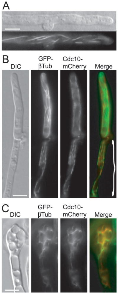
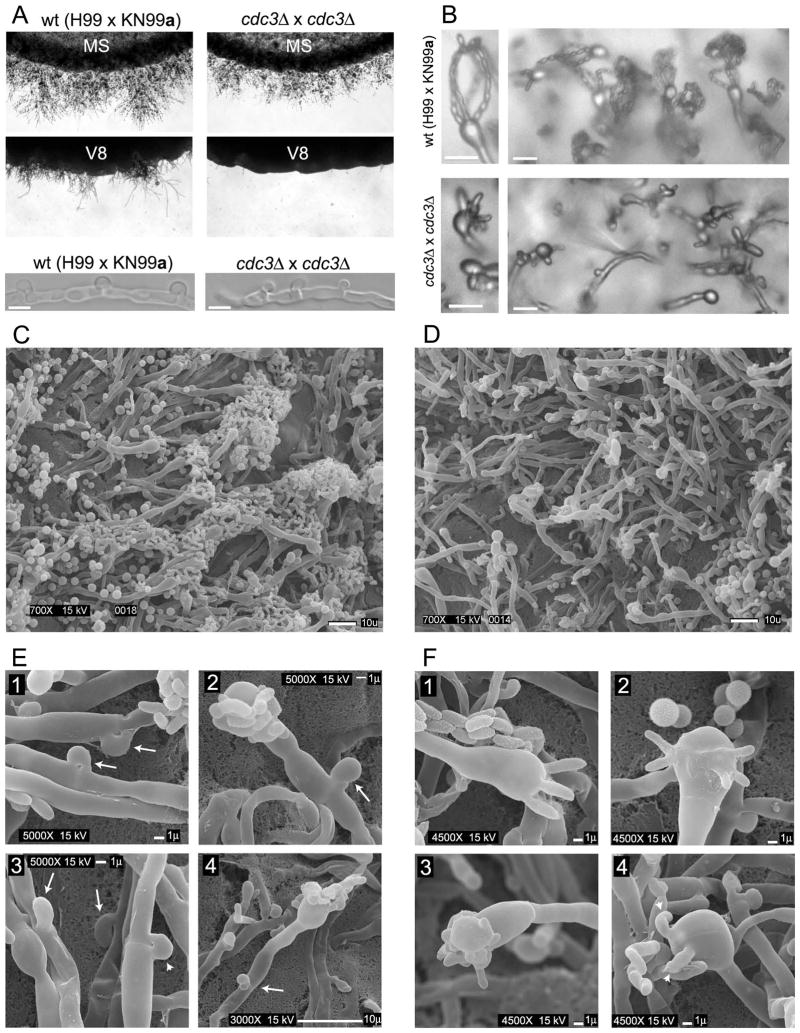
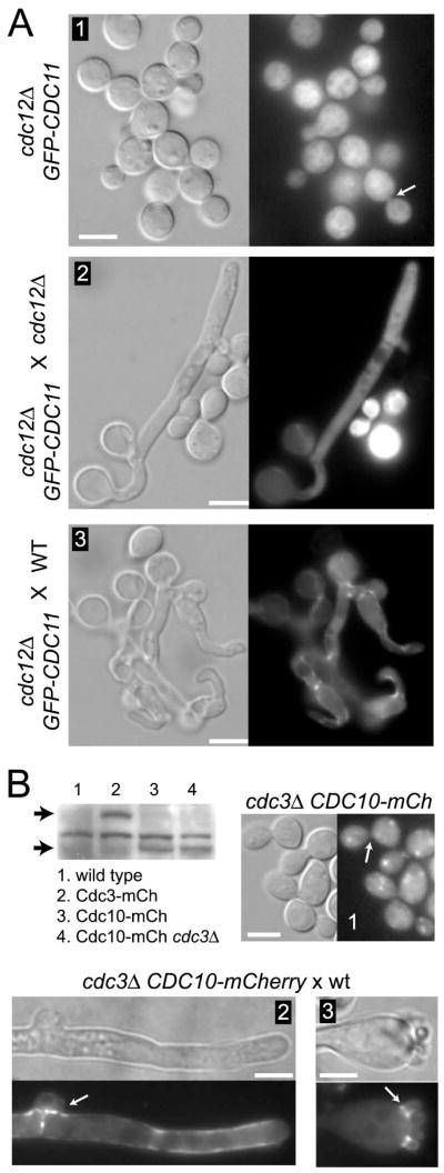
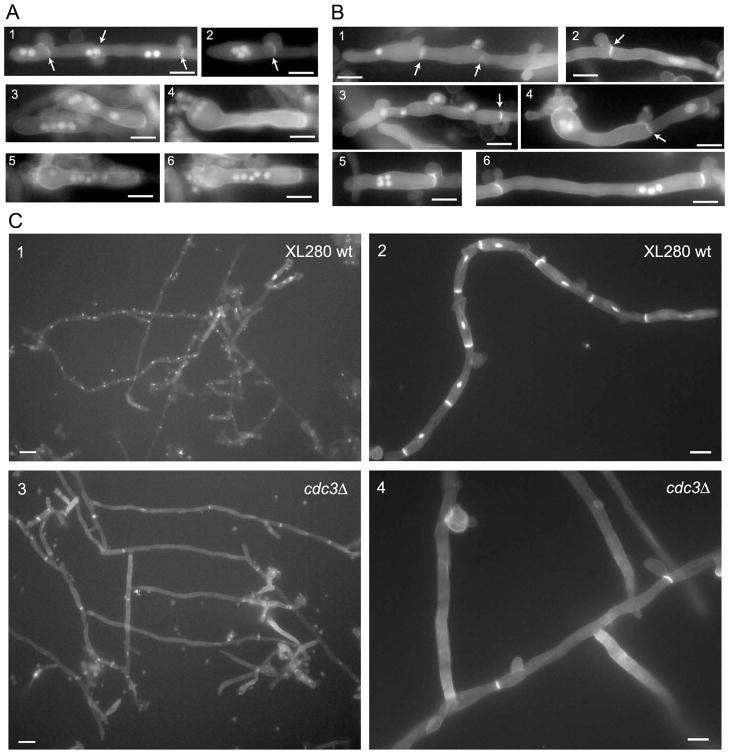
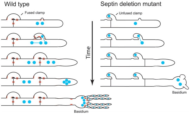
Septins are conserved, cytoskeletal GTPases that contribute to cytokinesis, exocytosis, cell surface organization and vesicle fusion by mechanisms that are poorly understood. Roles of septins in morphogenesis and virulence of a human pathogen and basidiomycetous yeast Cryptococcus neoformans were investigated. In contrast to a well-established paradigm in S. cerevisiae, Cdc3 and Cdc12 septin homologues are dispensable for growth in C. neoformans yeast cells at 24 degrees C but are essential at 37 degrees C. In a bilateral cross between septin mutants, cells fuse but the resulting hyphae exhibit morphological abnormalities, including lack of properly fused specialized clamp cells and failure to produce spores. Interestingly, post-mating hyphae of the septin mutants have a defect in nuclear distribution. Thus, septins are essential for the development of spores, clamp cell fusion and also play a specific role in nuclear dynamics in hyphae. In the post-mating hyphae the septins localize to discrete sites in clamp connections, to the septa and the bases of the initial emerging spores. Strains lacking CDC3 or CDC12 exhibit significantly reduced virulence in a Galleria mellonella model of infection. Thus, C. neoformans septins are vital to morphology of the hyphae and contribute to virulence.


Figures

Figure 1
Figure 1
Phylogenetic tree showing septin proteins from C. neoformans and other fungi: Cn – Cryptococcus neoformans (bold), Cc – Coprinopsis cinerea, Um – Ustilago maydis, Sc – Saccharomyces cerevisiae, An – Aspergillus nidulans. Shaded areas divide proteins into four distinct groups represented by Cdc3, Cdc10, Cdc11, and Cdc12 in S. cerevisiae. Currently, there is no strong evidence that Cns5 is a septin. The scale bar indicates a branch length corresponding to 1 amino-acid substitution/site.
Figure 2
Figure 2
Septin mutants are not viable at 37°C, possess a defect in septation, and exhibit sensitivity to cell wall damaging agents. (A) Serial dilutions of septin deletion strains (Liu et al., 2008) were spotted on YPD and on YPD with 1 M sorbitol and grown for 2 days at the indicated temperatures. B. Strains from A were grown in YPD liquid medium at either 24°C or 37°C and the morphology was evaluated using DIC microscopy. (C) Strains from A were grown on either an YPD medium or on YPD medium supplemented with 1 M sorbitol at 37°C for two days. Next, the plates were shifted to room temperature and examined over 5 days. (D) Septin deletion strains generated in this study were examined for growth on medium supplemented with 0.06% SDS (cdc3Δ) and on medium supplemented with 0.5 mg/ml caffeine (cdc3Δ and cdc12Δ). Cells were grown at either room temperature or 30°C (SDS medium only). (E) The wild type cells (KN99a, panels 1, 2, 3) and the cdc3Δ cdc12Δ double mutant (panels 4, 5, 6) were examined with TEM. CW, cell wall; PS, primary septum; SS, secondary septum. Panels 3 and 6 are the enlargements of panels 2 and 5, respectively. (F) Nuclei (DAPI) and the chitin (calcofluor white) were examined in the wild type strain (KN99a) and in the cdc12Δ mutant at three temperatures as indicated. Arrows indicate cells with multiple nuclei. See text for details. Scale bars represent 5 μm.
Figure 3
Figure 3
Septin mutant strains are less virulent in a heterologous host. An α cdc3Δ mutant strain was injected into larvae of Galleria mellonella. The larvae were incubated at 24°C to eliminate the influence of the non-permissive temperature. A significant (p < 0.01) decrease in virulence was observed for the α cdc3Δ mutant strain as compared to wild type (α H99) strain. (B) The a cdc12Δ mutant and the a cdc3Δ cdc12Δ double mutant were significantly less virulent than the wild type a KN99 strain (p = 0.005, p = 0.023) and not virulent when compared to the PBS control (p = 0.57, p = 0.3).
Figure 4
Figure 4
(A) Cdc10-mCherry was visualized in yeast cells. An arrow shows a fluorescent patch signal in an unbudded cell, and the arrowhead shows a septin ring that is split during cytokinesis (B–F) Localization of Cdc10-mCherry in dikaryotic post-mating hyphae. A strain expressing Cdc10-mCherry was crossed to a strain of opposite mating type that was wild type or also expressed Cdc10-mCherry. (B) Initial zygotes were examined after 2 days of mating. Cdc10-mCherry was expressed by mating type a (panel 2), or both mating types (panel 1 and C, D, E). (C) Cdc10-mCherry accumulates on the curvatures in the cortex of “pioneer” hypha (indicated by arrows). (D) Cdc10-mCherry localizes to the clamp cell. Panel 1 shows a hypha containing two clamp cells, one of which (indicated by white outline) is magnified in panel 2. Septa formed just beneath the clamp cell and between the clamp cell and the hypha are clearly seen in the DIC image (panel 1). Cdc10-mCherry accumulates on both sides of septa as two puncta in the center. It also outlines the point of fusion between the hypha and the clamp cell (See also Figure 9). Cdc10-mCherry localizes to the base of the basidium (panel 3, arrow), medial point in the septum (panel 3, arrowhead), and to the septa at points of constriction of the hypha (panel 4). (E) Cdc10-mCherry is visible as a ring in the center of the hyphal septum. (F) A strain expressing Cdc10-mCherry was crossed to a strain expressing GFP-βTub. Cdc10-mCherry localizes to the bases of emerging spores on the basidium (panels: 2, 4, 6) but not to the connection points between individual spores (panel 6). Localization of GFP-βTub is shown in panel 3. Scale bars represent 5 μm.
Figure 5
Figure 5
Septins occasionally co-localize with microtubules in the dikaryotic hyphae. (A) Two opposite mating type strains expressing Cdc10-mCherry were crossed. Septin filaments spanning the hypha were observed in a small subset of post-mating hyphae. (B and C) A strain expressing Cdc10-mCherry was crossed to a strain expressing GFP-βTub. A rare co-localization of Cdc10-mCherry and GFP-βTub was observed in parts of hyphae (demarcated in B merged image) and in basidia with large vacuoles (C). Scale bars represent 5 μm.
Figure 6
Figure 6
Cdc3 and Cdc12 are essential for proper morphology of dikaryotic hyphae. (A) A bilateral cross between two cdc3Δ mutant strains was evaluated for filamentation on either MS or V8 media and compared to the H99α x KN99a cross. A severe defect in filamentation of cdc3Δ was observed on V8 medium and only a moderate defect on MS medium. In the cdc3Δ mutant cross, the number of fused clamp cells was reduced in filaments formed on the MS medium (A, bottom 2 panels). (B) Basidia formed by the wild type cross were decorated by chains of spores (B, top), while none of the basidia formed by septin mutants had spore chains (B, bottom). (C–D) Scanning Electron Microscopy was performed to analyze hyphae from bilateral crosses between cdc12Δ mutants and a control cross between cdc12Δ and the wild type H99 strain. Hyphae from cdc12Δ x H99 cross were rich in basidia decorated with chains of spores (C), whereas cdc12Δ x cdc12Δ hyphae lacked spore chains (D). The majority of clamp cells from the cross with the wild type strain were fused (E, panel 1, arrows). In contrast most clamp cells from the cross with the cdc12Δ mutant were not fused (E, panel 3, arrows, fused clamp cell is indicated by an arrowhead) including clamp cells that were formed next to basidia (panels 2 and 4, arrows). Unlike wild type basidia (F, panel 1), most basidia from the cdc12Δ mutant cross had aberrant budded protrusions including atypical numbers (F, panels 2 and 4) and aberrant morphology (F, panel 3). Bars are 5 μm (A, B), or as indicated (C–F).
Figure 7
Figure 7
In the absence of Cdc3 or Cdc12 localization of other septins is disrupted. (A) In the cdc12Δ GFP-CDC11 mutant strain (LK177), GFP-Cdc11 is not localized to the bud neck (panel 1, arrow). When the strain LK177 is crossed to a cdc12Δ mutant, GFP-Cdc11 reveals a diffuse signal throughout the cytoplasm of the dikaryotic hyphae (panel 2). In contrast, when the strain LK177 is crossed to a wild-type, GFP-Cdc11 shows a wild type localization (panel 3). (B) Cdc10-mCherry is expressed in a cdc3Δ mutant strain as shown in the western blot but does not localize to the bud neck (panel 1, arrow). In the western blot, the two arrows indicate Cdc3-mCherry (lane 2) and Cdc10-mCherry (lane 3) proteins migrated near their predicted sizes (~81 kDa and ~64 kDa). The localization of Cdc10-mcherry is “rescued” when the cdc3Δ CDC10-mCherry mutant is crossed to a wild type strain (panel 2 and 3). Scale bars represent 5 μm.
Figure 8
Figure 8
Septins participate in nuclear distribution in hyphae. (A and B) Hyphae from a wild type α H99 x a KN99 cross (A) or from crosses between septin mutant strains (B, α cdc3Δ x a cdc3Δ, panels: 1, 4, and 6; α cdc3Δ cdc12Δ x a cdc3Δ cdc12Δ, panels: 2, 3, and 5) were stained with DAPI and calcofluor white to visualize nuclei and septa. (B) Dikaryotic hyphae from the crosses between septin deletion mutants have aberrant nuclear distribution. (C) Nuclei and septa were examined in hyphae resulting from monokaryotic fruiting of either a reference XL280 strain or a congenic cdc3Δ strain. See text for detailed description. Scale bars represent 5 μm (A, B and C panels 2 and 4) or 10 μm (C panels 1 and 3).
Figure 9
Figure 9
(A) A model showing localization of septins in the clamp cell and the basidium of the dikaryotic post-mating hyphae. Hypothetically septins mark the initial site of clamp cell formation. Subsequently, septins localize to the base of the outgrowing clamp cell. Possibly septins could also localize to the tip of the clamp cell as well as to the peg, which is a small “bud” growing from the hypha towards the tip of the clamp cell (Badalyan et al., 2004). Septins accumulate on both sides in the middle of the hyphal septum and the septum, which is formed between the hypha and the clamp cell. Septins mark the base of the basidium and form rings at the basal regions of the first set of four budded protrusions that subsequently give rise to four chains of spores. Whereas localizations to the septa and at points of fusion between clamp cells and the hypha appear stable, accumulation of septin complexes at bases of the basidia and initial budded protrusions are transient and likely happen during early stages of the development of the basidium. (B) Hypothetical model showing one possible scenario of defects in nuclear distribution and morphology in post-mating hypae of a septin mutant.

References

    1. Adams TH, Wieser JK, Yu JH. Asexual sporulation in Aspergillus nidulans. Microbiol Mol Biol Rev. 1998;62:35–54. - PMC - PubMed
    1. Alspaugh JA, Perfect JR, Heitman J. Cryptococcus neoformans mating and virulence are regulated by the G-protein alpha subunit GPA1 and cAMP. Genes Dev. 1997;11:3206–3217. - PMC - PubMed
    1. An H, Morrell JL, Jennings JL, Link AJ, Gould KL. Requirements of fission yeast septins for complex formation, localization, and function. Mol Biol Cell. 2004;15:5551–5564. - PMC - PubMed
    1. Badalyan SM, Polak E, Hermann R, Aebi M, Kues U. Role of peg formation in clamp cell fusion of homobasidiomycete fungi. J Basic Microbiol. 2004;44:167–177. - PubMed
    1. Bahn YS, Kojima K, Cox GM, Heitman J. Specialization of the HOG pathway and its impact on differentiation and virulence of Cryptococcus neoformans. Mol Biol Cell. 2005;16:2285–2300. - PMC - PubMed

Publication types

MeSH terms