. 2009 Nov 28:2:37.
doi: 10.1186/1756-6606-2-37.

Critical involvement of Rho GTPase activity in the efficient transplantation of neural stem cells into the injured spinal cord

Affiliations

Critical involvement of Rho GTPase activity in the efficient transplantation of neural stem cells into the injured spinal cord

Fujiki Numano et al. Mol Brain. .

Abstract

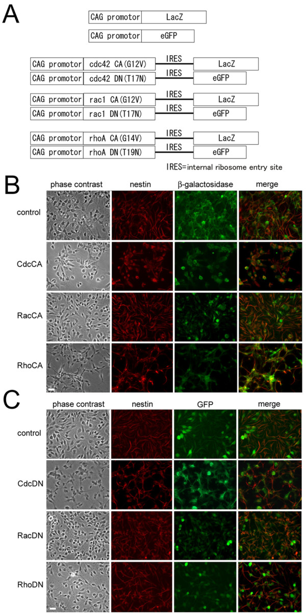
Background: Transplantation of neural stem/progenitor cells is a promising approach toward functional restoration of the damaged neural tissue, but the injured spinal cord has been shown to be an adverse environment for the survival, migration, and differentiation of the donor cells. To improve the efficiency of cell replacement therapy, cell autonomous factors in the donor cells should be optimized. In light of recent findings that Rho family GTPases regulate stem cell functions, genetic manipulation of Rho GTPases can potentially control phenotypes of transplanted cells. Therefore we expressed mutant forms of Rho GTPases, Rac, Rho, and Cdc42, in the neural stem/progenitor cells and examined their survival and migration after transplantation.

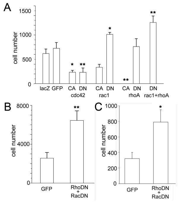
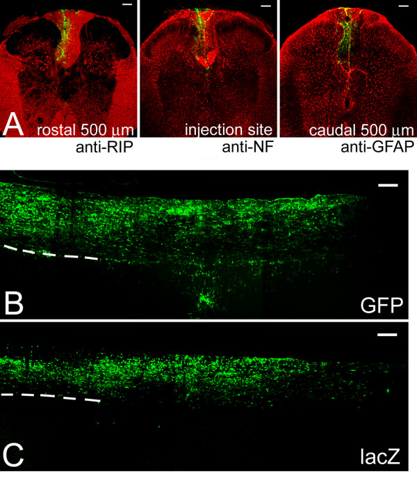
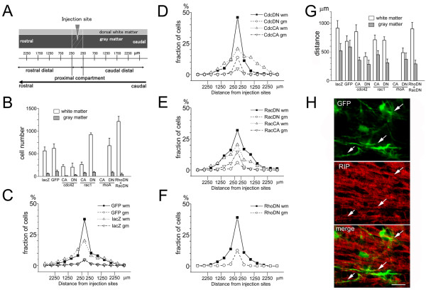
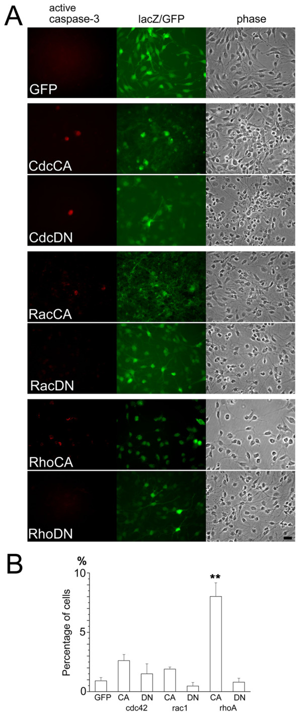
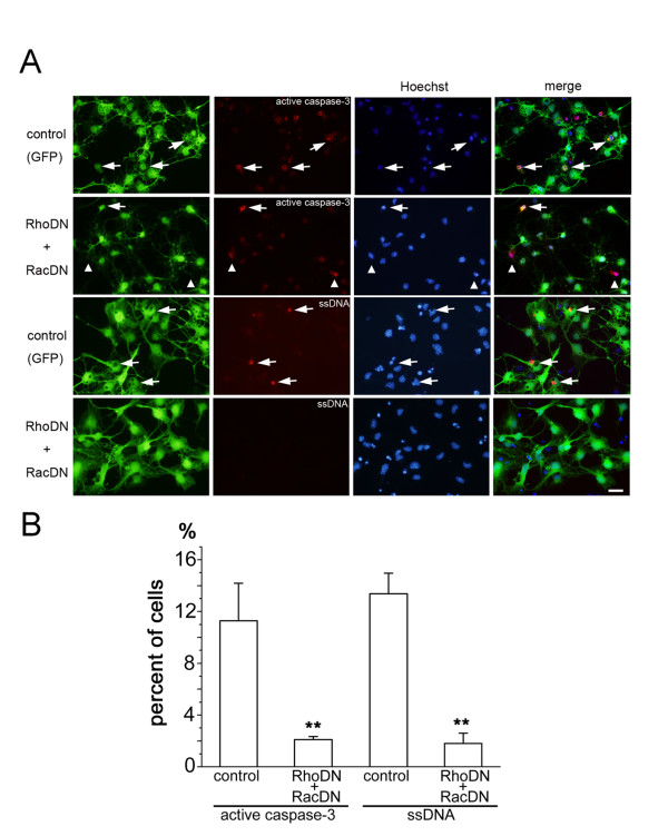
Results: Manipulation of the individual Rho GTPases showed differential effects on survival, with little variation in their migratory route and predominant differentiation into the oligodendroglial lineage. Combined suppression of both Rac and Rho activity had a prominent effect on promoting survival, consistent with its highly protective effect on drug-induced apoptosis in culture.

Conclusion: Manipulation of Rac and Rho activities fully rescued suppression of cell survival induced by the spinal cord injury. Our results indicate that precise regulation of cell autonomous factors within the donor cells can ameliorate the detrimental environment created by the injury.


Figures

Figure 1
Figure 1
Transplantation of NSPCs into the injured spinal cord. (A) Montages of confocal microscopic images of the intact and injured spinal cords immunostained with anti-GFP antibody to detect the distribution of the transplanted NSPCs. The sites of spinal cord transection (arrowhead) are clearly separated from the sites of transplantation of NSPCs (arrows). The fixed spinal cord sections were stained with both anti-GFP antibody (green) and anti-neurofilament-200 antibody (red). Bar, 200 μm. (B) Numbers of surviving cells 7 days after transplantation in the white and gray matter. Single sections containing the highest number of GFP-positive cells were selected and the total numbers of surviving cells in these sections were taken as a representation of the extent of surviving cells. In both the intact and injured spinal cords, there was a preference of transplanted cells to reside within the white matter (intact gray matter; 83.4 ± 28.9 cells, intact white matter; 398.5 ± 151.5 cells, t-test p < 0.05, injured gray matter; 8.2 ± 2.8 cells, injured white matter; 171.5 ± 28.5 cells, t-test p < 0.05). (C) Numbers of surviving cells 7 days after transplantation in the total area of the spinal cord sections. The difference between injured and intact spinal cords was statistically significant (intact; 481.9 ± 173.3 cells, injured; 179.7 ± 28.6 cells, t-test p < 0.05). (D) Normalized rostrocaudal distribution of transplanted cells in the intact and injured spinal cords. The proportion of transplanted cell numbers in each 500 μm section along the rostrocaudal axis was plotted. The difference of the cell distribution was statistically significant (Mann-Whitney-U test, p < 0.05).
Figure 2
Figure 2
Expression of mutated Rho GTPases in NSPCs. (A) Design of expression constructs of recombinant adenoviruses. DNA fragments coding three CA forms of Rho GTPases (G12V mutant of cdc42, G12V mutant of rac1, G14V mutant of rhoA) were placed under the control of a CAG promoter in combination with the reporter gene lacZ. DNA fragments coding three DN forms of Rho GTPases (T17N mutant of cdc42, T17N mutant of rac1, T19N mutant of rhoA) were also placed under the control of a CAG promoter in combination with the reporter gene GFP. Intervening internal ribosomal entry sites (IRES) support the expression of reporter genes lacZ and GFP in these constructs. (B and C) Infection of cultured NSPCs with recombinant adenoviruses of CA forms (B) or DN forms (C) of Rho GTPases. Nestin-positive NSPCs expressed comparative amounts of β-galactosidase/GFP 2 days after infection. Bar, 20 μm.
Figure 3
Figure 3
Transplantation of NSPCs expressing mutant forms of Rho GTPases. (A) Number of surviving cells in sections of spinal cords 7 days after transplantation. After transplantation of CA forms of cdc42 and rhoA, numbers of the surviving cells were significantly lower than the control cells expressing only lacZ (LacZ-positive control; 620.0 ± 90.9 cells, CdcCA; 235.6 ± 38.5 cells (p < 0.05), RacCA; 317.3 ± 60.5 cells, RhoCA; 0.0 cells (ANOVA, p < 0.01)). Although expression of the DN form of cdc42 reduced the number of surviving cells, expression of the DN form of rac1 increased the number of surviving cells (GFP-positive control; 723.3 ± 117.6 cells, CdcDN; 232.8 ± 88.8 cells (p < 0.01), RacDN; 1013.6 ± 39.7 cells (ANOVA, p < 0.05), RhoDN; 760.8 ± 162.9 cells). Double infection of adenoviruses expressing RacDN and RhoDN further enhanced the survival of transplanted NSPCs (1258.5 ± 133.6 cells (ANOVA, p < 0.01 in comparison with two types of control conditions)). (B) Increase in the total number of surviving NSPCs by expression of DN forms of both Rac and Rho. Complete serial sections of the transplanted spinal cords were examined and the total numbers of GFP-positive cells in two conditions were counted. A significant increase in surviving cells was observed in the condition of infecting two DN forms of Rho GTPases (control; 2565.2 ± 583.9 cells, RhoDN and RacDN; 6439 ± 990.6 cells, t-test p < 0.01). (C) Number of surviving cells in sections of spinal cords three weeks after transplantation. Double infection of adenoviruses expressing RacDN and RhoDN enhanced the survival of transplanted NSPCs (RacDN and RhoDN; 791.6 ± 156.9 cells, GFP-positive control; 320.8 ± 80.4 cells, t-test p < 0.05).
Figure 4
Figure 4
Migration pattern of NSPCs expressing DN forms of both Rho and Rac after transplantation into the intact spinal cords. (A) Distribution of the transplanted NSPCs expressing both RhoDN and RacDN at three different levels of the spinal cord. The sections were immunostained with specific markers for oligodendrocytes (RIP), neuronal axons (NF; neurofilament-200 antibody), and astrocytes (GFAP; anti-glial fibrillary acidic protein antibody). Transplanted cells are restricted to the dorsal white matter, where strong immunoreactivity of RIP and NFs was observed. Bar, 100 μm. (B and C) Rostrocaudal distribution of NSPCs expressing GFP (B) and LacZ (C) after spinal cord transplantation detected by immunostaining with anti-GFP antibody (B) or anti-β-galactosidase antibody (C). The dashed lines indicate the border between the white matter and the gray matter. Preferential localization of the immunoreactive cells in the white matter is evident. Bar, 200 μm.
Figure 5
Figure 5
Quantitative analysis of the migration pattern of NSPCs expressing mutant forms of Rho GTPases after transplantation. (A) Schematic representation of the compartments defined by their distance from the injection site. Cells were grouped by their localization either within the white matter or the gray matter. Spinal cords were further subdivided into the proximal compartment, rostral distal compartment, and caudal distal compartment. The proximal compartment is the area within 250 μm from the injection sites. (B) Number of cells in the white matter or the gray matter. Preferential localization of transplanted cells in the white matter was observed in all NSPCs expressing CA or DN forms of Rho GTPases, except NSPCs expressing RhoCA. No surviving cells were detected in transplantation of NSPCs expressing RhoCA. (C-F) Migratory profiles of transplanted NSPCs expressing reporter genes (C) or mutant forms of Rho GTPases cdc42 (D), rac1 (E), and rhoA (F). In spite of the differences in the total number of surviving cells, the overall profiles of cell migration were similar in cells expressing mutant Rho GTPases. (G) Mean migratory distance of surviving NSPCs 7 days after transplantation. Cells expressing mutant forms of Rho GTPases showed a similar extent of migration along the rostrocaudal axis except for RhoCA. (H) Morphology and differentiated phenotype of transplanted cells expressing both RhoDN and RacDN. Ramified processes extended from the GFP-positive cell bodies and these structures were immunopositive with an oligodendrocyte marker RIP (arrows). Bar, 20 μm.
Figure 6
Figure 6
Differential responses of NSPCs expressing mutated forms of Rho GTPases after withdrawal of bFGF. (A) Anti-activated caspase-3 immunostaining of cultured NSPCs infected with recombinant adenoviruses expressing mutated forms of Rho GTPases. Activated caspase-3 immunoreactivity was observed in a small fraction of NSPCs (red). Similar extent of adenovirus infection was confirmed by immunostaining with anti-β-galactosidase antibody (green; CA mutations) or anti-GFP antibody (green; DN mutations). Bar, 20 μm. (B) Quantification of cells immunopositive with anti-activated caspase-3. Percentage of immunopositive cells is presented. There was a significant increase of cell death in cells expressing RhoCA (GFP control; 0.9 ± 0.3%, CdcCA; 2.6 ± 0.5%, CdcDN; 1.5 ± 0.6%, RacCA; 1.9 ± 0.2%, RacDN; 0.5 ± 0.3%, RhoCA; 8.0 ± 1,1% (ANOVA, p < 0.01), RhoDN; 0.8 ± 0.3%).
Figure 7
Figure 7
Effects of suppressing Rho and Rac activity in staurosporine-induced cell death. (A) Anti-activated caspase-3 and anti-ssDNA immunostaining of cultured NSPCs infected with recombinant adenoviruses expressing both RhoDN and RacDN. Higher proportions of anti-activated caspase-3 or ssDNA immunopositive cells (red) were observed in the control condition (arrows). Several GFP-negative cells expressed active caspase-3 (arrow heads). Cells were counterstained with Hoechst 33342 dye to visualize nuclei (blue). Bar, 20 μm. (B) Quantification of immunopositive cells for anti-activated caspase-3 or anti-ssDNA antibodies colocalized with GFP. Reduction of immunopositive cells was statistically significant (anti-activated caspase-3; p < 0.01, anti-ssDNA; p < 0.01, Mann-Whitney test).
Figure 8
Figure 8
Enhancement of cell survival in the injured spinal cord by suppression of Rho and Rac activity. (A) Distribution of transplanted stem cells expressing both RhoDN and RacDN 7 days after transplantation. Cells expressing both RhoDN and RacDN (green) survived in the injured spinal cord (arrow). The sections were stained with anti-neurofilament-200 antibody (red). Arrowhead indicates the site of transection. Bar, 200 μm. (B) Quantification of cells present in the injured spinal cords 7 days after transplantation of NSPCs expressing RhoDN and RacDN. There was a significant increase of surviving cell numbers after transplantation of DN forms of Rho and Rac (control; 179.7 ± 28.6 cells (n = 6), RhoDN and RacDN; 420.6 ± 72.2 cells (n = 5), t-test p < 0.01).

Similar articles

Cited by

References

    1. Gage FH. Mammalian neural stem cells. Science. 2000;287:1433–1438. doi: 10.1126/science.287.5457.1433. - DOI - PubMed
    1. McKay RD. Stem cell biology and neurodegenerative disease. Philos Trans R Soc Lond B Biol Sci. 2004;359:851–856. doi: 10.1098/rstb.2004.1472. - DOI - PMC - PubMed
    1. Johe KK, Hazel TG, Muller T, Dugich-Djordjevic MM, McKay RD. Single factors direct the differentiation of stem cells from the fetal and adult central nervous system. Genes Dev. 1996;10:3129–3140. doi: 10.1101/gad.10.24.3129. - DOI - PubMed
    1. Okabe S, Forsberg-Nilsson K, Spiro AC, Segal M, McKay RD. Development of neuronal precursor cells and functional postmitotic neurons from embryonic stem cells in vitro. Mech Dev. 1996;59:89–102. doi: 10.1016/0925-4773(96)00572-2. - DOI - PubMed
    1. Brustle O, Jones KN, Learish RD, Karram K, Choudhary K, Wiestler OD, Duncan ID, McKay RD. Embryonic stem cell-derived glial precursors: a source of myelinating transplants. Science. 1999;285:754–756. doi: 10.1126/science.285.5428.754. - DOI - PubMed

Publication types

MeSH terms