. 2009 Dec;85(6):890-6.
doi: 10.1016/j.ajhg.2009.11.008.

Loss-of-function mutations in the human ortholog of Chlamydomonas reinhardtii ODA7 disrupt dynein arm assembly and cause primary ciliary dyskinesia

Affiliations

Loss-of-function mutations in the human ortholog of Chlamydomonas reinhardtii ODA7 disrupt dynein arm assembly and cause primary ciliary dyskinesia

Philippe Duquesnoy et al. Am J Hum Genet. 2009 Dec.

Abstract

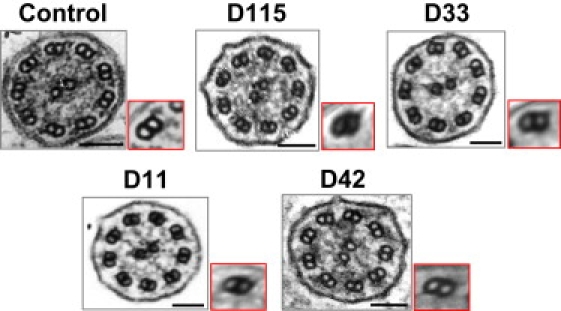
Cilia and flagella are evolutionarily conserved structures that play various physiological roles in diverse cell types. Defects in motile cilia result in primary ciliary dyskinesia (PCD), the most prominent ciliopathy, characterized by the association of respiratory symptoms, male infertility, and, in nearly 50% of cases, situs inversus. So far, most identified disease-causing mutations involve genes encoding various ciliary components, such those belonging to the dynein arms that are essential for ciliary motion. Following a candidate-gene approach based on data from a mutant strain of the biflagellated alga Chlamydomonas reinhardtii carrying an ODA7 defect, we identified four families with a PCD phenotype characterized by the absence of both dynein arms and loss-of-function mutations in the human orthologous gene called LRRC50. Functional analyses performed in Chlamydomonas reinhardtii and in another flagellated protist, Trypanosoma brucei, support a key role for LRRC50, a member of the leucine-rich-repeat superfamily, in cytoplasmic preassembly of dynein arms.


Figures

Figure 1
Figure 1
LRRC50 Molecular Defects in Patients with PCD (A) Exonic organization of the human LRRC50 cDNA (top) and domain-organization model of the corresponding protein (bottom), on which are shown the mutations identified in the four families described in this study. The 12 exons are indicated by empty or hashed boxes, depicting translated or untranslated sequences, respectively. “CC,” coiled-coil domain; “Pro-rich,” proline-rich domain. (B) Mutations identified in families D115, D33, D11, and D42. The expected consequences of these sequence abnormalities (circled), which are detailed in the text, are shown. The intragenic deletion (del) found in patient D42_II1 (P) involves exons 2 and 3 and flanking intronic sequences, as identified through long-range PCR amplifications (right; C stands for control) followed by direct sequencing (bottom). The evolutionary conservation of the deleted exons is shown (dashed frame).
Figure 2
Figure 2
Function of LRRC50, as Inferred from Cross-Species Studies (A) Evolutionary conservation of the third LRR domain (conserved residues defining the LRR consensus are in bold). Leucine 175 is shown in yellow. (B) Predicted structure of the LRR region of CrODA7. Side chains are indicated for three rows of leucines that line the center of this beta barrel; Leu92 (the equivalent of human Leu175) is indicated (yellow arrow). Red, alpha-helix; blue, beta-sheet; green, turn; gray, random coil. (C) CrODA7 is primarily cytoplasmic, and requires Leu92 for its function. C1: Expression of HA-tagged ODA7 proteins in the oda7-1 mutant background is shown by a blot of whole cell protein probed with anti-HA. Tubulin is shown as a loading control. The beat frequency (Hz) of each strain, measured by strobe analysis, is shown below each lane. C2: The relative abundance of ODA7 is shown by blots of stoichiometric amounts of whole cells (WC) compared with deflagellated cell bodies (CB) and flagella (FL). More ODA7 is present in cell bodies than in a 50-fold excess of flagella. Other proteins for comparison include a chloroplast protein (cyt f) that localizes, therefore, only to the cytoplasm, an intraflagellar transport protein (IFT46) that is present in both the cytoplasm and the flagella, and an ODA subunit (IC2) that localizes mainly to the flagella. Data for blots of cell fractions probed with anti-IFT46, anti-cyt f, and anti-IC2 appeared in a previous publication, and an identical blot prepared from the same samples was probed with polyclonal anti-ODA7. C3: Rescue of ODA assembly is shown by a blot of flagellar proteins probed with anti-IC2. Dynein assembly is supported by the L92 (WT) and the L92A, but not the L92R-tagged product; very low levels of IC2 are present in flagella of the L92D strain (see text). Inner-row dynein subunit IC140 is shown as a loading control. (D) TbODA7 localizes to the cell body and is required for ODA formation. Upper panel: left, GFP signal for GFP:TbODA7; right, flagellum labeled with an anti-PFR2 marker antibody (red). Lower panel: left, DAPI staining merged with the phase-contrast picture; right, TbDNAI1, a component of ODAs, is still produced in TbODA7RNAi-induced cells but fails to be correctly incorporated into the flagellar skeleton. T, total; S, soluble fraction; C, cytoskeleton (detergent-resistant fraction). The PFR2 protein (probed with monoclonal antibody L13D6, 1:50) and BiP (probed with rabbit polyclonal antibody, 1:1000) are shown as loading and fractionation controls of the cytoskeleton and soluble fractions, respectively. One representative experiment of four independent experiments is shown.
Figure 3
Figure 3
Electron Micrographs of Respiratory Cilia from Patients with LRRC50 Mutations, Showing the Absence of Both Dynein Arms For each patient, the ten best-defined axonemal sections were selected, and the corresponding micrographs were submitted to computational TEM (composite images framed in red) for the improvement of IDA visualization, as described previously. Scale bars represent 0.1 μm.

References

    1. Haimo L.T., Rosenbaum J.L. Cilia, flagella, and microtubules. J. Cell Biol. 1981;91:125s–130s. - PMC - PubMed
    1. Satir P., Christensen S.T. Overview of structure and function of mammalian cilia. Annu. Rev. Physiol. 2007;69:377–400. - PubMed
    1. Basu B., Brueckner M. Cilia multifunctional organelles at the center of vertebrate left-right asymmetry. Curr. Top. Dev. Biol. 2008;85:151–174. - PubMed
    1. Afzelius B.A. A human syndrome caused by immotile cilia. Science. 1976;193:317–319. - PubMed
    1. Duriez B., Duquesnoy P., Escudier E., Bridoux A.M., Escalier D., Rayet I., Marcos E., Vojtek A.M., Bercher J.F., Amselem S. A common variant in combination with a nonsense mutation in a member of the thioredoxin family causes primary ciliary dyskinesia. Proc. Natl. Acad. Sci. USA. 2007;104:3336–3341. - PMC - PubMed

Publication types