. 2010 Feb 5;397(1):80-8.
doi: 10.1016/j.virol.2009.10.046. Epub 2009 Nov 26.

Myristylation and palmitylation of HSV-1 UL11 are not essential for its function

Affiliations

Myristylation and palmitylation of HSV-1 UL11 are not essential for its function

Nicholas L Baird et al. Virology. .

Abstract

All herpesviruses encode a homolog of the herpes simplex virus type-1 UL11 tegument protein. Deletion of UL11 disrupts virus envelopment, causes capsid accumulation within the cytoplasm, and reduces virus release. UL11 requires acylation with myristate and palmitate for membrane binding, lipid raft trafficking, and accumulation at the site of virus envelopment. Thus, it was predicted that acylation of UL11 would be necessary for efficient virion production, similar to HIV-1 Gag which requires myristylation for virus production. Accordingly, recombinant viruses were created to express UL11 derivatives that are not acylated, are partially acylated, or contain foreign acylation signals. Unexpectedly, the non-acylated UL11 rescued some growth defects of a UL11-null mutant, even though the unmodified protein was unstable. Furthermore, a myristylated and palmitylated chimera did not fully rescue the null virus. These results suggest that UL11 maintains some function(s) when not membrane-bound, and the sequence context of the acylations is important for UL11 function.


Figures

FIG. 1
FIG. 1
Relocalization of the UL11-coding sequence. (A) Stepwise strategy used to move the UL11-coding sequence into the UL35 locus. A recombinant of the KOS strain of HSV-1 that contains a bacterial artificial chromosome (“BAC,” 11.5 kbp) inserted between UL37 and UL38 was used (“KOSBAC”). In mutant Δ30–96, all the nucleotides that do not overlap with the essential UL12 gene were deleted, leaving the codons for the N-terminal 29 amino acids of UL11. Next, the codons at the UL35 locus were replaced with the entire UL11-coding sequence, creating mutant U1. (B) Vero cells were infected with the indicated viruses and harvested 24 h later. Equal numbers of infected cells were lysed in sample buffer, separated by SDS-PAGE, and proteins were transferred to nitrocellulose. The indicated proteins were detected by immunoblotting with the corresponding antibodies.
FIG. 2
FIG. 2
UL11 expression and virion incorporation. Vero cells were infected with the indicated viruses, and 24 h later, extracellular virions were collected from the media by centrifugation through a 30% (w/v) sucrose cushion. The virions and infected-cell lysates were separated by SDS-PAGE and transferred to nitrocellulose. The indicated proteins were detected using corresponding antibodies. Equal numbers of infected cells were loaded with VP5, the major capsid protein, serving as the loading control. To achieve approximately equal VP5 levels for the “Media” samples, additional cells (indicated as “2×” or “4×”) were infected, and the collected media were combined.
FIG. 3
FIG. 3
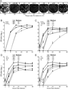
Growth properties of the recombinant viruses. (A) Plaque sizes of UL11 mutants. Confluent monolayers of Vero cells were infected with the indicated viruses, and 4 days later, the cells were stained with crystal violet and imaged. Plaque size was determined by measuring 10 randomly selected plaques and represented as a percent relative to U1. (B–E) Single-step growth curve analyses of the mutants. Vero cells were infected with indicated virus and then acid-washed to inactivate any input virus that had not fused with the cellular membrane. At the indicated times, samples were collected, and virus titers were determined by plaque assay. The results of at least two independent experiments are shown for each mutant. (B) Comparison of the wild-type KOS strain and the recombinant carrying the bacterial artificial chromosome (“KOSBAC”). (C) Comparison of mutants that lack full-length UL11 expression (“Δ30–96”) or express UL11 from the UL35 locus (“U1”). (D and E) Analysis of recombinant viruses that encode UL11-derivatives with altered acylation signals. Solid lines denote PFU associated with the cells, whereas dashed lines represent PFU released into the medium.
FIG. 4
FIG. 4
UL11 mutants. (A) Diagram of the wild-type and mutant forms of UL11 as expressed from the UL35 locus. The sites of myristylation (G) and palmitylation (CCC) are indicated with jagged lines denoting the fatty acid modifications. N-terminal extensions corresponding to the first 10 amino acids of v-Src [sCCC(−)] or Fyn (fUL11) are indicated with gray lines. Fatty-acid modifications are indicated and “+” indicates basic residues. “stop” marks the location of a 2-nucleotide change that created a stop codon in mutant Δ30–96 without altering the UL12 protein, and the dotted line represents the remaining UL11-coding sequence downstream from the introduced stop codon. The solid circle represents the GFP-tag on the C-terminus of Myr(−)GFP (B) Alignment of the UL11- and UL12-coding sequences. The two nucleotide substitution that introduces a stop codon in the UL11 sequence without altering UL12 is indicated.
FIG. 5
FIG. 5
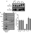
Myristylation and membrane-binding properties of UL11 derivatives. (A) Vero cells were infected with the indicated viruses and radiolabeled with either [35S]methionine/cysteine for 2.5 h or [3H]myristic acid for 30 min. All labeling periods were concluded at 9 hpi, at which time cell lysates were prepared. The UL11 derivatives were immunoprecipitated, resolved by SDS-PAGE, and visualized by autoradiography. (B) In parallel experiments, cells were labeled for 2.5 h with [35S]methionine/cysteine, scraped from the plates, and osmotically disrupted. The ability of the UL11 mutants to float to the upper regions of sucrose step-gradients during centrifugation was monitored. Representative autoradiographs are shown with the tops and bottoms of the gradients indicated. (C) Phosphorimager analysis was used to quantitate the amount of membrane-bound UL11 (top three fractions) relative to the total. The results from at least two independent experiments are shown.
FIG. 6
FIG. 6
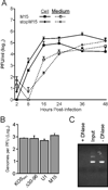
Further characterization of mutant M15. (A) The growth properties of M15 and stopM15 were compared, revealing that the 29-amino-acid, N-terminal peptide does not trans-compliment. The results from two independent experiments are shown. (B) To compare the specific infectivities of the indicated mutant virus stocks, samples were treated with DNase to eliminate any DNA that was not contained in virions, and viral DNA was subsequently purified. The numbers of genomes present in each sample were measured by quantitative PCR and normalized for PFUs. The limit of detection was 100 copies per PCR reaction. The results from at least two independent experiments are shown. (C) To ensure that the DNase treatments were effective, random PCR products were incubated with (+) or without (−) the enzyme. “Input” is the initial amount of PCR product in each sample. A representative agarose gel stained with ethidium bromide is shown.
FIG. 7
FIG. 7
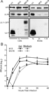
Addition of GFP stabilizes Myr(−)UL11 but does not enhance growth of the recombinant virus. Vero cells were infected with the indicated viruses. Cell and virus samples were harvested, and proteins were separated by SDS-PAGE. The indicated proteins were detected using the corresponding antibodies. (B) Single step growth curves were performed as described in the legend to Fig. 3.

Similar articles

Cited by

References

    1. Baines JD, Roizman B. The UL11 gene of herpes simplex virus 1 encodes a function that facilitates nucleocapsid envelopment and egress from cells. J. Virol. 1992;66:5168–5174. - PMC - PubMed
    1. Baird NL, Yeh PC, Courtney RJ, Wills JW. Sequences in the UL11 tegument protein of herpes simplex virus that control association with detergent-resistant membranes. Virology. 2008;374:315–321. - PMC - PubMed
    1. Britt WJ, Jarvis M, Seo JY, Drummond D, Nelson J. Rapid genetic engineering of human cytomegalovirus by using a lambda phage linear recombination system: demonstration that pp28 (UL99) is essential for production of infectious virus. J. Virol. 2004;78:539–543. - PMC - PubMed
    1. Desai P, DeLuca NA, Person S. Herpes simplex virus type 1 VP26 is not essential for replication in cell culture but influences production of infectious virus in the nervous system of infected mice. Virology. 1998;247:115–124. - PubMed
    1. Farnsworth A, Wisner TW, Johnson DC. Cytoplasmic residues of herpes simplex virus glycoprotein gE required for secondary envelopment and binding of tegument proteins VP22 and UL11 to gE and gD. J. Virol. 2007;81:319–331. - PMC - PubMed

Publication types