. 2010 Feb 1;338(1):63-73.
doi: 10.1016/j.ydbio.2009.11.025. Epub 2009 Nov 26.

miR-9a prevents apoptosis during wing development by repressing Drosophila LIM-only

Affiliations

miR-9a prevents apoptosis during wing development by repressing Drosophila LIM-only

Fernando Bejarano et al. Dev Biol. .

Abstract

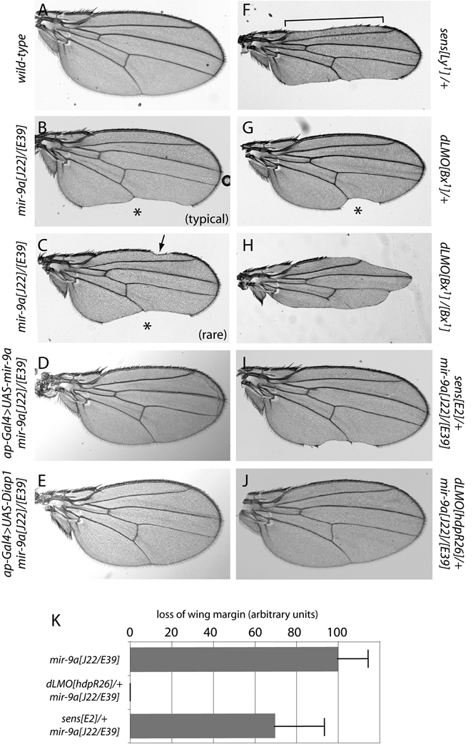
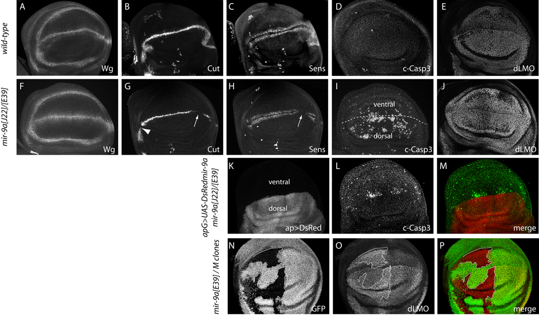
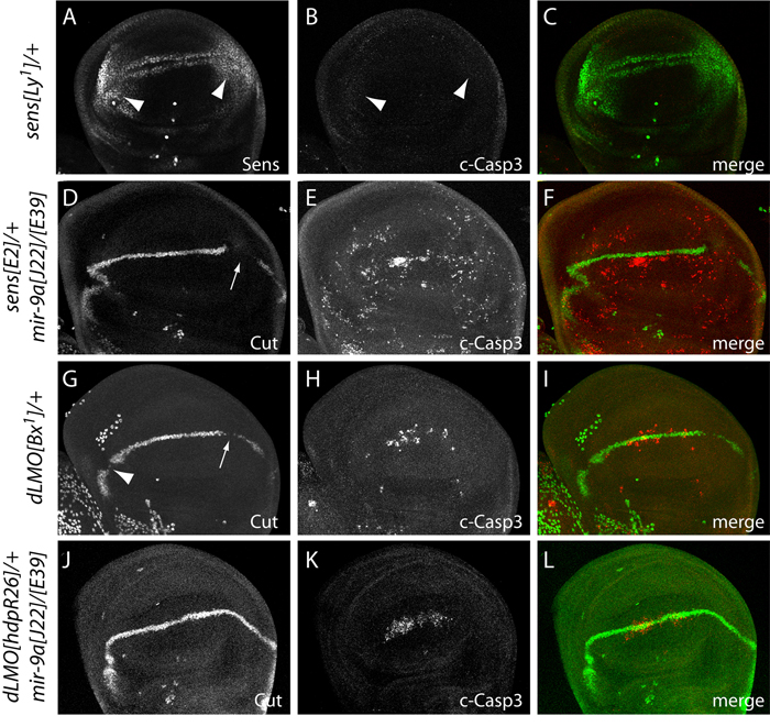
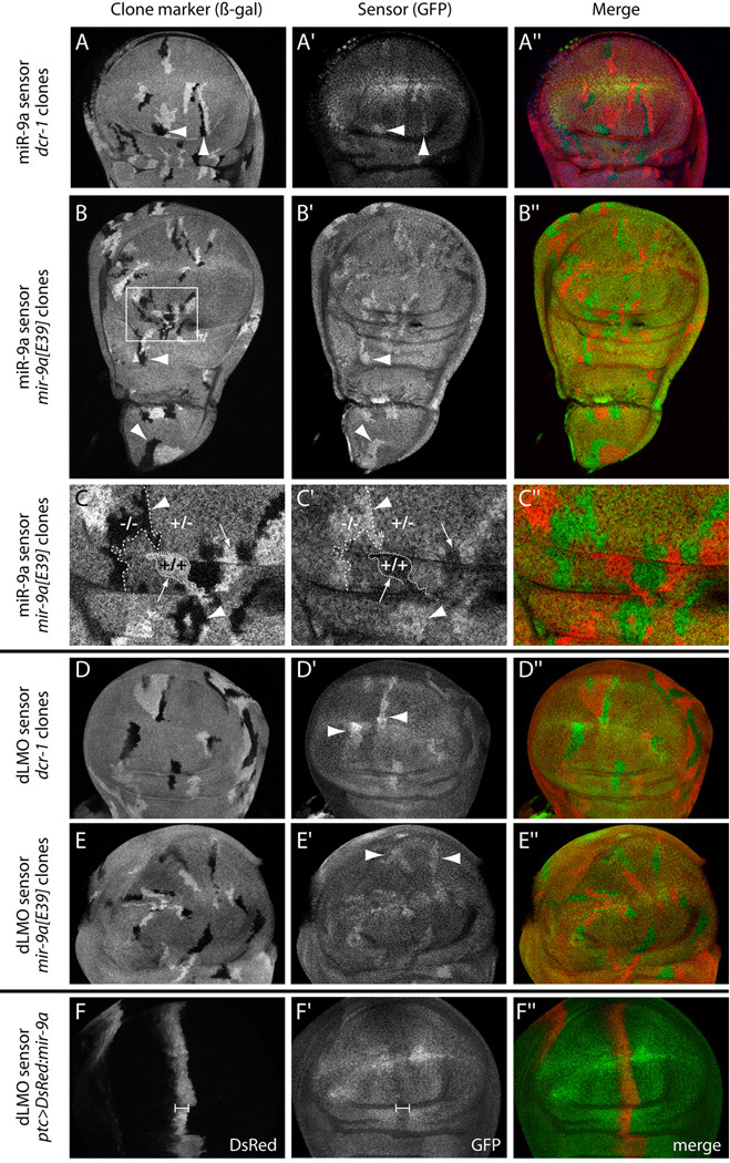
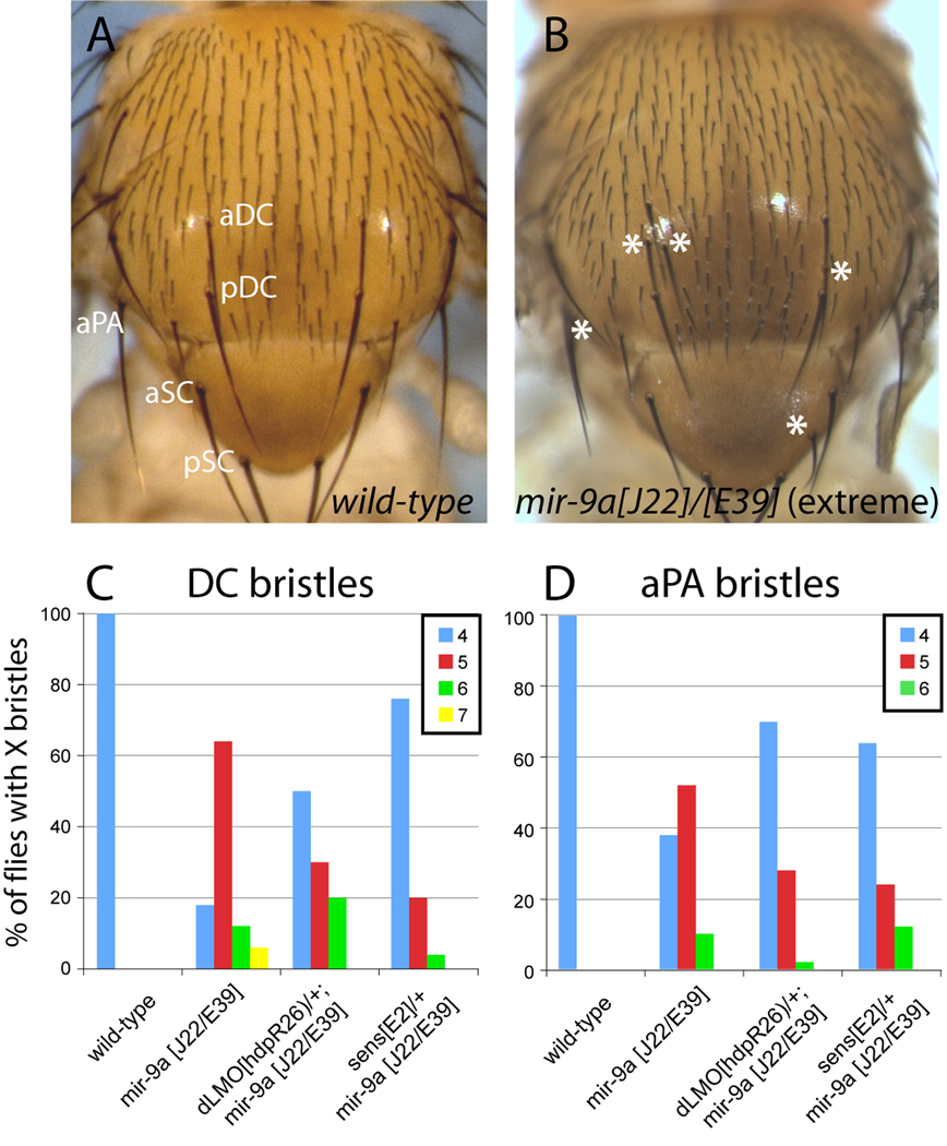
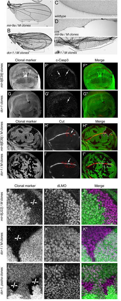
Loss of Drosophila mir-9a induces a subtle increase in sensory bristles, but a substantial loss of wing tissue. Here, we establish that the latter phenotype is largely due to ectopic apoptosis in the dorsal wing primordium, and we could rescue wing development in the absence of this microRNA by dorsal-specific inhibition of apoptosis. Such apoptosis was a consequence of de-repressing Drosophila LIM-only (dLMO), which encodes a transcriptional regulator of wing and neural development. We observed cell-autonomous elevation of endogenous dLMO and a GFP-dLMO 3'UTR sensor in mir-9a mutant wing clones, and heterozygosity for dLMO rescued the apoptosis and wing defects of mir-9a mutants. We also provide evidence that dLMO, in addition to senseless, contributes to the bristle defects of the mir-9a mutant. Unexpectedly, the upregulation of dLMO, loss of Cut, and adult wing margin defects seen with mir-9a mutant clones were not recapitulated by clonal loss of the miRNA biogenesis factors Dicer-1 or Pasha, even though these mutant conditions similarly de-repressed miR-9a and dLMO sensor transgenes. Therefore, the failure to observe a phenotype upon conditional knockout of a miRNA processing factor does not reliably indicate the lack of critical roles of miRNAs in a given setting.


Figures

Figure 1
Figure 1
Genetic interactions amongst mir-9a, senseless, and dLMO in wing morphology. Shown are wings of adult females, oriented with anterior to the top and posterior to the bottom. (A) Wild-type. (B) Transheterozygotes of mir-9a[J22]/[E39] null alleles exhibit completely penetrant notching along the posterior margin (asterisk). (C) About 10% of mir-9a[J22]/[E39] flies also show notching along the anterior wing margin (arrow). (D) Wing notching in mir-9a mutants can be rescued by reintroduction of miR-9a in the dorsal compartment using ap-Gal4. (E) Wing notching in mir-9a mutants can also be rescued by ectopic expression of Drosophila inhibitor of apoptosis 1 (Diap1). (F) Heterozygotes of the gain-of-function allele of senseless, Lyra[1], exhibit loss of both anterior (bracket) and posterior wing margin. (G) Heterozygotes of the gain-of-function allele of dLMO, Bx[1], show loss of only posterior margin (asterisk). (H) Bx[1] homozygotes exhibit loss of both anterior and posterior margin. (I) Heterozygosity for the null allele sens[E2] partially suppressed mir-9a wing notching. (J) Heterozygosity for the null allele dLMO[hdpR26] completely rescued mir-9a wing defects. (K) Quantification of wing margin loss; each bar depicts the mean and standard deviation across 50 female wings.
Figure 2
Figure 2
Cellular basis of mir-9a mutant phenotypes. All panels depict the wing pouch region of third instar wing imaginal discs stained for the indicated markers, oriented with anterior to the left, posterior to the right, ventral to the top and dorsal to the bottom. (A–E) Wildtype, (F–J) mir-9a[J22]/[E39], (K–M) ap-Gal4>UAS-DsRed-mir-9a in mir-9a[J22]/[E39], (N–P) mir-9a clones generated in hs-FLP; FRT80B M ubi-GFP/FRT80B mir-9a[J22]. The following panels are double stainings of the same tissue: B and C, G and H, F and I, K–M and N–P. (A, F) The expression of Wingless (Wg) is fairly normal in mir-9a mutants. (B, G) mir-9a mutants exhibit posterior breaks in Cut at the wing margin (arrow), and occasional anterior breaks (arrowhead). (C, H) mir-9a mutants exhibit breaks in wing margin-associated expression of Sens, particularly in the posterior compartment (arrow); the isolated stained cells are sensory organ precursors. (D, I) mir-9a mutants exhibit a high degree of apoptosis in the wing pouch as marked by cleaved Caspase-3 (c-Casp3), mostly in the dorsal compartment. (E, J) mir-9a mutants accumulate higher levels of dLMO. (K–M) Dorsal-specific expression of miR-9a suppresses the majority of the apoptotic defect of mir-9a mutants; a small amount of ectopic ventral apoptosis remains. (N–P) Clonal analysis using Minute technique demonstrates cell-autonomous elevation of dLMO in wing pouch cells lacking miR-9a.
Figure 3
Figure 3
Rescue of mir-9a phenotypes by heterozygosity for dLMO. (A–C) Strong ectopic accumulation of Sens in a heterozygote of the gain-of-function allele Ly[1] (A, arrowheads) is not associated with ectopic cell death (B, arrowheads). (D–F) Heterozygosity for the null allele sens[E2] did not rescue the Cut or apoptosis defects of the mir-9a mutant. (G–I) Female heterozygote of the dLMO gain-of-function allele Bx[1] exhibits breaks in wing margin expression of Cut and ectopic apoptosis, similar to mir-9a mutant discs (compare with Fig. 2G, I). (J–L) Female heterozygosity for the null allele dLMO[hdpR26] rescued continuity of Cut expression at the wing margin and strongly suppressed ectopic apoptosis caused by loss of mir-9a.
Figure 4
Figure 4
dLMO is a direct in vivo target of miR-9a in the Drosophila wing disc. (A–A") hs-FLP; tub-GFP-mir-9a/+; FRT82B, dcr-1[Q1147x]/FRT82B, arm-lacZ disc carrying somatic clones marked by the absence of β-galactosidase (arrowheads). dcr-1 homozygous mutant cells exhibit elevated levels of the miR-9a GFP sensor. (B–B") hs-FLP; tub-GFP-mir-9a/+; mir-9a[E39], FRT80B/arm-lacZ, FRT80B disc bearing β-gal-negative clones (arrowheads). mir-9a homozygous mutant cells exhibit increased levels of the miR-9a GFP sensor throughout the wing disc, in both the wing pouch and the presumptive notum. (C–C") Magnification of the region boxed in (B) highlights the sensitivity of the miR-9a sensor to mir-9a dosage. Homozygous mutant cells (−/−) exhibit highest sensor activity while homozygous wildtype twinspots (+/+) exhibit lowest sensor activity; the remaining heterozygous (+/−) cells express an intermediate level of miR-9a sensor. (D–D") hs-FLP; tub-GFP-dLMO 3' UTR/+, FRT82B, dcr-1[Q1147x]/FRT82B, arm-lacZ disc; dLMO sensor is upregulated in dcr-1 mutant cells. (E–E") hs-FLP; tub-GFP-dLMO 3'UTR/+; mir-9a[E39], FRT80B/arm-lacZ, FRT80B disc; dLMO sensor is upregulated in mir-9a mutant cells. (F–F") ptc-Gal4, tub-GFP-dLMO 3' UTR; UAS-DsRed-mir-9a disc; the dLMO sensor is suppressed in the domain of ectopic miR-9a (bracket).
Figure 5
Figure 5
Contribution of dLMO and Sens to ectopic neurogenesis in mir-9a mutants. (A) Notum of a wild-type animal shows an orderly array of microchaete sensory organ bristles and precise positioning of the larger macrochaete bristles; aDC and pDC: anterior and posterior dorsocentrals; aSC and pSC: anterior and posterior scutellars; aPA: anterior postalar bristle. (B) Notum of mir-9a[J22]/[E39] mutant exhibits slightly increased density of microchaete bristles and ectopic DC, aSC and aPA bristles. Note that an extreme phenotype is shown for illustration purposes; many mir-9a mutant animals exhibit no ectopic macrochaetes or only a single extra bristle across all positions. (C) Heterozygosity for sens substantially suppresses the ectopic DC phenotype of mir-9a mutants. (D) Heterozygosity for either dLMO or sens partially suppressed the ectopic aPA phenotype of mir-9a mutants. Bristle counts were performed for 50 females in each genotype.
Figure 6
Figure 6
dcr-1 and pasha clones do not phenocopy mir-9a clones. (A) Wing carrying large mir-9a[E39] clones induced with hs-FLP and the Minute technique exhibit notching along the posterior wing margin. (B) Wing carrying large dcr-1[Q1147x] mutant clones induced with hs-FLP and the Minute technique. The uneven surface of the wing prevented a flat mounting, and the asterisk indicates a section of the wing that folded over on itself. Nevertheless, no loss of margin tissue was observed. (C) Closeup of the wildtype posterior wing margin highlights a regular array of non-sensory bristles. (D) Magnification of the posterior wing margin in panel A (boxed region) illustrates substantial loss of tissue in wing bearing mir-9a clones. (E) Magnification of the posterior margin of the wing bearing dcr-1 clones in panel B (boxed region); the differentiation of posterior wing margin bristles is highly disturbed (dotted line), but the margin remained continuous. (F–L) Clonal analysis of wing pouch development. Mutant clones are marked by their absence of β-gal or GFP in the "clonal marker" channel; antigens of interest are shown in F'–L', and merged images are shown in F"–L". Panels F, G, I, K and L depict lacZ clonal markers while panels H and J used GFP markers; the choice of marker did not affect the results. The Minute technique ("M" genotype) was used in panels H, I, J and K. (F–G) Staining for cleaved caspase-3 in mir-9a and dcr-1 clones showed that both exhibit apoptotic cells. (H–I) mir-9a/M clones that overlap the middle region of the posterior wing margin reliably showed a break in Cut expression (H', arrow), whereas similarly positioned dcr-1/M clones frequently maintained Cut expression (I', arrow). (J–L) dLMO staining of ventral wing pouch clones of mir-9, dcr-1 and pasha mutant alleles; these are shown at higher magnification than panels F–I. Homozygous mutant clones are indicated by "−/− ". (J) mir-9a[E39]/M clones reliably exhibit strong upregulation of dLMO in the ventral wing pouch. (K) dcr-1[Q1147x]/M ventral wing clones do not upregulate dLMO. (L) dcr-1[Q1147x], pasha[KO] double mutant ventral clones express normal levels of dLMO.

References

    1. Aravin A, Lagos-Quintana M, Yalcin A, Zavolan M, Marks D, Snyder B, Gaasterland T, Meyer J, Tuschl T. The small RNA profile during Drosophila melanogaster development. Dev. Cell. 2003;5:337–350. - PubMed
    1. Asmar J, Biryukova I, Heitzler P. Drosophila dLMO-PA isoform acts as an early activator of achaete/scute proneural expression. Dev Biol. 2008;316:487–497. - PubMed
    1. Baek D, Villen J, Shin C, Camargo FD, Gygi SP, Bartel DP. The impact of microRNAs on protein output. Nature. 2008;455:64–71. - PMC - PubMed
    1. Bartel DP. MicroRNAs: target recognition and regulatory functions. Cell. 2009;136:215–233. - PMC - PubMed
    1. Bejarano F, Luque CM, Herranz H, Sorrosal G, Rafel N, Pham TT, Milan M. A gain-of-function suppressor screen for genes involved in dorsal-ventral boundary formation in the Drosophila wing. Genetics. 2008;178:307–323. - PMC - PubMed

Publication types

MeSH terms

LinkOut - more resources