

Efficient correction of inhomogeneous static magnetic field-induced distortion in Echo Planar Imaging
- PMID: 19944768
- PMCID: PMC2819607
- DOI: 10.1016/j.neuroimage.2009.11.044
Efficient correction of inhomogeneous static magnetic field-induced distortion in Echo Planar Imaging
Abstract
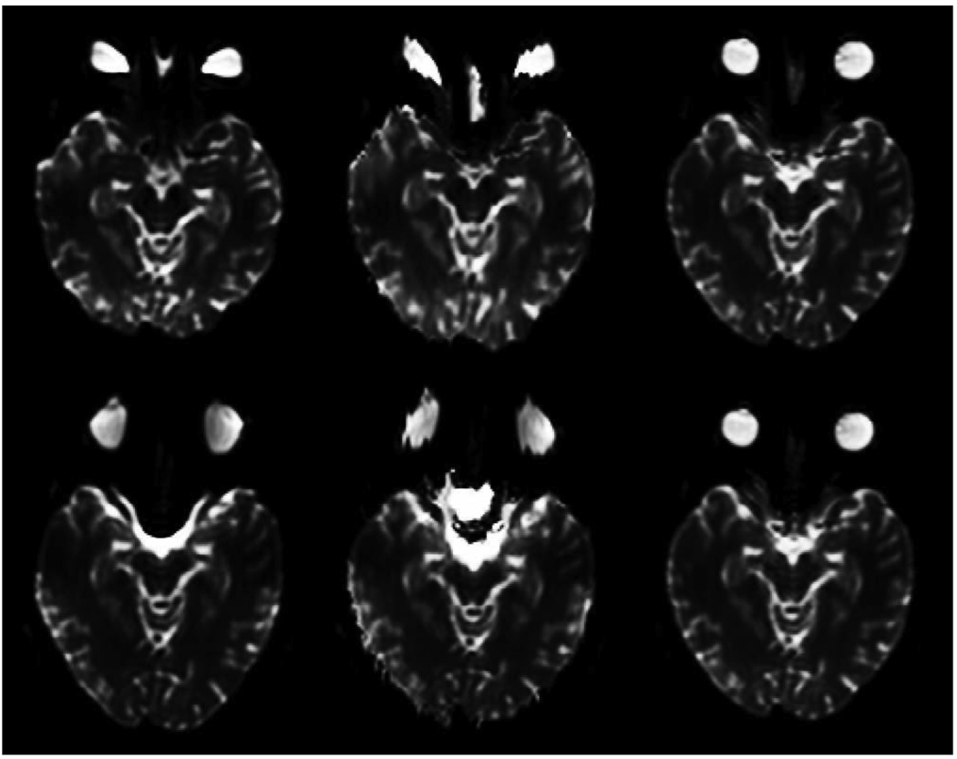
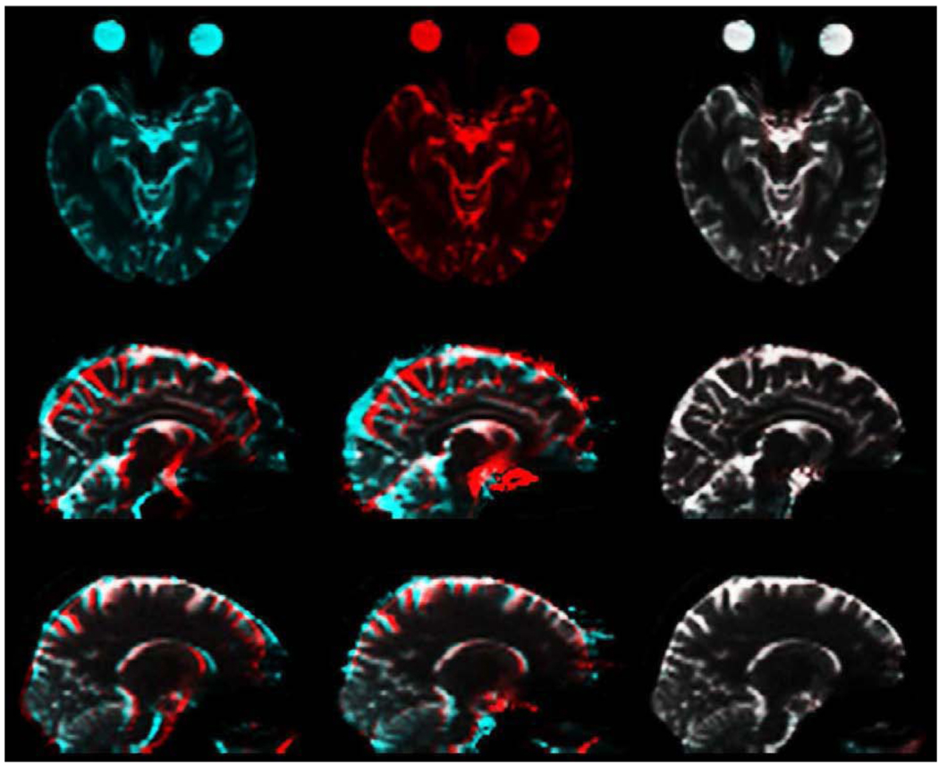
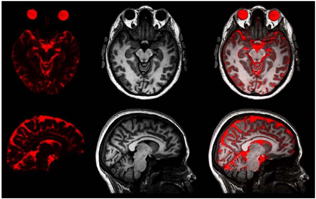
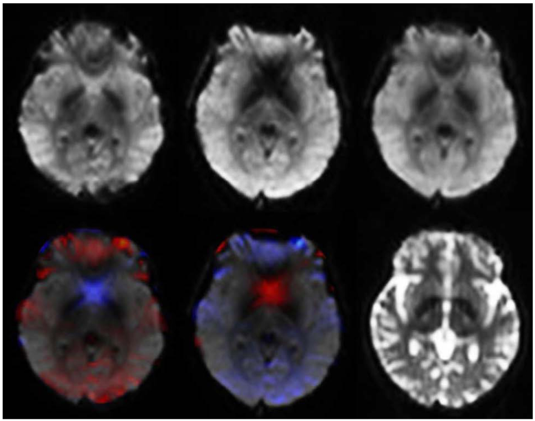
Single-shot Echo Planar Imaging (EPI) is one of the most efficient magnetic resonance imaging (MRI) acquisition schemes, producing relatively high-definition images in 100 ms or less. These qualities make it desirable for Diffusion Tensor Imaging (DTI), functional MRI (fMRI), and Dynamic Susceptibility Contrast MRI (DSC-MRI). However, EPI suffers from severe spatial and intensity distortion due to B(0) field inhomogeneity induced by magnetic susceptibility variations. Anatomically accurate, undistorted images are essential for relating DTI and fMRI images with anatomical MRI scans, and for spatial registration with other modalities. We present here a fast, robust, and accurate procedure for correcting EPI images from such spatial and intensity distortions. The method involves acquisition of scans with opposite phase encoding polarities, resulting in opposite spatial distortion patterns, and alignment of the resulting images using a fast nonlinear registration procedure. We show that this method, requiring minimal additional scan time, provides superior accuracy relative to the more commonly used, and more time consuming, field mapping approach. This method is also highly computationally efficient, allowing for direct "real-time" implementation on the MRI scanner. We further demonstrate that the proposed method can be used to recover dropouts in gradient echo (BOLD and DSC-MRI) EPI images.
Copyright (c) 2009 Elsevier Inc. All rights reserved.
Figures
References
-
- Andersson JL, Skare S, Ashburner J. How to correct susceptibility distortions in spin-echo echo-planar images: application to diffusion tensor imaging. Neuroimage. 2003 Oct;20:870–888. - PubMed
-
- Bernstein MA, King KF, Zhou XJ. Handbook of MRI Pulse Sequences. Burlington, MA: Elsevier Academic Press; 2004a.
-
- Bernstein MA, King KF, Zhou XJ. Handbook of MRI Pulse Sequences. Burlington, MA: Elsevier Academic Press; 2004b.
-
- Buxton R. Introduction to Functional Magnetic Resonance Imaging: Principles and Techniques. 2nd Edn. Cambridge University Press; 2009. Oct,
Publication types
MeSH terms
Grants and funding
LinkOut - more resources
Full Text Sources
Other Literature Sources
Medical
