. 2009 Nov 25;139(5):934-44.
doi: 10.1016/j.cell.2009.10.031.

TFB2 is a transient component of the catalytic site of the human mitochondrial RNA polymerase

Affiliations

TFB2 is a transient component of the catalytic site of the human mitochondrial RNA polymerase

Marina Sologub et al. Cell. .

Abstract

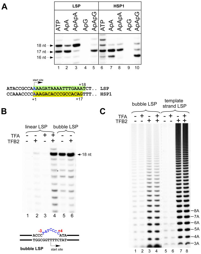
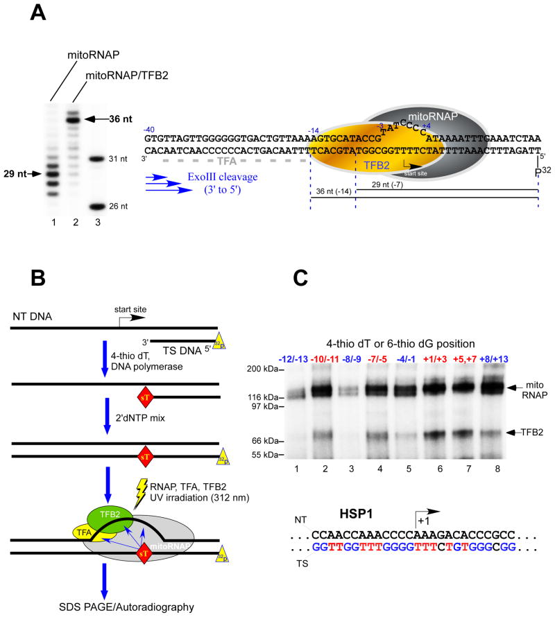
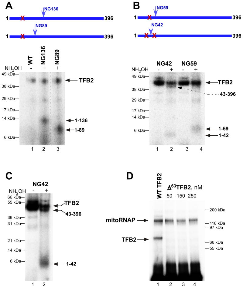
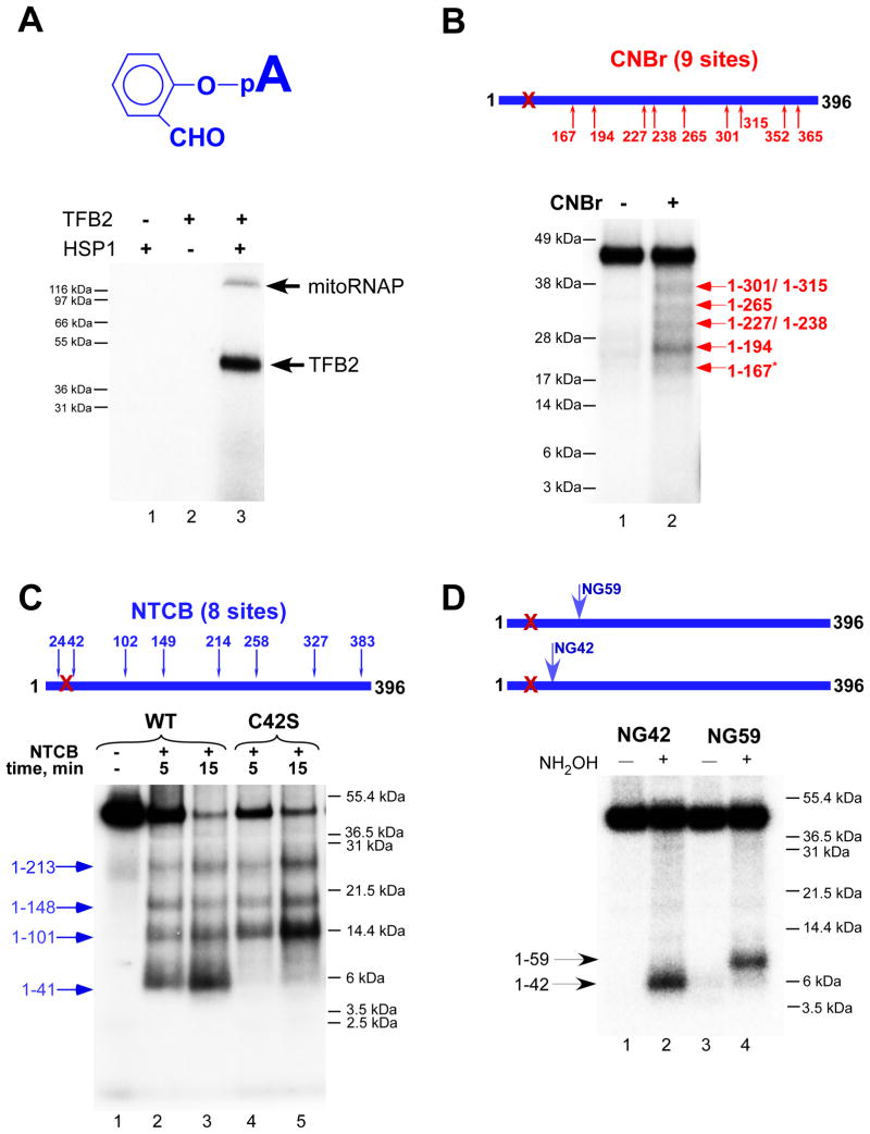
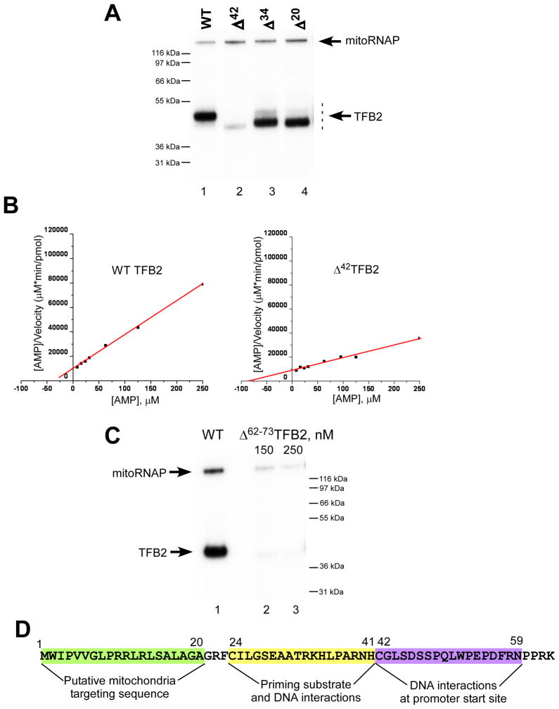
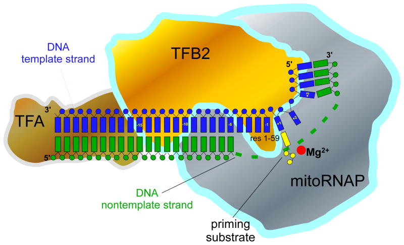
Transcription in human mitochondria is carried out by a single-subunit, T7-like RNA polymerase assisted by several auxiliary factors. We demonstrate that an essential initiation factor, TFB2, forms a network of interactions with DNA near the transcription start site and facilitates promoter melting but may not be essential for promoter recognition. Unexpectedly, catalytic autolabeling reveals that TFB2 interacts with the priming substrate, suggesting that TFB2 acts as a transient component of the catalytic site of the initiation complex. Mapping of TFB2 identifies a region of its N-terminal domain that is involved in simultaneous interactions with the priming substrate and the templating (+1) DNA base. Our data indicate that the transcriptional machinery in human mitochondria has evolved into a system that combines features inherited from self-sufficient, T7-like RNA polymerase and those typically found in systems comprising cellular multi-subunit polymerases, and provide insights into the molecular mechanisms of transcription regulation in mitochondria.


Figures

Figure 1
Figure 1. TFB2 is required for promoter melting
A. Top panel - Human mitochondrial in vitro transcription system. Transcription was performed with the nucleotide sets lacking CTP (LSP) or UTP (HSP1) for 30 min at 35°C. RNA transcripts in reactions using ATP (lanes 1 and 6) were treated with phosphatase to match their mobility to the mobility of the transcripts generated by incorporation of the RNA primers. Transcripts obtained using ApApG primers (lanes 5 and 10) served as the size markers as they could only anneal to the complementary sequence in the promoter DNA and thus generated 17 and 16 nt RNA products on LSP and HSP1, respectively. Lanes 1 and 6 are overexposed to compensate for the more efficient label incorporation in reactions containing RNA primers. Bottom panel - Sequences of the LSP and the HSP1 promoters aligned at their transcription start sites. Shaded boxes indicate transcribed regions in the presence of the limited nucleotide sets as described above. B. MitoRNAP can initiate transcription in the absence of TFA and TFB2 on bubble promoter templates. Transcription assays were performed on linear (lanes 1–4) and bubble (lanes 5,6) templates containing the LSP promoter in the presence of ApApA primer, GTP, ATP and UTP for 30 min. C. TFB2 stimulates A-ladder production on bubble and single-stranded templates. The bubble LSP promoter template (lanes 1–4) and the template strand of the LSP promoter (lanes 5–8) were transcribed in the presence of ATP (0.3 mM) and [α-32P]ATP for 30 min.
Figure 2
Figure 2. TFB2 makes direct interactions with the promoter DNA
A. Exo III footprints of the TFB2/mitoRNAP complex Left panel. The bubble LSP template was prepared by annealing of 32P-labeled template strand with the NT DNA and preincubated with the mitoRNAP (lane 1) or with the TFB2/mitoRNAP complex (lane 2) for 10 min at room temperature prior to the treatment with Exo III as described in Experimental Procedures. Products of the reaction were resolved using 15% PAGE containing 6M urea. DNA size markers (lane 3) were obtained by 5′ end labeling of 26 nt and 31 nt long oligonucleotides having sequence identical to the 5′ end of the template DNA strand. Right panel. Schematics of the mitochondrial IC. Major DNA products (36 nt and 29 nt) generated by Exo III cleavage are indicated with the black lines below the DNA sequence. Grey dash line indicates TFA footprint (−38 to −15) as determined in (Gaspari et al., 2004). B. Schematics of the synthesis of 32P-labeled templates containing photo crosslinking reagents. The 5′ end 32P-labeled DNA primer annealed to the NT DNA strand was extended by DNAP I in the presence of the photo reactive probe (4-thio dTMP). Upon the probe incorporation, the DNAP I was allowed to complete the primer extension by providing dNTPs mixture. The labeled DNA templates were then incubated with mitoRNAP and transcription factors to form the IC, followed by UV irradiation to activate the probe. C. DNA crosslinking of the IC. ICs (50 nM) were UV-irradiated for 10 min at 30°C and the products of the reaction resolved in 4–12 % Bis-Tris MES-SDS gel. Positions of the photoreactive probes, 4-thio dTMP (red letters) and 6-thio dGMP (blue letters) in the template strand of the HSP1 promoter are indicated.
Figure 3
Figure 3. Mapping of TFB2-DNA interactions at the promoter start site
A. The crosslinking region is located in the N-terminal part of TFB2. Crosslinking was performed as described in Experimental Procedures using NG-less mitoRNAP, TBF2 mutants indicated and the HSP1 template containing 4-thio dTMP at positions from +1 to +3 and internal [α-32P] dCMP label. After UV irradiation, the complexes were first treated with DNAse I and then with hydroxylamine for 6h at 43°C, and resolved using 4–12% Bis-Tris MES-SDS gel. B, C. Fine mapping of TFB2 crosslink with hydroxylamine. Crosslinking was performed as above using NG42 and NG59 TFB2 mutants. The products of the hydroxylamine reaction (5h at 43°C) were resolved using 4–12% Bis-Tris MES-SDS (B) or 10% Bis-Tris MES-SDS gel (C). D. Cross-linking efficiency of Δ 63TFB2 mutant. Crosslinking reaction was performed as described above with the WT (lane 1) or Δ 63 TFB2 mutant (lane 2–4). The products of the reaction were treated with DNAse I prior to their resolution in 4–12% Bis-Tris MES-SDS gel.
Figure 4
Figure 4. TFB2 interacts with the priming substrate in the IC
A. Catalytic autolabeling with 2-hydroxybenzaldehyde AMP depends on the functional activity of the IC. Autolabeling with 2-hydroxybenzaldehyde AMP (top panel) was performed with the reaction mixtures that lacked TFB2 (lane 1), DNA template (lane 2) or with the IC (lane 3) after the crosslinking (bottom panel). Reaction products were resolved using 4–12% Bis-Tris MES-SDS gel. B. Mapping of TFB2 crosslink with CNBr. Crosslinking reactions containing WT TFB2 were treated with CNBr for 10 min at 37°C and the products resolved as indicated above. The pattern of labeled bands observed corresponds to a nested set of N-terminal fragments that extend to the CNBr cleavage sites indicated. The asterisk indicates resistant to CNBr cleavage methionine-serine pair. C. Mapping of TFB2 crosslink with NTCB. Crosslinking reactions containing WT (lanes 1–3) or mutant C42S TFB2 (lanes 4,5) were treated with NTCB for the time indicated at 37°C and resolved using SDS PAGE as above. D. Mapping of TFB2 crosslink with hydroxylamine. Crosslinking reactions containing NG59 (lanes 1,2) and NG42 TFB2 mutants (lanes 3,4) were treated with 0.2 M hydroxylamine for 5 h at 43°C. Reaction products were resolved using 10% Bis-Tris MES-SDS gel.
Figure 5
Figure 5. The ICs formed with Δ42TFB2 mutant have low affinity to the priming nucleotide
A. Catalytic autolabeling of TFB2 deletion mutants. Reactions containing WT (lane 1), Δ42 (lane 2), Δ34 (lane 3) and Δ20 (lane 4) TFB2 were resolved using 4–12% Bis-Tris MES-SDS. B. Steady state kinetic experiments. Transcription reactions were performed using AGU-LSP promoter in the presence of 7–1000 μM AMP (priming substrate) and 50 μM GTP to produce pApG transcripts. The data are presented using Hanes-Woolf plots for WT (left panel) and Δ42 (right panel) TFB2. Kmapp was calculated using the data obtained in four independent experiments. C. Catalytic autolabeling using Δ62–73TFB2 mutant. Reactions containing WT (lane 1) and Δ62–73 (lanes 2 and 3) TFB2 were resolved using 4–12% Bis-Tris MES-SDS. D. Schematics of TFB2 interactions due to its unique N-terminal region. The amino acid sequence of the N-terminal part (residues 1–63) of H.s. TFB2 is indicated.
Figure 6
Figure 6
Schematics of the molecular organization of human mitochondrial transcription IC TFB2 contacts promoter DNA near the transcription start site and interacts with TFA, mitoRNAP and the priming nucleotide. The N-terminal region of TFB2 implicated in interactions with the first base pair of the RNA-DNA hybrid is shown in the vicinity of the mitoRNAP active site. We are thankful to Drs William T McAllister and Vadim G Nikiforov for fruitful discussion and critical reading of the manuscript. This work was supported in part by UMDNJ Foundation grant (to D.T.) and by National Institutes of Health Grants GM30317 (to A.M.) and GM38147 (to W.T.M).

Similar articles

Cited by

References

    1. Alam TI, Kanki T, Muta T, Ukaji K, Abe Y, Nakayama H, Takio K, Hamasaki N, Kang D. Human mitochondrial DNA is packaged with TFAM. Nucleic Acids Res. 2003;31:1640–1645. - PMC - PubMed
    1. Amiott EA, Jaehning JA. Mitochondrial transcription is regulated via an ATP “sensing” mechanism that couples RNA abundance to respiration. Mol Cell. 2006a;22:329–338. - PubMed
    1. Amiott EA, Jaehning JA. Sensitivity of the yeast mitochondrial RNA polymerase to +1 and +2 initiating nucleotides. J Biol Chem. 2006b;281:34982–34988. - PubMed
    1. Bonawitz ND, Clayton DA, Shadel GS. Initiation and beyond: multiple functions of the human mitochondrial transcription machinery. Mol Cell. 2006;24:813–825. - PubMed
    1. Chang DD, Clayton DA. Precise identification of individual promoters for transcription of each strand of human mitochondrial DNA. Cell. 1984;36:635–643. - PubMed

Publication types

MeSH terms