Comparative Study
. 2009 Nov 25;64(4):463-70.
doi: 10.1016/j.neuron.2009.10.015.

Classical MHCI molecules regulate retinogeniculate refinement and limit ocular dominance plasticity

Affiliations
Comparative Study

Classical MHCI molecules regulate retinogeniculate refinement and limit ocular dominance plasticity

Akash Datwani et al. Neuron. .

Abstract

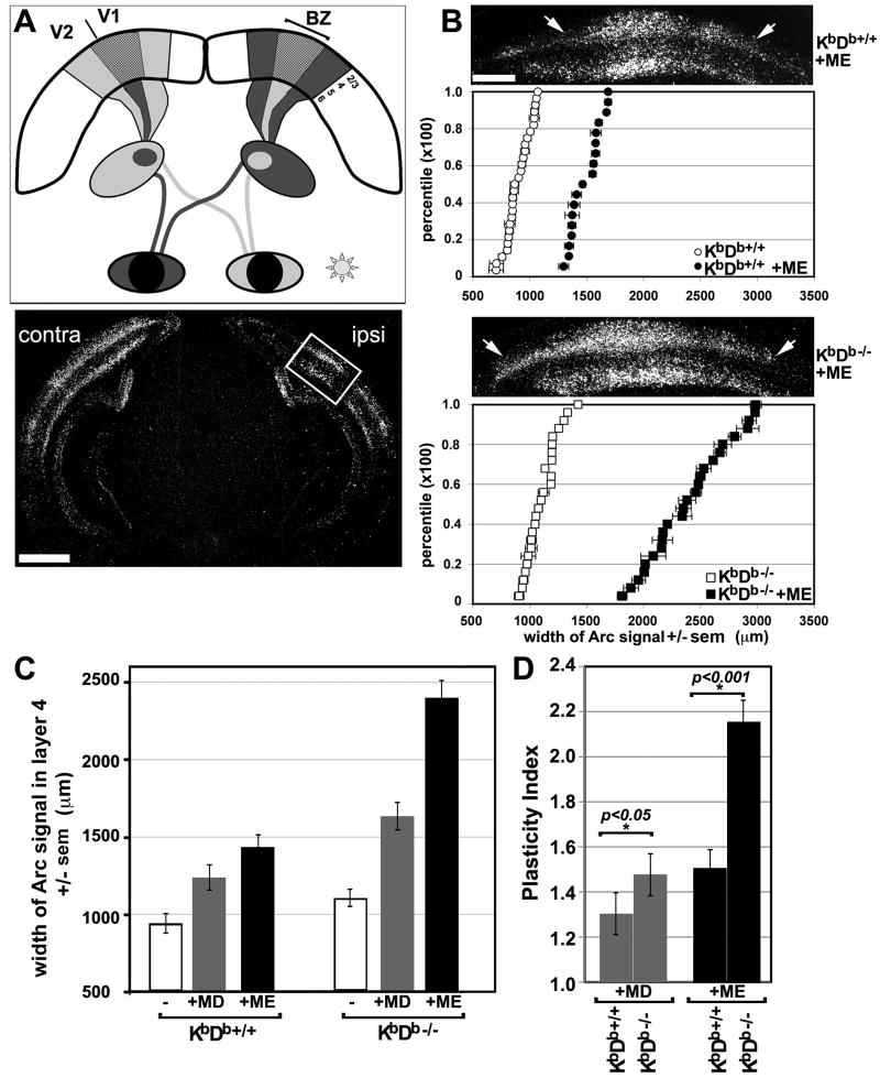
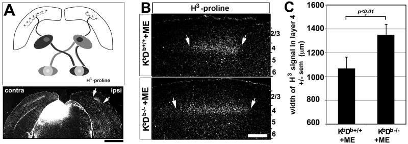
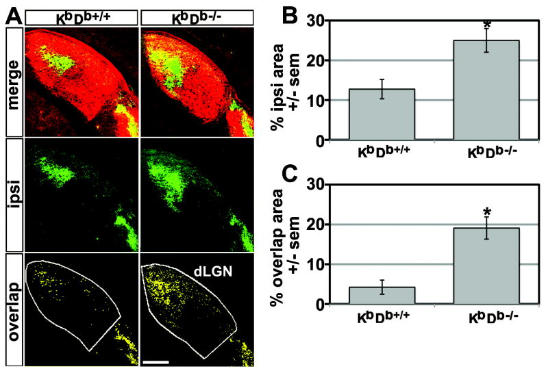
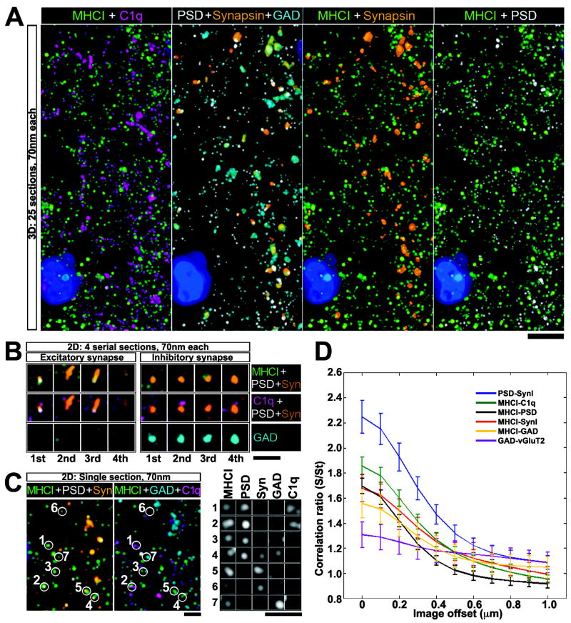
Major histocompatibility complex class I (MHCI) genes were discovered unexpectedly in healthy CNS neurons in a screen for genes regulated by neural activity. In mice lacking just 2 of the 50+ MHCI genes H2-K(b) and H2-D(b), ocular dominance (OD) plasticity is enhanced. Mice lacking PirB, an MHCI receptor, have a similar phenotype. H2-K(b) and H2-D(b) are expressed not only in visual cortex, but also in lateral geniculate nucleus (LGN), where protein localization correlates strongly with synaptic markers and complement protein C1q. In K(b)D(b-/-) mice, developmental refinement of retinogeniculate projections is impaired, similar to C1q(-/-) mice. These phenotypes in K(b)D(b-/-) mice are strikingly similar to those in beta2 m(-/-)TAP1(-/-) mice, which lack cell surface expression of all MHCIs, implying that H2-K(b) and H2-D(b) can account for observed changes in synapse plasticity. H2-K(b) and H2-D(b) ligands, signaling via neuronal MHCI receptors, may enable activity-dependent remodeling of brain circuits during developmental critical periods.


Figures

Figure 1
Figure 1. Enhanced ocular dominance plasticity in visual cortex of KbDb-/- mutant mice
(A) Schematic of mouse visual system. Binocular zone (BZ), located between primary (V1) and secondary (V2) visual cortex, receives input from both eyes. Arc mRNA induction was used to map cortical neurons driven by stimulation of one eye (Experimental procedures and (Tagawa et al., 2005)). Below: darkfield autoradiograph of Arc in situ hybridization in a coronal section from a P34 WT (KbDb+/+) mouse; 30 minutes of visual stimulation upregulates Arc mRNA in neurons of layers 2-4 and 6 of visual cortex (layer 5 neurons only express very low levels of Arc mRNA). Box indicates region of Arc mRNA upregulation driven by ipsilateral eye stimulation within the BZ; broad induction of Arc mRNA is present throughout V1 and V2 contralateral to the stimulated eye, Scale = 900μm. (B) Ipsilateral eye representation in cortex expands more in KbDb-/- than in WT (KbDb+/+) mice following monocular enucleation (ME) during the critical period (P22-31). Top: In situ hybridization for Arc mRNA in KbDb+/+ (upper) and KbDb-/- (lower) visual cortex ipsilateral to the remaining eye; arrows indicate borders of signal in layer 4. Below: cumulative histograms of mean width of Arc induction in layer 4 ± sem for KbDb+/+ (upper) and KbDb-/- (lower) mice reared with normal vision (open symbols) or mice that received ME (filled symbols). Note increased width of Arc induction following ME in both genotypes. (KbDb+/++ME: 1426.3 ± 89.7 μm, n = 18 mice vs. KbDb+/+ normal vision: 945 ± 58.3 μm, n = 2 8 mice; P < 0.05; KbDb-/-+ME: 2404.6 ± 105.1 μm, n = 25 mice; P = 0.0017). Note also that width of Arc induction in KbDb-/- mutant mice reared with normal visual experience (open squares) is slightly larger than that of normally-reared KbDb+/+ mice (open circles): KbDb-/-: 1111 ± 50.8 μm, n = 25 mice; KbDb+/+: 945 ± 58.3 μm n = 2 8 mice. Each symbol represents the average of several scanned sections from a single animal ± sem. Scale = 400μm. (C) Average width of Arc induction in layer 4 for normally reared KbDb+/+ or KbDb-/- mice (open bar) vs. mice receiving monocular visual deprivation from P25-34 (MD: gray bar, n = 7) or from P22-31 (ME: black bar, n = 25). (D) Plasticity Index (see text) reveals greater OD plasticity in KbDb-/- than in KbDb+/+ visual cortex by MD and ME, (*) statistical significance determined by one-way ANOVA. Error bars = standard error of mean (sem) in B and C, and root mean square error (RMSE) in D.
Figure 2
Figure 2. Enhanced thalamocortical plasticity in KbDb-/- mutant mice
(A) Schematic of connections in mouse visual system: thalamocortical axon terminals innervate layer 4 of cortex. Below, transneuronal transport of H3–proline following intraocular injection reveals (age P34; Experimental procedures) a broad contralateral signal (left), and smaller ipsilateral patch (right) in layer 4. Scale=1500μm. (B) Higher magnification of representative sections from KbDb+/+ (top) and KbDb-/- (bottom) mice. Arrows indicate measurement borders used. Scale = 450μm. (C) Population averages for width ± sem of layer 4 label for each genotype after ME. P<0.05; (Mann-Whitney U test).
Figure 3
Figure 3. Incomplete segregation of RGC inputs to dLGN in KbDb-/- mutant mice
(A) RGC projections labeled by intraocular injections of CTB AF594 (red) into the contralateral (contra) eye and CTB AF488 (green) into the ipsilateral (ipsi) eye. Top: Merged fluorescent micrographs of dLGN from KbDb+/+ and KbDb-/- mice at P34. Middle (green pixels): ipsilateral eye projection pattern (intensity threshold = 60% of maximum). Bottom (yellow pixels): overlapping pixels from ipsilateral and contralateral eye projections each at an intensity threshold 60% of maximum (Figure S3 and Experimental procedures). Note ectopic patches of ipsilateral eye projections not eliminated during development in KbDb-/- mice. Scale = 150μm. (B, C) Mean % of dLGN area ± sem of ipsilateral eye projection and mean overlapping pixels in both channels. More ipsilateral territory as well as overlapping pixels are present in mutant dLGN: KbDb+/+ = 5 mice, KbDb-/- = 5 mice, P< 0.05 (two-tailed t-test).
Figure 4
Figure 4. MHCI localization in relation to synaptic proteins during period of retinogeniculate refinement
(A) AT micrographs reconstructed from 25 serial ultrathin sections (each 70nm thick) of P7 LGN showing MHCI immunostaining (green) in relation to known synaptic markers (Syn=synapsin (orange), PSD=postsynaptic density-95 (white), GAD=glutamic acid decarboxylase 65/67 (cyan)), as well as to C1q=complement protein C1q (magenta). DAPI stain of nuclei, blue. Scale = 5μm. (B) Four serial sections of 2 different synapsin positive puncta: Left example is characteristic of an excitatory synapse (close apposition of presynaptic marker synapsin with postsynaptic excitatory synapse marker PSD-95); Right example is characteristic of an inhibitory synapse (overlap of synapsin with GAD and absence of PSD-95). MHCI and C1q are closely associated with both types of synapses. (C) Single ultrathin section, showing colocalization between MHCI puncta and PSD, Synapsin, GAD and C1q. Bottom right, zoomed in view of puncta numbered on left, showing immunofluoresence signal in separate channels, Scale = 2μm. Note colocalization of signal for MHCI, PSD and C1q at puncta #1 and 2. (D) Cross correlations showing pairwise comparisons of degree of spatial overlap between puncta immunostained for the various markers (Experimental procedures and Figure S4 online). Synapsin vs. PSD95 shows strongest correlation, and GAD vs. vGluT2 the weakest. MHCI is more correlated with C1q than with other markers. Si/St = Cr, the correlation ratio between two channels as a multiple of their baseline correlation. Cr = 1 indicates no correlation, Cr ≫ 1 indicates high correlation, Cr < 1 indicates negative correlation.

References

    1. Antonini A, Fagiolini M, Stryker MP. Anatomical correlates of functional plasticity in mouse visual cortex. J Neurosci. 1999;19:4388–4406. - PMC - PubMed
    1. Atwal JK, Pinkston-Gosse J, Syken J, Stawicki S, Wu Y, Shatz C, Tessier-Lavigne M. PirB is a functional receptor for myelin inhibitors of axonal regeneration. Science (New York, N Y. 2008;322:967–970. - PubMed
    1. Bjartmar L, Huberman AD, Ullian EM, Renteria RC, Liu X, Xu W, Prezioso J, Susman MW, Stellwagen D, Stokes CC, et al. Neuronal pentraxins mediate synaptic refinement in the developing visual system. J Neurosci. 2006;26:6269–6281. - PMC - PubMed
    1. Butts DA, Kanold PO, Shatz CJ. A burst-based “Hebbian” learning rule at retinogeniculate synapses links retinal waves to activity-dependent refinement. PLoS biology. 2007;5:e61. - PMC - PubMed
    1. Cang J, Kalatsky VA, Lowel S, Stryker MP. Optical imaging of the intrinsic signal as a measure of cortical plasticity in the mouse. Visual neuroscience. 2005;22:685–691. - PMC - PubMed

Publication types

MeSH terms