Comparative Study
. 2009 Nov 25;64(4):484-97.
doi: 10.1016/j.neuron.2009.09.027.

DSCAM and DSCAML1 function in self-avoidance in multiple cell types in the developing mouse retina

Affiliations
Comparative Study

DSCAM and DSCAML1 function in self-avoidance in multiple cell types in the developing mouse retina

Peter G Fuerst et al. Neuron. .

Abstract

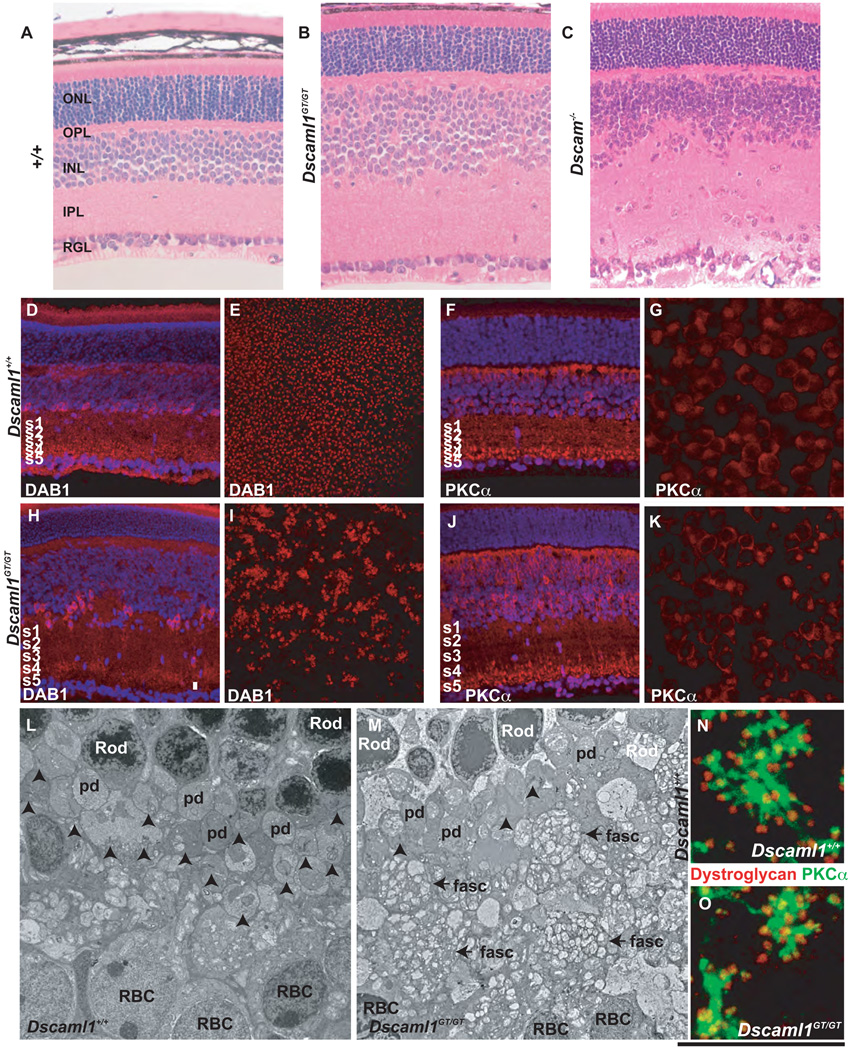
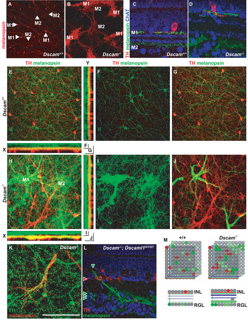
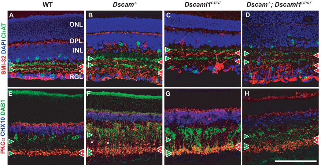
DSCAM and DSCAM-LIKE1 (DSCAML1) serve diverse neurodevelopmental functions, including axon guidance, synaptic adhesion, and self-avoidance, depending on the species, cell type, and gene family member studied. We examined the function of DSCAM and DSCAML1 in the developing mouse retina. In addition to a subset of amacrine cells, Dscam was expressed in most retinal ganglion cells (RGCs). RGCs had fasciculated dendrites and clumped cell bodies in Dscam(-/-) mice, suggesting a role in self-avoidance. Dscaml1 was expressed in the rod circuit, and mice lacking Dscaml1 had fasciculated rod bipolar cell dendrites and clumped AII amacrine cell bodies, also indicating a role in self-avoidance. Neurons in Dscam or Dscaml1 mutant retinas stratified their processes appropriately in synaptic laminae in the inner plexiform layer, and functional synapses formed in the rod circuit in mice lacking Dscaml1. Therefore, DSCAM and DSCAML1 function similarly in self-avoidance, and are not essential for synaptic specificity in the mouse retina.


Figures

Figure 1
Figure 1. Dscam and Dscaml1 are expressed in distinct populations of retinal neurons
A-D, In-situ hybridization was performed using anti-sense Dscam probes at E12.5, E14.5 E17 and P6 (inset in D is dual labeling with probes to Dscam and Brn3b). Labeling was observed in all, or nearly all, cells in the retinal ganglion cell layer (RGL) at all ages, as well as a subset of cells in the inner nuclear layer (INL) at P6. Labeling was not observed in the neuroblast layer (NBL) or outer nuclear layer (ONL). E, In-situ hybridization was performed using anti-sense Dscaml1 probes, at E14.5 no labeling was observed. F, Section of Dscaml1GT/+ retina labeled with β-gal antibodies. Staining was observed in the inner and outer portion of the inner nuclear layer, the outer plexiform layer, and photoreceptors. G and H, Sections of P20 wild type and Dscaml1GT/GT retina were stained with a polyclonal antibody to the extracellular domain of DSCAML1. DSCAML1 immunoreactivity was observed in the outer plexiform layer (OPL) and inner plexiform layer (IPL). DSCAML1 immunoreactivity was not detected in the Dscaml1GT/GT retina. I and J, Section of Dscaml1GT/+ or DscamGT/GT retina stained with antibodies to β-gal and PKCα, a marker of RBCs (I) or DISABLED1 (DAB1), a marker of AII amacrine cells (J). K, Dscam is expressed in retinal ganglion cells and some amacrine cells where as Dscaml1 is expressed in AII amacrine cells, RBCs and photoreceptors. The scale bar in (J) is equivalent to 450 µm in A, 750 µm in B, 900 µm in C, 144 µm in D, 675 µm in E, 108 µm in F, 90 µm in G and H, 79 µm in I and J and 31.6 µm in Ii and 27.6 µm in Ji.
Figure 2
Figure 2. Dscam is required for retinal ganglion cell spatial patterning but does not confer cell type identity
Retinal ganglion cell populations were labeled in adult wild type and Dscam−/− retinas using the antibody SMI-32 to non-phosphorylated neurofilament (A and D; alpha RGCs), anti-melanopsin (B and E; melanopsin-positive RGCs), and the Mito-Y transgene (C and F; Mito-Y positive RGCs). A and D, Alpha RGCs are aggregated in the periphery of the Dscam−/− retina compared to the wild type retina, arrowheads denote cell bodies in the central retina of wild type mice. B and E, Melanopsin-positive RGCs are densely aggregated in the Dscam−/− retina compared to wild type. The dendrites of melanopsin-positive RGCs form fascicles in the Dscam−/− retina, whereas they arborize in wild type. C and F, Mito-Y positive RGCs are aggregated and fasciculated in the Dscam−/− retina. G–L, Pair-wise labeling of adult wild type and Dscam−/− retinas with antibodies to melanopsin, non-phosphorylated neurofilament (SMI-32), and the Mito-Y transgene (N>3). RGC aggregates and fascicles in the Dscam−/− retina were composed primarily of a single RGC type (H, J and L). M, RGC cell types form mosaics in the wild type retina and aggregate and fasciculate with cells of the same type in the Dscam−/− retina. The scale bar in (H) is equivalent to 280 µm in A-–F and 387.5 µm in G–L.
Figure 3
Figure 3. Dscaml1 is required for mosaic patterning of AII amacrine cells and dendritic morphology of rod bipolar cells
A–C, Sections of wild type, Dscaml1GT/GT and Dscam−/− retinas stained with hematoxylin and eosin. The inner nuclear and inner plexiform layers of the Dscaml1GT/GT and Dscam−/− retinas are expanded and disorganized compared to wild type. The Dscam−/− RGL was also disorganized. No histological differences were detected between wild type and Dscaml1GT/+ retinas (data not shown). Sections or whole retinas from wild type and Dscaml1GT/GT mice were stained with antibodies to DAB1, a marker of AII amacrine cells (D, E, H and I) or PKCα, a marker of RBCs (F, G, J and K); (N≥3). D and H, AII amacrine cells in sections of wild type and Dscaml1GT/GT retina. AII amacrine cells are clumped and vertically disorganized in the Dscaml1GT/GT inner nuclear layer compared to the regular distribution in the wild type retina. E and I, AII amacrine cells are distributed in a mosaic across the surface of the inner nuclear layer of the wild type retina, but clump in the Dscaml1GT/GT retina. F and J, Rod bipolar cells in sections of wild type and Dscaml1GT/GT retinas. The Dscaml1GT/GT retina has an increased number of RBCs compared to wild type, but the synaptic termini of both wild type and Dscaml1GT/GT RBCs are stratified in ON vitreal portion of the inner plexiform layer. G and K, No difference was observed when comparing the spacing of Dscaml1GT/GT and wild type RBCs, although they are more abundant in the Dscaml1GT/GT retina. L and M, Transmission electron microscopy was used to examine the structure of the Dscaml1GT/GT outer plexiform layer (N=4 mice of each genotype). The size of photoreceptor peduncles was reduced in the Dscaml1GT/GT outer plexiform layer and fewer ribbon synapses were observed (arrows). Fascicles of rod bipolar dendrites (fasc) were also observed in the mutant retina. N and O, Wild type and Dscaml1GT/GT retinas were stained with antibodies to PKCα and dystroglycan, a marker of the rod presynapse. Rod bipolar cell dendrites extend less far into the OPL in the Dscaml1GT/GT retina. The scale bar in the lower right corner of Figure 3 is equivalent to 156 µm in A–C, 204 µm in D, F, H and J, 664 µm in E and I, 68 µm in G and K, 12.75 mm in L and M and 13.5 µm in N and O.
Figure 4
Figure 4. Analysis of patterning of DA and AII cells
Wild type and Dscaml1GT/GT retinas were stained with antibodies to label DA cells (TH) and AII cells (DAB1). A–C, Spatial organization of DA and AII amacrine cells form mosaics in the wild type retina. AII cells are wrapped by neurites from DA cells (arrows). Images are shown as the merge (A), and the TH (B) and DAB1 (C) signals individually. D–F, AII amacrine cells require Dscaml1GT/GT for normal organization and clump in its absence. DA neurites are absent from areas lacking AII cells (E). G, H, DRP analysis was performed comparing distribution of AII amacrine cells in the wild type (G) and Dscaml1GT/GT retina (H). In the wild type retina an exclusion zone is observed (reduction below average spacing), whereas strong aggregation is observed in the Dscaml1GT/GT retina (above average spacing). I, J, DRP analysis was performed comparing distribution of DA cells in the wild type (I) and Dscaml1GT/GT retina (J). DA cells had an exclusion zone in both wild type and Dscaml1GT/GT retina. The horizontal lines in G-J indicate average cell densities. The scale bar below (F) is equivalent to 132 mm.
Figure 5
Figure 5. Confocal projection of ipRGCs and DA cells
Wild type and Dscam−/− retinas were stained with antibodies to TH and melanopsin to detect DA cells and ipRGCs, respectively. A, Anti-melanopsin stains two populations of ipRGC, bright M1 cells and dim M2 cells in the wild type retina. B, In the Dscam−/− retina M1 and M2 cells aggregate and fasciculate separately. C and D, Cross-sections of wild type and Dscam−/− retina stained with antibodies to melanopsin, TH and ChAT. C, In the wild type retina ChAT labels the neurites of cholinergic amacrine cells in the ON and OFF portion of the INL, while M1 ipRGC dendrites costratify with DA neurites immediately below the INL and M2 dendrites stratify in the ON portion of the INL proximal to the RGL. D, This lamination pattern is preserved in the Dscam−/− retina. E, A confocal Z-projection from the RGL through the IPL to the amacrine cell bodies in the INL in the wild type retina. F, The bottom half of the Z-stack contains ganglion cell bodies and the M2 dendrites in the ON portion of the IPL. G, The top half contains dopaminergic amacrine cell bodies and M1 dendrites. H–J, In the Dscam−/− retina, melanopsin-positive dendrites stratify in two bands, with the IPL proximal band costratifying with DA neurites. I, Dim M2 cells stratify dendrites in the ON portion of the IPL. J, Bright M1 cell dendrites project to the IPL immediately below the INL. K, Cofasciculation of DA cell neurites and M1 dendrites is observed in the Dscam−/− retinas. L, In Dscam−/−; Dscaml1−/− double mutant retinas, ipRGC dendrites are fasciculated in the ON portion of the IPL, while dopaminergic amacrine neurites are fasciculated in the OFF portion of the IPL proximal to the INL; however, colamination in S1 is very disorganized. M, Dscam is required for mosaic patterning of DA and ipRGCs but gross synaptic lamination is intact. The measurement bar in panel (K) is equivalent to 496.8 µm in A and B, 64.1 µm in C and D, 193.75 µm in E-J and 132 µm in K.
Figure 6
Figure 6. Synaptic lamination is preserved in the Dscam or Dscaml1 null retina
A–D, Retina sections were stained with antibodies to ChAT, to detect cholinergic Starburst amacrine cells, and SMI-32, to detect alpha RGCs. The location of ON and OFF cholinergic bands are used to demarcate synaptic layers S2 and S4. Paired cholinergic bands (green arrowheads) were observed in wild type and mutant retinas, although their lamination was not as compact in the Dscam−/−; Dscaml1−/− retina. A small number of cholinergic amacrine cell neurites were laminated in an ectopic plexiform layer within the INL of the Dscam−/−; Dscaml1−/− retina. Alpha RGCs stratify dendrites on the RGL proximal side of cholinergic bands (read arrow heads). Stratification proximal to cholinergic bands was observed in all genotypes, with dendrite fascicles observed in the ON portion of the Dscam−/− and Dscam−/−; Dscaml1−/− retina. E–H, Retina sections were stained with antibodies to CHX10, to label bipolar cells, PKCa, to label RBCs and DAB1, to label AII amacrine cells. The axon terminals of RBCs were laminated in S4-S5 of all genotypes. Varicosities were observed in RBC axons in the Dscam−/− and Dscam−/−; Dscaml1−/− retina. AII neurites were stratified in S1/S2 and S4/S5 of all genotypes and also colaminated with the axonal terminals of RBCs in all genotypes. AII amacrine cells were observed to stratify neurites in an ectopic plexiform layer within the INL and to also project neurites past RBC terminals, towards the RGL in the Dscam−/−; Dscaml1−/− retina. The scale bar in (H) is equivalent to 134.6 mm for all panels except D, and H, in which it is equivalent to 123 mm.
Figure 7
Figure 7. Rod bipolar and AII amacrine cells remain synaptically coupled in the Dscaml1 null retina
A, Schematic of the rod visual circuit. Light responses in the rods leads to synaptic activation of RBCs. Rod bipolar cells form a dyad synapse with A17 amacrine cells, which are inhibitory, and AII amacrine cells. AII amacrine cells form an inhibitory glycinergic synapse with OFF cone bipolar cells and RGCs, and gap junction connections with ON cone bipolar cell terminals, which transmit visual information to ON RGCs. Dscaml1 is expressed in three cell types within this circuit; rods, RBCs, and AII amacrine cells. B, C, Sections of wild type and Dscaml1GT/GT retina were stained with antibodies to disabled (DAB1), to label AII amacrine cells, and PKCα, to label RBCs (N=3). Colamination of RBC axonal terminals and AII amacrine cell neurites was observed in the innerplexiform layer proximal to the retinal ganglion cell layer. Colamination was preserved in the Dscaml1GT/GT retina, even in rare cases where the rod bipolar axon terminals were ectopically localized within the inner plexiform layer. D, E, Wild type and Dscaml1GT/GT retinas were imaged by transmission electron microscopy (N=4). Rod bipolar cells, AII amacrine cells and A17 amacrine cells form a three-part synapse, the dyad synapse, which was observed in both wild type and Dscaml1GT/GT retinas. Dscaml1GT/GT synapses had an over abundance of vesicles and rudimentary synaptic ribbons in bipolar cell terminals. F, Photomicrograph of paired recording shown with fluorescence images of the rod bipolar (green) and AII (red) superimposed over a transmitted light image of the retinal slice. In the wild type retina, depolarization of the presynaptic rod bipolar from −50 to −10 mV elicits an inward Ca current (i) and evokes an EPSC in the postsynaptic AII (ii is an individual EPSC; iii is the average of 7 responses). G, The same paired recording analyses performed in Dscaml1GT/GT mice, again showing the presynaptic bipolar cell Ca current (i), an individual EPSC (ii), and the average EPSC (iii), note the slower decay phase of the postsynaptic response. The scale bar below panel (E) is equivalent to 76 µm in B and C and 1.2 µm in D and E, 32.4 µm.
Figure 8
Figure 8. Rods and rod bipolar cells remain synaptically coupled in the Dscaml1GT/GT retina
A, B, Sections of wild type and Dscaml1GT/GT retinas were stained with antibodies to PKCα, to label RBCs, Bassoon, a marker of the rod presynapse, and Cacna1s, a component of the RBC postsynapse. Presynaptic and postsynaptic markers were paired in both the wild type and Dscaml1GT/GT retina, although the number of synapses was reduced in the mutant retina. C, D, Sections of wild type and Dscaml1GT/GT retinas were stained with antibodies to PKCα, to label RBCs, and calbindin a marker of horizontal cells (N=2). The dendrites of both wild type and Dscaml1GT/GT RBCs colaminated with horizontal cell axons. E–H, Transmission electron microscopy was used to examine the anatomy of the Dscaml1GT/GT ribbon synapses. Examples of ribbon synapses from wild type (E and F) and Dscaml1GT/GT (G and H) retinas showing the photoreceptor ribbon (R) apposed to a horizontal cell (H) and invaginating bipolar cells (B, typically an ON bipolar dendrite flanked by darker OFF bipolar processes). An increase in floating ribbons was seen in the mutant (H). I–K, Electroretinography (ERG) was performed on wild type, Dscaml1GT/GT, and Dscam−/− mice (N≥5). Rod bipolar cells receive input from rods in the Dscaml1GT/GT mice, as demonstrated by the robust B wave, reflecting postsynaptic bipolar cell activation, which had a greater amplitude compared to wild type and Dscam−/− mice. The scale bar below panel (H) is equivalent to 32.4 µm in A and B, 40.9 µm in C and D and 478 nm in E–H.

Comment in

References

    1. Agarwala KL, Ganesh S, Tsutsumi Y, Suzuki T, Amano K, Yamakawa K. Cloning and functional characterization of DSCAML1, a novel DSCAM-like cell adhesion molecule that mediates homophilic intercellular adhesion. Biochem Biophys Res Commun. 2001;285:760–772. - PubMed
    1. Amano K, Fujii M, Arata S, Tojima T, Ogawa M, Morita N, Shimohata A, Furuichi T, Itohara S, Kamiguchi H, et al. DSCAM deficiency causes loss of pre-inspiratory neuron synchroneity and perinatal death. J Neurosci. 2009;29:2984–2996. - PMC - PubMed
    1. Andrews GL, Tanglao S, Farmer WT, Morin S, Brotman S, Berberoglu MA, Price H, Fernandez GC, Mastick GS, Charron F, Kidd T. Dscam guides embryonic axons by Netrin-dependent and -independent functions. Development. 2008;135:3839–3848. - PMC - PubMed
    1. Chen BE, Kondo M, Garnier A, Watson FL, Puettmann-Holgado R, Lamar DR, Schmucker D. The molecular diversity of Dscam is functionally required for neuronal wiring specificity in Drosophila. Cell. 2006;125:607–620. - PubMed
    1. Coombs J, van der List D, Wang GY, Chalupa LM. Morphological properties of mouse retinal ganglion cells. Neuroscience. 2006;140:123–136. - PubMed

Publication types